Podcast
Questions and Answers
What is the correct order of development after fertilization?
What is the correct order of development after fertilization?
- Morula, blastocyst, gastrula, bilaminar disc (correct)
- Zygote, morula, blastocyst, bilaminar disc
- Zygote, morula, gastrula, embryo
- Morula, blastocyst, bilaminar disc, gastrula
Which structure forms as a longitudinal groove within the epiblast during gastrulation?
Which structure forms as a longitudinal groove within the epiblast during gastrulation?
- Amnion
- Chorion
- Primitive streak (correct)
- Neural folds
What is the role of the neural crest during embryonic development?
What is the role of the neural crest during embryonic development?
- Contributing to structures like the brain
- Emerging at the junction with surface ectoderm (correct)
- Differentiating into the three germ cell layers
- Forming the bilateral elevations called neural folds
When does gastrulation occur in humans?
When does gastrulation occur in humans?
Which cells migrate towards the primitive streak to form the mesoderm?
Which cells migrate towards the primitive streak to form the mesoderm?
What is the purpose of the bilaminar disc during embryonic development?
What is the purpose of the bilaminar disc during embryonic development?
What determines the size of the lens vesicle?
What determines the size of the lens vesicle?
What can cause the induction of a smaller lens vesicle?
What can cause the induction of a smaller lens vesicle?
What is the lining of the lens vesicle made up of?
What is the lining of the lens vesicle made up of?
Which structure promotes primary lens fiber formation?
Which structure promotes primary lens fiber formation?
What is responsible for the regression of the hyaloid artery?
What is responsible for the regression of the hyaloid artery?
Which clinical anomaly arises from incomplete regression of the hyaloid artery?
Which clinical anomaly arises from incomplete regression of the hyaloid artery?
Which cell type contributes to the formation of the corneal endothelium?
Which cell type contributes to the formation of the corneal endothelium?
What is the role of tight junctions formed by the corneal endothelium?
What is the role of tight junctions formed by the corneal endothelium?
Which structure surrounds and nourishes the lens during early gestation in dogs?
Which structure surrounds and nourishes the lens during early gestation in dogs?
What contributes to the formation of the anterior chamber, including the pupillary membrane?
What contributes to the formation of the anterior chamber, including the pupillary membrane?
From which layer of the optic cup do the iris and ciliary body develop?
From which layer of the optic cup do the iris and ciliary body develop?
What is responsible for the folding of the inner layer of the optic cup, representing early anterior ciliary processes?
What is responsible for the folding of the inner layer of the optic cup, representing early anterior ciliary processes?
What is the main result of the differential growth during the last trimester of human gestation?
What is the main result of the differential growth during the last trimester of human gestation?
Which cells develop first in the retina according to the text?
Which cells develop first in the retina according to the text?
What is the function of primary vitreous according to the text?
What is the function of primary vitreous according to the text?
What happens at around day 45 of gestation in dogs regarding retinal development?
What happens at around day 45 of gestation in dogs regarding retinal development?
Which tissue/tissues are derived from neural crest tissues according to the text?
Which tissue/tissues are derived from neural crest tissues according to the text?
What is one important role of retinal pigment epithelium (RPE) in development?
What is one important role of retinal pigment epithelium (RPE) in development?
Where do neural crest cells migrate beneath during the eye development process?
Where do neural crest cells migrate beneath during the eye development process?
What is the role of the mesoderm in eye development according to the text?
What is the role of the mesoderm in eye development according to the text?
What structure arises from the optic vesicle within the forebrain during eye development?
What structure arises from the optic vesicle within the forebrain during eye development?
What may cause colobomas in the iris, choroid, or optic nerve during eye development?
What may cause colobomas in the iris, choroid, or optic nerve during eye development?
Which structure plays a significant role in determining the size of the palpebral fissure and orbital structures?
Which structure plays a significant role in determining the size of the palpebral fissure and orbital structures?
What develops from mesenchyme in the optic fissure around day 25 of gestation?
What develops from mesenchyme in the optic fissure around day 25 of gestation?
What is the precursor to the vitreous base and lens zonules?
What is the precursor to the vitreous base and lens zonules?
Which component has been identified in developing zonules and Descemet's membrane?
Which component has been identified in developing zonules and Descemet's membrane?
What contributes to lens expansion?
What contributes to lens expansion?
What can lead to a flattened area inaccurately referred to as a lens coloboma?
What can lead to a flattened area inaccurately referred to as a lens coloboma?
What does Doppler ultrasound biomicroscopy document related to hyaloid regression between birth and postnatal day 13 in mice?
What does Doppler ultrasound biomicroscopy document related to hyaloid regression between birth and postnatal day 13 in mice?
Where do axons from developing ganglion cells traverse during their development?
Where do axons from developing ganglion cells traverse during their development?
What is Bergmeister's papilla a remnant of?
What is Bergmeister's papilla a remnant of?
Where do glial cells form a sheath as they surround the hyaloid artery?
Where do glial cells form a sheath as they surround the hyaloid artery?
What initiates myelinization of the optic nerve and progresses toward the eye?
What initiates myelinization of the optic nerve and progresses toward the eye?
What is the origin of glial cells surrounding the optic nerve and forming part of the lamina cribrosa?
What is the origin of glial cells surrounding the optic nerve and forming part of the lamina cribrosa?
Flashcards are hidden until you start studying
Study Notes
Tertiary Vitreous
- Forms as a thick accumulation of collagen fibers between the lens equator and the optic cup, known as Drualt's bundle
- Precursor to the vitreous base and lens zonules
- Early lens zonular fibers appear to be continuous with the inner membrane of the non-pigmented epithelial layer
- Elastin and emulin have been identified in developing zonules and Descemet's membrane
- Traction of zonules contributes to lens expansion, and localized absence of zonules can lead to a flattened area of the lens, inaccurately referred to as a lens coloboma
Optic Nerve
- Axons from developing ganglion cells traverse through vacuolated cells in the inner wall of the optic stalk
- Glial cells form a sheath around the hyaloid artery
- As the artery regresses, the space between it and the glial sheath expands
- Bergmeister's papilla is a remnant of glial cells around the hyaloid artery
- Glial cells migrate into the optic nerve and establish the primitive optic disc
- Glial cells surrounding the optic nerve and those forming part of the lamina cribrosa originate from the inner layer of the optic stalk, derived from neural ectoderm
- Subsequently, a mesenchymal portion of the lamina cribrosa develops from neural crest origin
- Myelinization of the optic nerve initiates at the chiasm, progresses toward the eye, and reaches the optic disc after birth
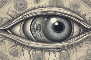
Ocular Embryology and Congenital Malformations
- Gastrulation and neurulation:
- Fertilization → zygote → morula (cluster of 12-16 cells)
- Morula becomes blastocyst, which contains a fluid-filled cavity
- Cells of the blastocyst form embryo proper and extraembryonic tissues (amnion and chorion)
- Embryo bilaminar disc: hypoblast and epiblast
- Gastrulation: process where blastocyst becomes gastrula, forming mesodermal germ layer
- Day 10 in dogs, day 7 in mice, and day 15-20 in humans
- Primitive streak forms as a longitudinal groove within epiblast (future ectoderm)
- Epiblast cells migrate towards primitive streak, invaginate to form mesoderm
- Results in the 3 classic germ cell layers: Ectoderm, mesoderm, and endoderm
- Gastrulation proceeds in cranial-to-caudal progression, with cranial surface ectoderm proliferating to form bilateral elevations called neural folds (forming the brain)
Formation of the Optic Vesicle and Optic Cup
- Optic vesicle development starts with the emergence of optic sulci, which are paired outgrowths of the forebrain neural ectoderm
- Optic sulci are paired evaginations of forebrain neural ectoderm, around day 13 of gestation in dogs
- Concurrent with the closure of the neural tube, the optic sulci transform into optic vesicles
- Optic vesicle enlarges and approaches the basal lamina underlying the surface ectoderm
- Optic vesicle and optic stalk invaginate through growth and infolding
- Surface ectoderm in contact with optic vesicle thickens to form lens placode, which then invaginates with underlying neural ectoderm
Lens Formation
- Surface ectoderm becomes competent to respond to lens inducers
- Anterior neural plate produces inductive signals to give this area of ectoderm a "lens-forming bias"
- Optic vesicle then adheres to lens placode, ensuring alignment of lens and retinal in the visual axis
- Lens placode invaginates, forming a hollow sphere = lens vesicle
- Size of lens vesicle is determined by contact area of optic vesicle with surface ectoderm and ability of surface ectoderm to respond to induction of growth signals
- Abnormal orientation of optic vesicle can cause induction of a smaller lens vesicle
- Lens vesicle detachment leads to anterior chamber formation
- Induction of small lens vesicle that does not separate normally from surface ectoderm can lead to anterior segment dysgenesis
Vascular Development
- Hyaloid artery is the termination of the primitive ophthalmic artery, which branches off the internal ophthalmic artery
- Remains within the optic cup following closure of the optic fissure
- Branches round the posterior lens capsule and anastomoses with vessels in the pupillary membrane
- Hyaloid vascular network forms around the lens, providing nutrition to the lens and anterior segment
- Venous drainage occurs near the equatorial lens area, with no discrete hyaloid vein present
- Ciliary body begins to produce aqueous humor, which nourishes the lens, and the hyaloid artery starts to regress
- Regression of the hyaloid vasculature allows for the development of retinal vessels, primarily mediated by vascular endothelial growth factor (VEGF)-A
Development of the Cornea and Anterior Chamber
- After detachment of the lens vesicle, the anterior margins of the optic cup extend beneath the surface ectoderm, initiating corneal development
- Surface ectoderm above the optic cup secretes a thick matrix (primary stroma)
- Mesenchymal neural crest cells migrate between the surface ectoderm and the optic cup, using the basal lamina of the lens vesicle as a substrate
- Induction of corneal endothelium requires the presence of an adjacent lens vesicle
- Dehydration leads to a 50% reduction in stromal thickness, aided by the corneal endothelium, which develops tight junctions and forms Descemet's membrane
- Transparency of the cornea is achieved by the end of gestation in dogs, with subsequent postnatal changes in corneal thickness observed after eyelid opening
Development of the Iris, Ciliary Body, and Iridocorneal Angle
- The two layers of the optic cup are neuroectoderm in origin
- The iris and ciliary body develop from the anterior aspect of the optic cup, while the retina develops from the posterior aspect
- Optic vesicle is organized with all cell apices directed towards the center, so during invagination, apices of inner and outer epithelial layers become adjacent
- Both pigmented and nonpigmented epithelial cells develop apical cilia and show increased Golgi complexes and vesicles around day 40 of gestation in dogs
- Nonpigmented and pigmented epithelium of the iris and ciliary body originates from the neural ectoderm of the optic cup
- Stroma comes from neural crest cell origin (mesenchymal tissue)
- Differential growth of optic cup epithelial layers causes folding of the inner layer, representing early anterior ciliary processes
Retina and Optic Nerve Development
- The infolding of the neuroectodermal optic vesicle leads to the formation of a bilayered optic cup
- The outer layer of the optic cup, which originates from the optic vesicle, becomes cuboidal and develops melanin granules, forming the retinal pigment epithelium (RPE)
- The inner layer of the optic cup gives rise to the neurosensory retina
- Bruch's membrane, the basal lamina of the RPE, appears during this time, coinciding with the development of the choriocapillaris
- RPE cells adopt a hexagonal shape by day 45 in dogs and develop microvilli that interdigitate with projections from photoreceptors
- At the time of lens placode induction, the retinal primordium consists of an outer nuclear zone and an inner marginal (anuclear) zone
- Retinal histiogenesis involves cell proliferation in the nuclear zone, migration into the marginal zone, and differentiation into inner and outer neuroblastic layers
Studying That Suits You
Use AI to generate personalized quizzes and flashcards to suit your learning preferences.