Podcast
Questions and Answers
Which part of the gastrointestinal tract is derived from the foregut?
Which part of the gastrointestinal tract is derived from the foregut?
- Duodenum (correct)
- Appendix
- Ascending colon
- Rectum
The midgut is supplied by which artery?
The midgut is supplied by which artery?
- Superior mesenteric artery (correct)
- Renal artery
- Celiac artery
- Inferior mesenteric artery
The esophagus develops primarily from the ventral portion of the respiratory diverticulum.
The esophagus develops primarily from the ventral portion of the respiratory diverticulum.
False (B)
What are the two main parts of the stomach formed during its development?
What are the two main parts of the stomach formed during its development?
The liver and gall bladder develop from an outgrowth of __________ at the distal end of the foregut.
The liver and gall bladder develop from an outgrowth of __________ at the distal end of the foregut.
Match the following structures to their respective developmental origin:
Match the following structures to their respective developmental origin:
What is the function of the lesser omentum?
What is the function of the lesser omentum?
Which ligament connects the liver and the abdominal wall?
Which ligament connects the liver and the abdominal wall?
The duodenum is primarily formed from the midgut.
The duodenum is primarily formed from the midgut.
Flashcards
Foregut development
Foregut development
Development of the part of the gut supplied by the Celiac artery, including esophagus, stomach, pancreas, liver, and proximal duodenum.
Midgut development
Midgut development
Development of the part of the gut supplied by the Superior mesenteric artery, including parts of the small and large intestine.
Hindgut development
Hindgut development
Development of the part of the gut supplied by the Inferior mesenteric artery, including parts of the large intestine.
Esophagus development stages
Esophagus development stages
Signup and view all the flashcards
Stomach rotation
Stomach rotation
Signup and view all the flashcards
Duodenum formation
Duodenum formation
Signup and view all the flashcards
Liver development
Liver development
Signup and view all the flashcards
Pancreas development
Pancreas development
Signup and view all the flashcards
Lesser Omentum
Lesser Omentum
Signup and view all the flashcards
Study Notes
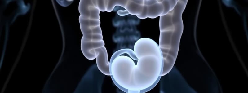
Embryology of the Gastrointestinal Tract (GIT)
- GIT originates from the endoderm.
- Divided into four main subdivisions:
- Pharyngeal gut (buccopharyngeal membrane to tracheobronchial diverticulum)
- Foregut
- Midgut
- Hindgut
Foregut Development
- Supplied by the Coeliac artery.
- Forms key organs including:
- Lung buds
- Esophagus
- Stomach
- Pancreas
- Liver and extrahepatic biliary system
- Proximal duodenum to the bile duct entrance
Midgut Development
- Supplied by the Superior mesenteric artery.
- Includes structures such as:
- Duodenum (distal to bile duct entrance)
- Ascending colon
- Jejunum
- Ileum
- Caecum
- Appendix
- Proximal two-thirds of the transverse colon
Hindgut Development
- Supplied by the Inferior mesenteric artery.
- Comprises:
- Distal one-third of the transverse colon to upper anal canal
- Descending colon
- Sigmoid colon
- Rectum
- Upper part of anal canal
Esophagus Development
- Begins development in the 4th week.
- Respiratory diverticulum appears at the foregut's ventral wall.
- Esophagotracheal septum partitions the respiratory diverticulum into dorsal (esophagus) and ventral portions.
Esophagus Structure
- Initially short, elongates as heart and lungs descend.
- Consists of a muscular coat:
- Upper two-thirds: striated muscle
- Lower one-third: smooth muscle
- Innervation from vagus nerve and splanchnic plexus.
Duodenum Formation
- Represents the terminal part of the foregut and cephalic part of the midgut.
- C-shaped due to stomach rotation, moving to the right side of the abdominal cavity.
Stomach Development
- Appears in the 4th week as fusiform dilation of foregut.
- Undergoes two rotations:
- 90° clockwise around its longitudinal axis.
- Key structures formed:
- Greater curvature (dorsal side)
- Lesser curvature (ventral side)
- Innervation follows stomach rotation: left vagus becomes anterior, right vagus becomes posterior.
Stomach Attachments
- Attached to the ventral and dorsal walls via:
- Ventral mesogastrium (forming lesser omentum)
- Dorsal mesogastrium (forming gastrosplenic and lienorenal ligaments)
Lesser Omentum
- Connects liver to lesser curvature of the stomach and duodenum.
- Contains important structures such as the common bile duct and hepatic artery.
Falciform Ligament
- Connects liver to the abdominal wall.
- Free margin contains umbilical vein, which obliterates postnatally to become round ligament of the liver (ligamentum teres).
Liver and Gallbladder Development
- Begins in the 3rd week as an outgrowth from endodermal epithelium at the foregut’s distal end.
- Liver bud penetrates the septum transversum and divides into:
- Pars cystica (forms gallbladder)
- Pars hepatica (forms liver)
- Proximal portions form bile duct.
Pancreas Development
- Develops from the endodermal lining of the duodenum.
- Consists of two buds:
- Dorsal pancreatic duct (in dorsal mesentery)
- Ventral pancreatic duct
- Rotation of the duodenum causes the ventral duct to move dorsally.
Pancreas Structure
- Main pancreatic duct formed from the proximal ventral duct and distal dorsal duct.
- Ventral pancreatic duct forms the uncinate process and inferior head of the pancreas.
- Proximal duct may obliterate or persist as the accessory pancreatic duct of Santorini.
Studying That Suits You
Use AI to generate personalized quizzes and flashcards to suit your learning preferences.


