Podcast
Questions and Answers
¿Cuál de las siguientes estructuras se origina del tronco arterioso?
¿Cuál de las siguientes estructuras se origina del tronco arterioso?
- Ventrículo derecho
- Tronco de la arteria pulmonar (correct)
- Seno venoso
- Aurícula izquierda
¿Qué forma el ventrículo primitivo principalmente?
¿Qué forma el ventrículo primitivo principalmente?
- El ventrículo izquierdo (correct)
- La aurícula derecha
- El tronco pulmonar
- El ventrículo derecho
¿Cuál es la función de la región del bulbo cordis?
¿Cuál es la función de la región del bulbo cordis?
- Formar el infundíbulo del ventrículo derecho (correct)
- Conectar las aurículas con los ventrículos
- Generar el conducto arterioso
- Formar el seno venoso
¿De dónde se origina la aorta descendente?
¿De dónde se origina la aorta descendente?
¿Qué estructura forma el aurícula primitiva?
¿Qué estructura forma el aurícula primitiva?
¿Qué arterias se forman a partir del 1er arco aórtico?
¿Qué arterias se forman a partir del 1er arco aórtico?
¿Cuál de las siguientes afirmaciones es correcta sobre el 4to arco aórtico?
¿Cuál de las siguientes afirmaciones es correcta sobre el 4to arco aórtico?
¿De qué se deriva el conducto arterioso?
¿De qué se deriva el conducto arterioso?
¿Qué estructura no se forma a partir de los arcos aórticos?
¿Qué estructura no se forma a partir de los arcos aórticos?
¿Qué arco aórtico da origen a la arteria carótida común primitiva?
¿Qué arco aórtico da origen a la arteria carótida común primitiva?
¿Cuál es la función principal del conducto venoso en el sistema circulatorio embrionario?
¿Cuál es la función principal del conducto venoso en el sistema circulatorio embrionario?
¿Qué estructura del corazón se relaciona directamente con el foramen oval?
¿Qué estructura del corazón se relaciona directamente con el foramen oval?
¿Qué tipo de sangre es transportada a través de la vena umbilical?
¿Qué tipo de sangre es transportada a través de la vena umbilical?
¿Cuál de las siguientes estructuras no está involucrada en el flujo de sangre en el sistema circulatorio embrionario?
¿Cuál de las siguientes estructuras no está involucrada en el flujo de sangre en el sistema circulatorio embrionario?
¿Qué ocurre con el conducto venoso después del nacimiento?
¿Qué ocurre con el conducto venoso después del nacimiento?
¿Qué porción del septo interventricular es más pequeña?
¿Qué porción del septo interventricular es más pequeña?
¿Cuál es la función principal del septo aortopulmonar?
¿Cuál es la función principal del septo aortopulmonar?
¿De qué componente se deriva la porción muscular del septo interventricular?
¿De qué componente se deriva la porción muscular del septo interventricular?
¿Qué estructura se forma por la fusión de las crestas truncales?
¿Qué estructura se forma por la fusión de las crestas truncales?
¿Cuál es el origen de las válvulas semilunares?
¿Cuál es el origen de las válvulas semilunares?
¿Cuál es el primer centro hematopoyético que se desarrolla durante el embarazo?
¿Cuál es el primer centro hematopoyético que se desarrolla durante el embarazo?
¿Cuál de las siguientes estructuras NO se forma a partir del mesodermo esplácnico?
¿Cuál de las siguientes estructuras NO se forma a partir del mesodermo esplácnico?
¿Cuál es el gen maestro implicado en el desarrollo del corazón?
¿Cuál es el gen maestro implicado en el desarrollo del corazón?
En qué momento comienza la circulación intraembrionaria del embrión?
En qué momento comienza la circulación intraembrionaria del embrión?
¿Qué parte del corazón es responsable de la formación del seno venoso?
¿Qué parte del corazón es responsable de la formación del seno venoso?
¿Cuál es la función principal del campo cardiogénico secundario?
¿Cuál es la función principal del campo cardiogénico secundario?
¿Qué estructura se forma a partir de la fusión de los centros cardiogénicos primario y secundario?
¿Qué estructura se forma a partir de la fusión de los centros cardiogénicos primario y secundario?
¿Cuál es la estructura que transporta sangre oxigenada desde la placenta al seno venoso?
¿Cuál es la estructura que transporta sangre oxigenada desde la placenta al seno venoso?
¿Cuál de las siguientes estructuras ocupa la posición más craneal en el tubo cardíaco endotelial?
¿Cuál de las siguientes estructuras ocupa la posición más craneal en el tubo cardíaco endotelial?
¿Qué estructura se encuentra entre el endocardio y el miocardio en el corazón en desarrollo?
¿Qué estructura se encuentra entre el endocardio y el miocardio en el corazón en desarrollo?
¿Qué ocurre con el mesocardio dorsal durante el plegamiento del tubo cardíaco?
¿Qué ocurre con el mesocardio dorsal durante el plegamiento del tubo cardíaco?
¿Qué parte del corazón se forma a partir de las células mesoteliales del órgano proepicárdico?
¿Qué parte del corazón se forma a partir de las células mesoteliales del órgano proepicárdico?
¿Cómo contribuyen los pliegues del embrión en el desarrollo del corazón?
¿Cómo contribuyen los pliegues del embrión en el desarrollo del corazón?
¿De qué elementos se forman los cojines endocárdicos?
¿De qué elementos se forman los cojines endocárdicos?
Durante el desarrollo del corazón, ¿hacia dónde se mueve el bulbo cordis?
Durante el desarrollo del corazón, ¿hacia dónde se mueve el bulbo cordis?
¿Cuál es la característica notable de la convexidad del lazo cardíaco durante su formación?
¿Cuál es la característica notable de la convexidad del lazo cardíaco durante su formación?
¿Cuál es la función del septum primum en el desarrollo cardíaco?
¿Cuál es la función del septum primum en el desarrollo cardíaco?
¿Dónde se localiza el foramen primum?
¿Dónde se localiza el foramen primum?
¿Cuál es la principal diferencia entre el septum primum y el septum secundum?
¿Cuál es la principal diferencia entre el septum primum y el septum secundum?
¿Qué proceso es necesario para la formación del foramen secundum?
¿Qué proceso es necesario para la formación del foramen secundum?
Flashcards
Umbilical Vein
Umbilical Vein
Carries oxygen-rich blood from the placenta to the fetus.
Venous Duct Function
Venous Duct Function
Shunts blood from the umbilical vein to the inferior vena cava, bypassing the liver.
Venous Duct Location
Venous Duct Location
Located in the liver.
Foramen Ovale
Foramen Ovale
Signup and view all the flashcards
Ductus Arteriosus
Ductus Arteriosus
Signup and view all the flashcards
Umbilical Arteries
Umbilical Arteries
Signup and view all the flashcards
Inferior Vena Cava
Inferior Vena Cava
Signup and view all the flashcards
Ascending Aorta
Ascending Aorta
Signup and view all the flashcards
Pulmonary Artery
Pulmonary Artery
Signup and view all the flashcards
Pulmonary Vein
Pulmonary Vein
Signup and view all the flashcards
Oxygen Concentration Gradient
Oxygen Concentration Gradient
Signup and view all the flashcards
Seno venosus
Seno venosus
Signup and view all the flashcards
Truncus Arteriosus
Truncus Arteriosus
Signup and view all the flashcards
Bulbus Cordis
Bulbus Cordis
Signup and view all the flashcards
Primitive Ventricle
Primitive Ventricle
Signup and view all the flashcards
Primitive Atrium
Primitive Atrium
Signup and view all the flashcards
Interventricular Septum
Interventricular Septum
Signup and view all the flashcards
Membranous Portion (IV septum)
Membranous Portion (IV septum)
Signup and view all the flashcards
Muscular Portion (IV septum)
Muscular Portion (IV septum)
Signup and view all the flashcards
Aortopulmonary Septum
Aortopulmonary Septum
Signup and view all the flashcards
Semilunar Valves
Semilunar Valves
Signup and view all the flashcards
Aortic Arches
Aortic Arches
Signup and view all the flashcards
1st Aortic Arch
1st Aortic Arch
Signup and view all the flashcards
2nd Aortic Arch
2nd Aortic Arch
Signup and view all the flashcards
3rd Aortic Arch
3rd Aortic Arch
Signup and view all the flashcards
4th Aortic Arch (Right)
4th Aortic Arch (Right)
Signup and view all the flashcards
4th Aortic Arch (Left)
4th Aortic Arch (Left)
Signup and view all the flashcards
Right Subclavian Artery
Right Subclavian Artery
Signup and view all the flashcards
Left Subclavian Artery
Left Subclavian Artery
Signup and view all the flashcards
6th Aortic Arch (Right/Left)
6th Aortic Arch (Right/Left)
Signup and view all the flashcards
Endocardial Cushions
Endocardial Cushions
Signup and view all the flashcards
Septum primum
Septum primum
Signup and view all the flashcards
Foramen primum
Foramen primum
Signup and view all the flashcards
Foramen secundum
Foramen secundum
Signup and view all the flashcards
Septum secundum
Septum secundum
Signup and view all the flashcards
Foramen ovale
Foramen ovale
Signup and view all the flashcards
Hematopoiesis in Embryo
Hematopoiesis in Embryo
Signup and view all the flashcards
Yolk Sac Hematopoiesis
Yolk Sac Hematopoiesis
Signup and view all the flashcards
Heart Development Origin
Heart Development Origin
Signup and view all the flashcards
Primary Cardiogenic Field
Primary Cardiogenic Field
Signup and view all the flashcards
NKX2-5 Gene
NKX2-5 Gene
Signup and view all the flashcards
Primitive Ventricle
Primitive Ventricle
Signup and view all the flashcards
Primitive Atrium
Primitive Atrium
Signup and view all the flashcards
Liver Fetal Hematopoiesis
Liver Fetal Hematopoiesis
Signup and view all the flashcards
Extraembryonic Circulation
Extraembryonic Circulation
Signup and view all the flashcards
Cardiogenic Mesoderm
Cardiogenic Mesoderm
Signup and view all the flashcards
Hematopietic Centers
Hematopietic Centers
Signup and view all the flashcards
Circulation in Embryo
Circulation in Embryo
Signup and view all the flashcards
Secondary Cardiogenic Field
Secondary Cardiogenic Field
Signup and view all the flashcards
Endocardial Heart Tubes
Endocardial Heart Tubes
Signup and view all the flashcards
Cardiac Jelly
Cardiac Jelly
Signup and view all the flashcards
Heart Tube Positioning
Heart Tube Positioning
Signup and view all the flashcards
Truncus Arteriosus
Truncus Arteriosus
Signup and view all the flashcards
Bulbus Cordis
Bulbus Cordis
Signup and view all the flashcards
Aortic Sac
Aortic Sac
Signup and view all the flashcards
Seno Venosus Horns
Seno Venosus Horns
Signup and view all the flashcards
Seno Venosus Inputs
Seno Venosus Inputs
Signup and view all the flashcards
Epicardium origin
Epicardium origin
Signup and view all the flashcards
Cardiac Tube Folding - Dorsal Mesocardium
Cardiac Tube Folding - Dorsal Mesocardium
Signup and view all the flashcards
Cardiac Tube Folding - Bulbus Cordis Movement
Cardiac Tube Folding - Bulbus Cordis Movement
Signup and view all the flashcards
Cardiac Loop Convexity
Cardiac Loop Convexity
Signup and view all the flashcards
Cardiac Tube Folding - Ventricle Movement
Cardiac Tube Folding - Ventricle Movement
Signup and view all the flashcards
Cardiac Tube Folding - Atrium Location
Cardiac Tube Folding - Atrium Location
Signup and view all the flashcards
Study Notes
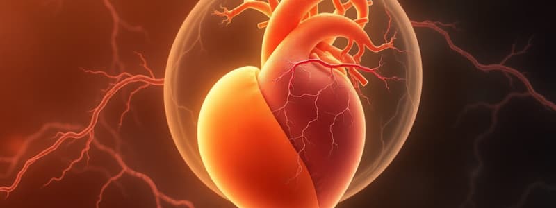
Flujo Sanguíneo Embrionario
- El flujo sanguíneo embrionario inicia con una mayor concentración de oxígeno y disminuye a medida que procede.
- Vena umbilical: Es una de las estructuras con mayor concentración de oxígeno.
- Conducto venoso: Se origina en el hígado, permite que la sangre proveniente de la vena umbilical pase directamente a la vena cava inferior. Luego, se transforma en el ligamento venoso.
- Vena cava inferior: Recibe sangre de las venas umbilicales.
- Aurícula derecha: Recibe sangre de la vena cava inferior.
- Foramen oval: Permite el paso de sangre desde la aurícula derecha a la aurícula izquierda
- Aurícula izquierda: Recibe sangre.
- Ventrículo izquierdo: Bombea sangre.
- Aorta ascendente: Se deriva del segmento caudal de la aorta dorsal.
- Arteria pulmonar: Se forman en el tronco arterioso.
- Conducto arterioso: Permite el paso de sangre de la aorta ascendente a la arteria pulmonar, luego se transforma en el ligamento arterioso.
- Arteria descendente: Se deriva del segmento caudal de la aorta dorsal.
- Arterias umbilicales: Transportan sangre de regreso a la placenta.
Studying That Suits You
Use AI to generate personalized quizzes and flashcards to suit your learning preferences.


