Podcast
Questions and Answers
What initiates each cardiac cycle as described in the context?
What initiates each cardiac cycle as described in the context?
- Pressure changes in the aorta
- Contraction of the atria before the ventricles
- Rapid contraction of the ventricles
- Spontaneous generation of action potential in the sinus node (correct)
During the second segment CD of the cardiac cycle, what is the primary change occurring in the left ventricle?
During the second segment CD of the cardiac cycle, what is the primary change occurring in the left ventricle?
- The left ventricle starts to relax (correct)
- Atrial contraction is occurring simultaneously
- LV pressure peak at 160 mmHg
- The left ventricle is in a state of diastole
According to the pressure readings, what is the systolic pressure of the left ventricle during the cardiac cycle?
According to the pressure readings, what is the systolic pressure of the left ventricle during the cardiac cycle?
- Rises even higher than 130 mmHg (correct)
- Peak of 100 mmHg
- Between 80 mmHg and 120 mmHg
- Approximately 130 mmHg
What is the function of the AV bundle in the cardiac cycle?
What is the function of the AV bundle in the cardiac cycle?
How do the pressure changes in the aorta compare to those in the left atrium during the cardiac cycle?
How do the pressure changes in the aorta compare to those in the left atrium during the cardiac cycle?
Who devised the original ECG lead system?
Who devised the original ECG lead system?
What does the tracing in an ECG display?
What does the tracing in an ECG display?
What characterizes the resultant cardiac vector in an ECG?
What characterizes the resultant cardiac vector in an ECG?
What is the shape of the triangle formed by the directional electrical forces in an ECG?
What is the shape of the triangle formed by the directional electrical forces in an ECG?
In what way can the configuration of the ECG vary?
In what way can the configuration of the ECG vary?
Which statement about Einthoven's triangle is incorrect?
Which statement about Einthoven's triangle is incorrect?
What does the term 'standard limb leads' refer to?
What does the term 'standard limb leads' refer to?
How does the pattern of the ECG change within an individual?
How does the pattern of the ECG change within an individual?
What event does the QRS wave signify in the cardiac cycle?
What event does the QRS wave signify in the cardiac cycle?
When does the QRS complex typically appear in relation to the P wave?
When does the QRS complex typically appear in relation to the P wave?
What physiological process follows the depolarization indicated by the QRS wave?
What physiological process follows the depolarization indicated by the QRS wave?
What does the T wave represent in the cardiac cycle?
What does the T wave represent in the cardiac cycle?
How does the timing of the T wave relate to ventricular contraction?
How does the timing of the T wave relate to ventricular contraction?
Which of the following is NOT directly caused by the electrical activity associated with the QRS complex?
Which of the following is NOT directly caused by the electrical activity associated with the QRS complex?
At what point in the cardiac cycle does the QRS complex initiate?
At what point in the cardiac cycle does the QRS complex initiate?
Which of the following statements about the QRS complex is false?
Which of the following statements about the QRS complex is false?
If the ejection fraction increases, what happens to the end-systolic volume?
If the ejection fraction increases, what happens to the end-systolic volume?
What physiological event results from a decrease in the firing rate of the SA node?
What physiological event results from a decrease in the firing rate of the SA node?
During which phase of the pressure volume loop do the aortic valves close and the ventricular pressure starts to decrease?
During which phase of the pressure volume loop do the aortic valves close and the ventricular pressure starts to decrease?
What is the primary representation of the t-wave in an electrocardiogram?
What is the primary representation of the t-wave in an electrocardiogram?
When the semilunar valves close, which of the following accurately describes this event?
When the semilunar valves close, which of the following accurately describes this event?
Which option does NOT represent a phase within the cardiac cycle?
Which option does NOT represent a phase within the cardiac cycle?
What does a high pulse pressure indicate in relation to cardiac function?
What does a high pulse pressure indicate in relation to cardiac function?
Which of the following best describes what occurs during the c-wave?
Which of the following best describes what occurs during the c-wave?
What is the normal increase in left ventricular volume during the phase referred to as end-systolic volume?
What is the normal increase in left ventricular volume during the phase referred to as end-systolic volume?
At what volume does the end-diastolic volume (EDV) typically reach before significant cardiac activity occurs?
At what volume does the end-diastolic volume (EDV) typically reach before significant cardiac activity occurs?
What electrical event is represented by the P wave in an electrocardiogram?
What electrical event is represented by the P wave in an electrocardiogram?
What happens to the atrial pressure curve following atrial contraction?
What happens to the atrial pressure curve following atrial contraction?
How much blood flow contributes to the ventricle during left ventricular volume increase?
How much blood flow contributes to the ventricle during left ventricular volume increase?
During which phase is the left ventricular end-diastolic volume measured?
During which phase is the left ventricular end-diastolic volume measured?
What is the volume of the left ventricular end-systolic volume mentioned in the content?
What is the volume of the left ventricular end-systolic volume mentioned in the content?
What primarily causes the increase in left ventricular volume to about 120 ml?
What primarily causes the increase in left ventricular volume to about 120 ml?
Flashcards are hidden until you start studying
Study Notes
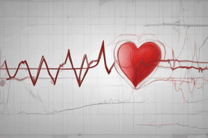
Wiggers Diagram and Cardiac Cycle
- The Wiggers diagram illustrates pressure changes in the aorta, left ventricle, and left atrium during the cardiac cycle.
- Aortic pressure is shown with upper dotted lines, ventricular pressure with a solid red line, and atrial pressure with lower dotted lines.
- The cardiac cycle starts with an action potential generated by the sinoatrial (SA) node, activating both atria and then the ventricles via the AV bundle.
Pressure Changes During Systole
- Systolic pressure in the left ventricle can reach approximately 130 mmHg.
- Aortic pressure reaches around 120 mmHg with further ventricle contraction, leading to blood ejection into the aorta.
- End diastolic volume (EDV) in the left ventricle can peak at 150 ml after venous return, having a normal increase of around 70 ml.
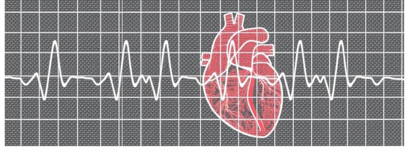
Electrical Activity and ECG Waves
- The P wave corresponds to atrial depolarization, causing a slight rise in atrial pressure.
- The QRS complex appears approximately 0.16 seconds after the P wave, marking ventricular depolarization and the onset of ventricular contraction.
- The T wave indicates ventricular repolarization and relaxation occurs slightly before ventricular contraction ends.
Electrophysiology and Limb Leads
- The electrocardiogram (ECG) displays the heart's electrical activity and varies by individual and lead placement.
- Willem Einthoven developed the standard limb leads system for ECG in the early 20th century.
- Cardiac vector in ECG represents the overall electrical force, lying within an equilateral triangle known as Einthoven’s triangle.
Cardiac Output and Ejection Fraction
- An increase in ejection fraction is associated with a decrease in end-systolic volume, reflecting a more efficient cardiac output.
- Bradycardia refers to a decreased firing rate of the SA node leading to reduced heart rate.
Phases of the Pressure-Volume Loop
- In the pressure-volume loop, the phase where the aortic valves close and ventricular pressure declines marks the transition from systole to diastole.
- During late ventricular contraction, a small rise in atrial pressure, known as the c-wave, results from ventricular contraction exerting force on the mitral valve, causing it to bulge into the atrium.
Studying That Suits You
Use AI to generate personalized quizzes and flashcards to suit your learning preferences.