Podcast
Questions and Answers
What is the total range of motion (ROM) for the elbow joint?
What is the total range of motion (ROM) for the elbow joint?
- 50-150 degrees
- 0-130 degrees
- 30-130 degrees
- 0-150 degrees (correct)
Which joint serves as a pivot joint in the elbow complex?
Which joint serves as a pivot joint in the elbow complex?
- Distal radioulnar joint
- Radiocapitellar joint
- Ulnohumeral joint
- Proximal radioulnar joint (correct)
What is the normal valgus carrying angle for females?
What is the normal valgus carrying angle for females?
- 15-20 degrees
- 20-25 degrees
- 10-15 degrees (correct)
- 5-10 degrees
Which structure attaches to the sublime tubercle on the ulna?
Which structure attaches to the sublime tubercle on the ulna?
In an extended elbow, what percentage of the axial load goes through the radioulnar joint?
In an extended elbow, what percentage of the axial load goes through the radioulnar joint?
How much internal rotation does the elbow joint exhibit?
How much internal rotation does the elbow joint exhibit?
What is the significance of the absence of cartilage in the lateral 120 degrees of the radial head?
What is the significance of the absence of cartilage in the lateral 120 degrees of the radial head?
What anatomical feature provides the fulcrum for forearm lever function at the elbow?
What anatomical feature provides the fulcrum for forearm lever function at the elbow?
What structure provides optimal stability to the elbow with appropriate tension during repair?
What structure provides optimal stability to the elbow with appropriate tension during repair?
Which muscle is NOT considered a dynamic stabilizer of the elbow?
Which muscle is NOT considered a dynamic stabilizer of the elbow?
What percentage of valgus stability is contributed by the radial head?
What percentage of valgus stability is contributed by the radial head?
Where does the LCL arise from in relation to the elbow anatomy?
Where does the LCL arise from in relation to the elbow anatomy?
What is the primary function of the annular ligament at the elbow?
What is the primary function of the annular ligament at the elbow?
Which nerve innervates the biceps and brachialis at the elbow?
Which nerve innervates the biceps and brachialis at the elbow?
What anatomical feature is essential for preventing excessive valgus stress on the elbow joint?
What anatomical feature is essential for preventing excessive valgus stress on the elbow joint?
During elbow extension, which structure contributes the greatest stability?
During elbow extension, which structure contributes the greatest stability?
What is the primary function of the coronoid tip in the elbow joint?
What is the primary function of the coronoid tip in the elbow joint?
At what angle of flexion is the elbow capsule maximally distended?
At what angle of flexion is the elbow capsule maximally distended?
Which component of the medial collateral ligament is the most crucial for resisting valgus stresses?
Which component of the medial collateral ligament is the most crucial for resisting valgus stresses?
What is the distal attachment point of the biceps brachii?
What is the distal attachment point of the biceps brachii?
What occurs if there is a loss of 50% or more of coronoid height?
What occurs if there is a loss of 50% or more of coronoid height?
Which muscle attaches 11 mm distal to the tip of the coronoid?
Which muscle attaches 11 mm distal to the tip of the coronoid?
What is the function of the lateral ulnar collateral ligament (LUCL)?
What is the function of the lateral ulnar collateral ligament (LUCL)?
In which scenario might maximal elbow flexion be limited?
In which scenario might maximal elbow flexion be limited?
Flashcards
Elbow Hinge Joint
Elbow Hinge Joint
The joint between the ulna and humerus, allowing for flexion and extension.
Coronoid Fossa
Coronoid Fossa
Part of the distal humerus that receives the coronoid process during flexion.
Coronoid Tip Stability
Coronoid Tip Stability
The coronoid process helps prevent posterior dislocation of the elbow.
Elbow Capsule Tension
Elbow Capsule Tension
Signup and view all the flashcards
Anterior Capsule Attachment
Anterior Capsule Attachment
Signup and view all the flashcards
Ulnohumeral Joint Stability
Ulnohumeral Joint Stability
Signup and view all the flashcards
MCL (Medial Collateral Ligament)
MCL (Medial Collateral Ligament)
Signup and view all the flashcards
MCL Function in Flexion
MCL Function in Flexion
Signup and view all the flashcards
Elbow Joint Components
Elbow Joint Components
Signup and view all the flashcards
Elbow Function
Elbow Function
Signup and view all the flashcards
Functional Elbow ROM
Functional Elbow ROM
Signup and view all the flashcards
Normal Carrying Angle
Normal Carrying Angle
Signup and view all the flashcards
Elbow Loading
Elbow Loading
Signup and view all the flashcards
Humerus Spiral Groove
Humerus Spiral Groove
Signup and view all the flashcards
Distal Humerus Flare
Distal Humerus Flare
Signup and view all the flashcards
Radiohumeral Articulation
Radiohumeral Articulation
Signup and view all the flashcards
Lateral Collateral Ligament (LCL)
Lateral Collateral Ligament (LCL)
Signup and view all the flashcards
Accessory Collateral Ligament
Accessory Collateral Ligament
Signup and view all the flashcards
Annular Ligament
Annular Ligament
Signup and view all the flashcards
Radiocapitellar Joint
Radiocapitellar Joint
Signup and view all the flashcards
Musculocutaneous Nerve
Musculocutaneous Nerve
Signup and view all the flashcards
Radial Nerve
Radial Nerve
Signup and view all the flashcards
Elbow Stability
Elbow Stability
Signup and view all the flashcards
Secondary static stabilizers in the elbow
Secondary static stabilizers in the elbow
Signup and view all the flashcards
Study Notes
Elbow Biomechanics
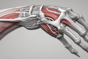
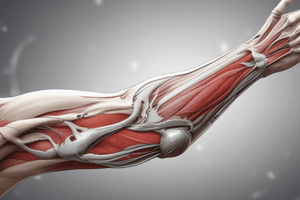
- The elbow joint is composed of the ulnohumeral, radiocapitellar, and proximal radioulnar joints.
- It plays a crucial role in daily activities, acting as a lever arm for positioning the hand and as a fulcrum for forearm movement.
- Functional range of motion (ROM) for flexion/extension is 0-150 degrees, and 50 degrees for supination/pronation.
- Normal carrying angle ranges from 5-10 degrees in males and 10-15 degrees in females. This carrying angle decreases with flexion.
- In an extended elbow, 40% of weight is through the ulnohumeral joint, and 60% is through the radiohumeral joint.
Osteology & Arthrology
- The humerus shaft has a spiral groove posteriorly, containing the radial nerve, roughly 13 cm proximal to the trochlea's articular surface.
- The distal humerus flare includes the medial and lateral epicondyles, contributing to half of the elbow joint's structure.
- The trochlea is spool-shaped and medially located, while the capitellum is laterally positioned.
- The sublime tubercle on the ulna is a site where the anterior bundle of the medial ulnar collateral ligament attaches distally
- The distal humerus comprises medial and lateral columns.
Arthrology - Axis and Alignment
- The joint surface of the elbow is anteriorly tilted approximately 30 degrees relative to the humerus shaft.
- The elbow exhibits a 6-degree valgus rotation.
- The axis of rotation is centrally located at the trochlea and capitellum, passing through the anteroinferior medial epicondyle.
Joint Type
- The radiohumeral articulation is a pivot joint.
- The radial head is covered by cartilage for approximately 240 degrees. The lateral 120 degrees lacks cartilage.
- The ulnohumeral articulation is a hinge joint.
- The coronoid fossa on the distal humerus receives the coronoid tip during deeper flexion.
- The coronoid tip acts as a buttress, preventing posterior dislocations.
- The elbow capsule is maximally distended at 70-80 degrees of flexion.
- The distal anterior capsule attachment is 6 mm distal to the coronoid tip.
- The coronoid is within the joint cavity.
Muscles of the Elbow
- Flexors: Biceps (radial tuberosity attachment), Brachialis (11mm distal to coronoid attachment), Brachioradialis
- Extensors: Triceps
Ligaments & Stability of Elbow
- The ulnohumeral joint (coronoid) is a primary static stabilizer. Loss of 50% or more of coronoid height can lead to instability.
- The medial collateral ligament (MCL) is composed of anterior, posterior, and transverse bundles, offering resistance to valgus and distracting forces.
- The MCL's origin is the anteroinferior aspect of the medial epicondyle, and insertion is the sublime tubercle and medial coronoid process.
- The anterior bundle of the MCL is crucial for resisting valgus stresses, while the radial head is another significant stabilizer.
- The posterior bundle forms the cubital tunnel floor, and is the primary restraint during maximal flexion.
Lateral Collateral Ligament Complex (LCL)
- The LCL comprises the radial collateral ligament (RCL) and the lateral ulnar collateral ligament (LUCL).
- It's a primary restraint for varus and external stress during full elbow motion.
- Origin is the posterior lateral epicondyle.
- Insertion is the radial collateral ligament on the supinator crest of the proximal ulna.
- The accessory collateral ligament contributes to lateral elbow stability.
- The annular ligament stabilizes the proximal radioulnar joint.
Secondary Static Stabilizers
- The radiocapitellar joint functions as a constraint for valgus stress.
- The radial head contributes approximately 30% of valgus stability.
- The capsule's most crucial role is at 0-30 degrees of flexion and pronation.
- Dynamic stabilizers include muscles crossing the elbow joint, such as anconeus, brachialis, triceps, and biceps. These muscles provide compressive stability during movement.
Nerves of the Elbow
- Musculocutaneous nerve: originates from the lateral cord of the brachial plexus and exits laterally, distal to the biceps tendon. It innervates the biceps and brachialis.
- Radial nerve: originates from the posterior cord of the brachial plexus and runs between the brachialis and brachioradialis. This nerve is found superficially to the joint capsule, located at the radiocapitellar joint. Runs through the spiral groove.
- Median nerve: originates from the medial/lateral cord of the brachial plexus and runs with the brachial artery from lateral to medial. It is superficial to the brachialis muscle and provides innervation to elbow.
- Ulnar nerve: originates from the medial cord of the brachial plexus. It passes medial to the brachial artery, pierces the medial intermuscular, and enters the posterior compartment. It then passes posterior to the medial epicondyle, through the cubital tunnel. Provides innervation to the elbow.
Blood Supply of Elbow
- The brachial artery is centrally located in the upper arm, entering the cubital fossa laterally.
- It splits into the radial and ulnar arteries at the elbow level.
- Key branches include the superior and inferior ulnar collateral and nutrient/muscular branches, and the supratrochlear.
Kinematics
- Flexion/extension axis: center of trochlea.
- Pronation/supination axis: capitellum through radial head to distal ulna.
- Large joint reaction forces due to short lever arms contribute to elbow degenerative changes.
Free Body Diagram
- Free body diagrams demonstrate elbow inefficiencies, using sum M = 0, and equations like 5B = 15W (B=3W).
- Static loads are close to body weight.
- Dynamic loads exceed body weight.
Arthrodesis
- Optimal unilateral arthrodesis: 90° flexion, 0-7° valgus.
- Bilateral arthrodesis: one elbow 110° flexion (feeding), one elbow 65° flexion (perineal hygiene).
Studying That Suits You
Use AI to generate personalized quizzes and flashcards to suit your learning preferences.