Podcast
Questions and Answers
Which three of the following are eukaryotic kingdoms?
Which three of the following are eukaryotic kingdoms?
- Animals (correct)
- Fungi (correct)
- Protoctists (correct)
- Plants
- Bacteria
Prokaryotic organisms are always multicellular.
Prokaryotic organisms are always multicellular.
False (B)
What distinguishes eukaryotic organisms from prokaryotic organisms?
What distinguishes eukaryotic organisms from prokaryotic organisms?
Eukaryotic organisms have a nucleus with a distinct membrane, while prokaryotic organisms do not.
______ organisms can be multicellular or single-celled and are made up of cells that contain a nucleus with a distinct membrane.
______ organisms can be multicellular or single-celled and are made up of cells that contain a nucleus with a distinct membrane.
What is the process by which cells develop the structure and characteristics needed to carry out their functions?
What is the process by which cells develop the structure and characteristics needed to carry out their functions?
Which of the following are examples of specialised cells in animals? (Select all that apply)
Which of the following are examples of specialised cells in animals? (Select all that apply)
Microvilli are hair-like projections that can move, unlike cilia. (True/False)
Microvilli are hair-like projections that can move, unlike cilia. (True/False)
Magnification is calculated using the equation: Magnification = ______________.
Magnification is calculated using the equation: Magnification = ______________.
Match the following microscope components with their descriptions:
Match the following microscope components with their descriptions:
What is a commonly used stain to stain cheek cells?
What is a commonly used stain to stain cheek cells?
What is a commonly used stain to stain onion cells?
What is a commonly used stain to stain onion cells?
What should be done to prevent the dehydration of tissue on a slide?
What should be done to prevent the dehydration of tissue on a slide?
What should be done to get clearer images when they are unclear or blurry under the microscope?
What should be done to get clearer images when they are unclear or blurry under the microscope?
Match the unit conversions correctly:
Match the unit conversions correctly:
Study Notes
Cell Structure
- Living organisms can be classified into five kingdoms: Animals, Plants, Fungi, Protoctists, and Prokaryotes.
- Animals, plants, fungi, and protoctists are eukaryotic organisms, characterized by having cells with a nucleus and distinct membrane.
- Eukaryotic cells can be multicellular or single-celled.
Eukaryotic Organisms: Animals & Plants
- Animals:
- Multicellular
- Cells contain a nucleus with a distinct membrane
- No cell walls made of cellulose
- No chloroplasts (cannot carry out photosynthesis)
- Feed on organic substances made by other living things
- Store carbohydrates as glycogen
- Have nervous coordination
- Able to move from place to place
- Plants:
- Multicellular
- Cells contain a nucleus with a distinct membrane
- Cell walls made of cellulose
- Contain chloroplasts (can carry out photosynthesis)
- Feed by photosynthesis
- Store carbohydrates as starch or sucrose
- No nervous coordination
Eukaryotic Organisms: Fungi & Protoctists
- Fungi:
- Usually multicellular, but some single-celled (e.g., yeast)
- Cells contain a nucleus with a distinct membrane
- Cell walls made of chitin
- No chloroplasts (cannot carry out photosynthesis)
- Feed by secreting extracellular digestive enzymes and absorbing digested molecules (saprotrophic nutrition)
- Some are parasitic and feed on living material
- Store carbohydrates as glycogen
- No nervous coordination
- Protoctists:
- Very diverse kingdom
- Mainly microscopic and single-celled
- Cells contain a nucleus with a distinct membrane
- Some have features making them more like animal cells (e.g., Plasmodium) or plant cells (e.g., green algae)
- Feed in various ways (photosynthesis or heterotrophy)
- No nervous coordination
- Examples: amoeba, Paramecium, Plasmodium, Chlorella
Prokaryotic Organisms
- Prokaryotes:
- Always single-celled
- No nucleus (nuclear material in cytoplasm)
- Much smaller than eukaryotic cells
- No chloroplasts or mitochondria
- Bacteria are prokaryotic organisms
- Bacteria:
- Microscopic single-celled organisms
- Possess cell wall (made of peptidoglycan), cell membrane, cytoplasm, and ribosomes
- Lack a nucleus, but have a circular chromosome of DNA in the cytoplasm
- Plasmids are present in prokaryotes, containing extra genes
- Lack mitochondria, chloroplasts, and other membrane-bound organelles
- Examples: Lactobacillus, Pneumococcus### Specialised Cells
- Specialised cells have developed characteristics (adaptations) to perform specific functions
- Cells specialise through differentiation, which involves developing the necessary structure and characteristics to carry out their functions
- Examples of specialised cells in animals include:
- Sperm cells
- Egg cells
- Ciliated epithelial cells
- Sperm cells are highly specialised for reproduction, with adaptations to carry DNA to the egg cell
- Adaptations include a tail for propulsion and a special membrane to fuse with the egg cell
- Egg cells are also highly specialised for reproduction, with adaptations to be fertilised by a single sperm and develop into an embryo
- Adaptations include a large size and a special membrane to fuse with the sperm cell
- Ciliated epithelial cells are highly specialised for wafting bacteria and other particles trapped by mucus up to the throat or down to the stomach
- Adaptations include cilia, which are hair-like projections that can move to 'waft' mucus along
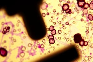
Microscopy
- Microscopy techniques have developed over time, increasing our understanding of cell structures and organelles
- The first light microscopes were developed in the 17th century, with scientists such as Anton van Leeuwenhoek and Robert Hooke using them to observe cells
- Robert Hooke observed the first cells (of a cork) in 1665 using a light microscope
- Light microscopes use light and lenses to form a magnified image of a specimen
- Modern light microscopes can achieve a maximum magnification of 1000-2000x
- Electron microscopes use beams of electrons to visualise specimens, with a much higher resolution and magnification than light microscopes
- Electron microscopes can achieve a maximum magnification of approximately 2,000,000x
- Electron microscopes have helped biologists understand many more subcellular structures, including mitochondria, chloroplasts, and ribosomes
Magnification Calculations
- Magnification is calculated using the equation: Magnification = Drawing size ÷ Actual size
- An equation triangle can be used to remember the formula: Magnification = Image size ÷ Actual size
- The formula can be rearranged to find actual size, image size, or magnification
- Magnification does not have units and is written as 'X' followed by a number (e.g. X 10 or X 5000)
Practical: Microscopy
-
Many biological structures are too small to be seen by the naked eye, making optical microscopes an essential tool for scientists
-
The key components of an optical microscope include:
- Eyepiece lens
- Objective lenses
- Stage
- Light source
- Coarse and fine focus
-
Other apparatus used in microscopy include:
- Forceps
- Scissors
- Scalpel
- Coverslip
- Slides
- Pipette
-
Specimens must be prepared on a microscope slide to be observed under a light microscope, with careful handling to avoid damaging the specimen and structures within it### Preparing and Using Microscopes
-
Stains may be required to make structures visible, depending on the type of tissue being examined. Commonly used stains include methylene blue for cheek cells and iodine for onion cells.
-
When using an optical microscope, always start with the low power objective lens to easily find what you are looking for in the field of view and prevent damage to the lens or coverslip.
-
To prevent dehydration of tissue, add a drop of water to the specimen (beneath the coverslip) to prevent cell damage.
-
To obtain clear images, switch to the lower power objective lens and use the coarse focus, and ensure the specimen sample is thin enough for light to pass through.
Measuring Cells and Structures
- Use a calibrated graticule to measure cells, cell structures, and organelles.
- An eyepiece graticule and stage micrometer are used to measure the size of the object when viewed under a microscope.
- The stage micrometer has three lines, and the eyepiece graticule has 100 division markings.
Producing Labelled Scientific Drawings
- Produce biological drawings of what you see under the microscope, following scientific rules.
- Drawings should be as large as possible, taking up at least half of the space available on the page.
- Limitations of biological drawings include inconsistencies in size and structure due to 3D cells being viewed on a 2D slide.
Limitations of Optical Microscopes
- Optical microscopes do not have the same magnification power as other types of microscopes, so some structures cannot be seen.
- The treatment of specimens when preparing slides can alter the structure of cells.
Using Units and Converting Between Them
- Convert units to ensure both measurements have the same unit before doing calculations.
- Remember that 1 mm = 1000 µm, and 1 µm = 0.001 mm.
- Use standard form to write very big or very small numbers using powers of 10.
- Standard form is written as a × 10n, where 1 ≤ a < 10, and n is the power of 10.
- Use standard form to convert between units, such as writing 1 metre in millimetres as 1 × 10³ mm.
- Examples of using standard form in conversion calculations include stating 45 centimetres in millimetres as 4.5 × 10² mm, and 250 micrometres in millimetres as 2.5 × 10⁻¹ mm.
Studying That Suits You
Use AI to generate personalized quizzes and flashcards to suit your learning preferences.
Description
Test your knowledge on cell structure, covering eukaryotic and prokaryotic organisms, specialised cells, and microscopy, specifically for Edexcel GCSE Biology students.