Podcast
Questions and Answers
What is the primary function of the P wave in an electrocardiogram?
What is the primary function of the P wave in an electrocardiogram?
- Represents the contraction of the ventricles
- Represents the relaxation of the atria
- Indicates repolarization of the ventricles
- Indicates depolarization of the atria (correct)
What is the result of the parasympathetic division releasing acetylcholine on heart rate?
What is the result of the parasympathetic division releasing acetylcholine on heart rate?
- Increases heart rate
- Decreases heart rate (correct)
- Has no effect on heart rate
- Stops the heart
What is the name of the node that acts as the heart's built-in pacemaker?
What is the name of the node that acts as the heart's built-in pacemaker?
- Purkinje fibers
- AV node
- SA node (correct)
- Bundle of His
What occurs during the diastole phase of the cardiac cycle?
What occurs during the diastole phase of the cardiac cycle?
What is the function of the QRS complex in an electrocardiogram?
What is the function of the QRS complex in an electrocardiogram?
What is the effect of norepinephrine and epinephrine on heart rate?
What is the effect of norepinephrine and epinephrine on heart rate?
What is the primary function of the T wave in an electrocardiogram?
What is the primary function of the T wave in an electrocardiogram?
What is the purpose of measuring changes in blood volume in the finger in this lab?
What is the purpose of measuring changes in blood volume in the finger in this lab?
What is the term for the recording of electrical changes in the cardiac muscle during the cardiac cycle?
What is the term for the recording of electrical changes in the cardiac muscle during the cardiac cycle?
What is the division of the autonomic nervous system that increases heart rate?
What is the division of the autonomic nervous system that increases heart rate?
What is the primary method of measuring blood volume in plethysmography?
What is the primary method of measuring blood volume in plethysmography?
What is the result of vasoconstriction on blood flow to a particular area?
What is the result of vasoconstriction on blood flow to a particular area?
What is the role of the sympathetic division in regulating blood flow?
What is the role of the sympathetic division in regulating blood flow?
What is the effect of cold temperatures on local blood vessels?
What is the effect of cold temperatures on local blood vessels?
What is the primary function of vasodilation in active skeletal muscle during exercise?
What is the primary function of vasodilation in active skeletal muscle during exercise?
What is the effect of metabolites and blood pH on local blood vessels?
What is the effect of metabolites and blood pH on local blood vessels?
What is the role of aldosterone in regulating blood vessel diameter?
What is the role of aldosterone in regulating blood vessel diameter?
What is the result of vasodilation on blood flow to a particular area?
What is the result of vasodilation on blood flow to a particular area?
What is the purpose of measuring pulse amplitude in plethysmography?
What is the purpose of measuring pulse amplitude in plethysmography?
What is the role of the smooth muscle in arteriole walls in regulating blood vessel diameter?
What is the role of the smooth muscle in arteriole walls in regulating blood vessel diameter?
The SA node is responsible for slowing down the heart rate.
The SA node is responsible for slowing down the heart rate.
The P wave indicates the depolarization of the ventricles.
The P wave indicates the depolarization of the ventricles.
Acetylcholine increases the rate of depolarization at the SA node.
Acetylcholine increases the rate of depolarization at the SA node.
The QRS complex represents the repolarization of the ventricles.
The QRS complex represents the repolarization of the ventricles.
The ECG measures changes in blood volume in the finger.
The ECG measures changes in blood volume in the finger.
The T wave represents the depolarization of the atria.
The T wave represents the depolarization of the atria.
Norepinephrine and epinephrine decrease heart rate.
Norepinephrine and epinephrine decrease heart rate.
The autonomic division has no influence on heart rate.
The autonomic division has no influence on heart rate.
The heart has its own built-in pacemaker node.
The heart has its own built-in pacemaker node.
Measuring changes in blood flow to the finger is not a part of this lab.
Measuring changes in blood flow to the finger is not a part of this lab.
The pulse transducer shines a beam of light through the skin in the finger and measures the light reflected by the oxygen in the blood.
The pulse transducer shines a beam of light through the skin in the finger and measures the light reflected by the oxygen in the blood.
Vasoconstriction occurs when the smooth muscle in the arteriole walls relax.
Vasoconstriction occurs when the smooth muscle in the arteriole walls relax.
The sympathetic division causes vasodilation of arterioles leading into the viscera and skin.
The sympathetic division causes vasodilation of arterioles leading into the viscera and skin.
During exercise, local vasoconstriction occurs in active skeletal muscle to reduce blood flow.
During exercise, local vasoconstriction occurs in active skeletal muscle to reduce blood flow.
The primary function of plethysmography is to measure the electrical changes in the cardiac muscle during the cardiac cycle.
The primary function of plethysmography is to measure the electrical changes in the cardiac muscle during the cardiac cycle.
The diameter of the arterioles has no effect on the amount of blood flowing to a particular body part.
The diameter of the arterioles has no effect on the amount of blood flowing to a particular body part.
Aldosterone is a hormone that causes vasodilation of arterioles.
Aldosterone is a hormone that causes vasodilation of arterioles.
Plethysmography involves measuring the changes in blood volume in the heart.
Plethysmography involves measuring the changes in blood volume in the heart.
The pulse amplitude is a direct measurement of the change in blood volume in the finger.
The pulse amplitude is a direct measurement of the change in blood volume in the finger.
Cold temperatures cause local vasodilation to increase blood flow to the skin or extremities.
Cold temperatures cause local vasodilation to increase blood flow to the skin or extremities.
Flashcards are hidden until you start studying
Study Notes
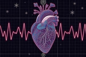
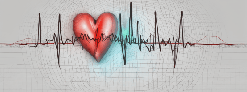
The ECG and the Heart
- The heart's mechanical activity (contraction and relaxation) is controlled by electrical activity (depolarization and repolarization events).
- An electrocardiogram (ECG) records electrical changes in cardiac muscle during the cardiac cycle.
- The P wave indicates depolarization of the atria, which leads to their contraction.
- The QRS complex indicates depolarization of the ventricles right before they contract to pump blood out of the heart.
- The T wave represents repolarization of the ventricles as they relax, allowing the heart to refill with blood.
Regulation of Heart Rate
- The heart has its own built-in pacemaker, the SA node.
- The autonomic division and hormones can influence the rate of depolarization at the SA node.
- Norepinephrine and epinephrine increase the rate of depolarization at the SA node, increasing heart rate under stress.
- Acetylcholine slows down the rate of depolarization at the SA node, resulting in a drop in heart rate.
Measuring Changes in Blood Volume
- Plethysmography records volume changes in the body.
- The volume of blood in tissues changes with the cardiac cycle, creating a pulse.
- A pulse transducer measures changes in blood volume in a body part, such as the finger.
- The pulse transducer shines a beam of light through the skin and measures the light reflected by the heme pigment in the blood.
Regulation of Blood Flow
- The amount of blood flowing to a particular body part depends on the diameter of the arterioles leading into that tissue.
- Vasoconstriction and vasodilation occur when the smooth muscle in the arteriole walls contracts and relaxes, respectively.
- External factors, such as the sympathetic division, and local factors, such as temperature and changes in metabolites, influence the smooth muscle of the arteries.
- Vasoconstriction and vasodilation can be influenced by various hormones, such as vasopressin (ADH) and aldosterone.
- Local factors, such as cold temperatures, can cause vasoconstriction to divert blood away from the cold skin or extremities.
- During exercise, local factors, such as carbon dioxide and metabolic acids, can cause vasodilation in active skeletal muscle.
Studying That Suits You
Use AI to generate personalized quizzes and flashcards to suit your learning preferences.