Podcast
Questions and Answers
Which of the following best describes a 'pixel' in digital imaging?
Which of the following best describes a 'pixel' in digital imaging?
- The depth of shades of grey.
- A measurement of image resolution.
- The smallest component of a matrix. (correct)
- The largest component of a matrix.
A larger pixel bit depth allows for a smaller number of shades of grey to be displayed in the image.
A larger pixel bit depth allows for a smaller number of shades of grey to be displayed in the image.
False (B)
What does a larger matrix size mean for the number of pixels in a given field of view?
What does a larger matrix size mean for the number of pixels in a given field of view?
higher number of pixels
The range of signal intensities an image receptor can accurately detect is referred to as its ______ ______.
The range of signal intensities an image receptor can accurately detect is referred to as its ______ ______.
Match the following digital imaging terms with their definitions:
Match the following digital imaging terms with their definitions:
In film terminology, what does 'FFD' stand for?
In film terminology, what does 'FFD' stand for?
In modern radiography practice, the term 'plain film' accurately describes the imaging process.
In modern radiography practice, the term 'plain film' accurately describes the imaging process.
In traditional film processing, what would happen to the undeveloped film if it was exposed to light before processing?
In traditional film processing, what would happen to the undeveloped film if it was exposed to light before processing?
In the context of film-screen radiography, markers, labels, and "______ ______" could be added to films to indicate the order of imaging.
In the context of film-screen radiography, markers, labels, and "______ ______" could be added to films to indicate the order of imaging.
What is the definition of an artifact in radiographic imaging?
What is the definition of an artifact in radiographic imaging?
Artifacts always enhance the diagnostic quality of radiographic images.
Artifacts always enhance the diagnostic quality of radiographic images.
Why is it becoming increasingly important to remove clothing and jewelry before radiographic imaging?
Why is it becoming increasingly important to remove clothing and jewelry before radiographic imaging?
Artifacts from patient ______ can reduce the quality of the image.
Artifacts from patient ______ can reduce the quality of the image.
Match the following radiographic practices with their impacts on image quality or process:
Match the following radiographic practices with their impacts on image quality or process:
In Computed Radiography (CR), what device is used to process the cassette?
In Computed Radiography (CR), what device is used to process the cassette?
In Computed Radiography, the tube and all equipment are modified, only the detectors will be the same.
In Computed Radiography, the tube and all equipment are modified, only the detectors will be the same.
What is the function of the aluminum backing in a CR cassette?
What is the function of the aluminum backing in a CR cassette?
A typical CR cassette contains a ______ ______ ______ (PSP) plate.
A typical CR cassette contains a ______ ______ ______ (PSP) plate.
Which layer of the PSP plate protects the phosphor layer?
Which layer of the PSP plate protects the phosphor layer?
The soft backing layer gives the PSP plate rigidity.
The soft backing layer gives the PSP plate rigidity.
What is the phosphor layer of a PSP plate usually made of?
What is the phosphor layer of a PSP plate usually made of?
The ______ layer of the PSP plate reflects light towards the photodetector during reading.
The ______ layer of the PSP plate reflects light towards the photodetector during reading.
Match the layers of the PSP plate with their functions.
Match the layers of the PSP plate with their functions.
What happens when the PSP plate is exposed to X-rays?
What happens when the PSP plate is exposed to X-rays?
All electrons liberated by X-ray exposure are trapped within the conduction band of the phosphor layer.
All electrons liberated by X-ray exposure are trapped within the conduction band of the phosphor layer.
What is the term for the image stored in the conduction band of the PSP plate before it is processed?
What is the term for the image stored in the conduction band of the PSP plate before it is processed?
The process of releasing trapped electrons in the PSP plate by using a laser light is called ______ ______.
The process of releasing trapped electrons in the PSP plate by using a laser light is called ______ ______.
Why is it important to store CR cassettes away from potential scatter radiation?
Why is it important to store CR cassettes away from potential scatter radiation?
Once a PSP plate has been 'cleaned' by the CR reader, it cannot be reused.
Once a PSP plate has been 'cleaned' by the CR reader, it cannot be reused.
What component of a CR reader converts the light energy released from the PSP plate into a digital signal?
What component of a CR reader converts the light energy released from the PSP plate into a digital signal?
Cassettes should be regularly "______" if not being regularly used.
Cassettes should be regularly "______" if not being regularly used.
Which of the following is an advantage of CR compared to traditional film-screen radiography?
Which of the following is an advantage of CR compared to traditional film-screen radiography?
Exposure indicators (EI) are universal across all CR systems, regardless of the vendor
Exposure indicators (EI) are universal across all CR systems, regardless of the vendor
Carestream uses what values for exposure index values?
Carestream uses what values for exposure index values?
Due to wide dynamic range, over or underexposed images are unlikely if the ______ is accurate.
Due to wide dynamic range, over or underexposed images are unlikely if the ______ is accurate.
To reduce scatter in digital imaging, which methods are most effective?
To reduce scatter in digital imaging, which methods are most effective?
Overexposing a patient is acceptable if it avoids repeat exposures, even if it violates the ALARA principle.
Overexposing a patient is acceptable if it avoids repeat exposures, even if it violates the ALARA principle.
What common language was required for the Picture Archiving and Communication Systems?
What common language was required for the Picture Archiving and Communication Systems?
Digital display stations always eliminate the chance of images being changed without authorization.
Digital display stations always eliminate the chance of images being changed without authorization.
What does RIS stand for?
What does RIS stand for?
______ is the term used when medical imaging files are accessed throughout a single facility, or from one facility to another.
______ is the term used when medical imaging files are accessed throughout a single facility, or from one facility to another.
Which of the following is a technique used for dynamic imaging?
Which of the following is a technique used for dynamic imaging?
Static imaging involves a continuous series of images of video.
Static imaging involves a continuous series of images of video.
Who invented the first fluoroscope?
Who invented the first fluoroscope?
______ or _______ beams of radiation allow visualisation of internal structure.
______ or _______ beams of radiation allow visualisation of internal structure.
Flashcards
Pixel
Pixel
The smallest component of a matrix in digital imaging.
Pixel bit depth
Pixel bit depth
The number of bits determining the precision in digitizing the analog signal, affecting shades of grey.
Matrix
Matrix
In digital imaging, this refers to the the number of pixels for a given field of view.
Dynamic Range
Dynamic Range
Signup and view all the flashcards
Resolution
Resolution
Signup and view all the flashcards
Image Artifact
Image Artifact
Signup and view all the flashcards
Computed Radiography (CR)
Computed Radiography (CR)
Signup and view all the flashcards
CR Reader
CR Reader
Signup and view all the flashcards
Photostimulable Phosphor (PSP) plate
Photostimulable Phosphor (PSP) plate
Signup and view all the flashcards
Protective Layer
Protective Layer
Signup and view all the flashcards
Phosphor Layer
Phosphor Layer
Signup and view all the flashcards
Reflective Layer in CR
Reflective Layer in CR
Signup and view all the flashcards
Exposure Indicator (EI)
Exposure Indicator (EI)
Signup and view all the flashcards
Dose Creep
Dose Creep
Signup and view all the flashcards
PACS (Picture Archiving and Communication System)
PACS (Picture Archiving and Communication System)
Signup and view all the flashcards
DICOM
DICOM
Signup and view all the flashcards
RIS (Radiology Information System)
RIS (Radiology Information System)
Signup and view all the flashcards
Teleradiography
Teleradiography
Signup and view all the flashcards
Dynamic Imaging
Dynamic Imaging
Signup and view all the flashcards
Fluoroscopy
Fluoroscopy
Signup and view all the flashcards
Angiography
Angiography
Signup and view all the flashcards
Angioplasty
Angioplasty
Signup and view all the flashcards
Cystography
Cystography
Signup and view all the flashcards
Collimation
Collimation
Signup and view all the flashcards
Study Notes
Digital Radiography Introduction
- Digital radiography (DR) includes digital imaging terminology, dynamic imaging, computed radiography (CR) systems, and PACS and RIS introduction.
Digital Terminology
- Pixel - the matrix's smallest component which determines resolution.
- Pixel bit depth - the amount of precision in digitizing the analogue signal, determining shades of grey.
- Matrix - a larger matrix size means a higher number of pixels for a given field of view.
- Dynamic Range - the range of signal intensities detectable by an image receptor and the detection of signals as very low to very high exposure intensities.
- Resolution - the ability of the image to demonstrate clear definition between adjacent areas of varying exposures.
Film Terminology and Practice
- Multiple exposures could be taken on a single film and can still be done with CR cassettes.
- Images had to be processed in a dark room to avoid fogging the entire image if undeveloped film was exposed to light.
- Busy departments required staff to operate dark rooms.
- Images were viewed in shared viewing areas instead of individual x-ray rooms.
- Markers, labels, and "red dots" were stickers added to films which indicated the imaging order.
- Radiographers had to be satisfied before patients could leave after imaging.
- Light boxes were in most hospital rooms for film viewing.
- FFD means film-focus distance.
- The term "plain film" no longer accurately describes radiography practice in NI due to the obsolescence of film.
Image Artefacts
- Artifacts are unwanted brightness on a radiographic image.
- Artifacts are detrimental as they hinder visibility of anatomy or pathology.
- Improper use of x-ray equipment can cause artifacts.
- Artifacts from patient clothing reduce image quality.
- With systems becoming more sensitive, removing clothing and jewelry is important to avoid obscuring areas of interest.
Computed Radiography
- CR systems can be added to existing radiographic equipment designed for film-screen imaging
- Only the detectors change; the tube and other equipment remain the same.
- The cassette is processed using a CR reader
- A typical CR cassette contains a photostimulable phosphor (PSP) plate.
- The cassette houses the PSP plate and is backed with aluminium for scatter reduction.
- The PSP plate has protective layers for static and dust reduction.
PSP Plate Composition
- Protective layer: a plastic layer that protects the phosphor layer
- Phosphor layer: made of barium fluorohalide with europium, and can be turbid or structured.
- Reflective layer: reflects light towards the photodetector during reading.
- Base layer: provides rigidity.
- Backing layer: protects the back of the plate.
PSP Plate Function
- When the PSP plate is hit by x-rays, electrons are removed from phosphor atoms.
- Around 50% of liberated electrons get trapped in the conduction band area of the phosphor layer.
- The phosphor atoms are ionized by x-rays.
- Approximately 50% of the freed electrons are trapped in the conduction band and this trapped energy forms the latent image.
- During processing, a laser releases the energy of trapped electrons, which is photostimulable luminescence.
CR Process
- A clean CR cassette is taken from storage.
- An x-ray exposure is made using the CR cassette.
- The radiographer retrieves the cassette and takes it to the reader.
- The radiographer checks the image while the patient waits.
- The CR reader cleans the PSP and releases the cassette.
- The CR reader opens the cassette, removes the PSP plate, and reads it
- The laser beam is projected onto the phosphor layer releasing trapped electrons
- Electrons return to their valence shells and emit energy as light,
- The light energy is directed via a light conducting material to the photodetector
- This light is converted to a digital signal using an analogue to digital convertor (ADC).
- The cleaned PSP plate is placed back into the cassette.
CR Practice
- Cassettes collect background radiation over time
- Requires storage away from potential scatter radiation
- Multiple rooms can share a single CR reader.
- More than one exposure can be made using a single cassette via use of lead to cover unexposed areas.
- Varying sizes available
- Cassettes require regular cleaning if not in use.
CR Image Extraction Process
- The image is stored in the cassette.
- The laser is used to extract the image during reading.
- During digitisation, the energy is liberated in the form of light.
- The digital signal is then converted into a matrix of grey-scale values.
CR Key Advantages
- Wide dynamic range
- Low running costs
- Cheap installation costs (when adapting film-screen room)
- Low maintenance costs
- Post-processing manipulation of images
- Faster processing than film-screen
Exposure Indicators & Values
- Exposure indicators (EI) represent the exposure level of the PSP plate.
- EI values are vendor-specific.
- Carestream uses exposure index values using logarithmic expressions.
- Optimal ranges should be displayed (e.g., 1500 to 1800.)
- A higher value indicates the PSP received more exposure than was expected for an area. A lower value means there was less exposure.
- Radiographers use EI values along with image appearance to determine image quality.
- Adjusting the image using contrast manipulation when the exposure indicator is outside of the expected range may result in suboptimal images that are not suitable for reporting.
Imaging Considerations
- Due to the wide dynamic range, over or underexposed images are unlikely if the technique is accurate.
- Radiographers must judge the need for repeats based on imaging appearance, using EI as a guide.
- Repeat exposures could result in the patient receiving double the intended dose.
- All forms of digital imaging are sensitive to low-energy radiation and, therefore, scatter
- Scatter can be reduced by collimation and the use of a grid.
- Cropping tools aren't effective at reducing scatter's impact.
- By law, radiographers adhere to the ALARA principle.
- Overexposing patients violates ALARA.
- Systems have recommended exposure values for average builds; these values should be reduced for smaller patients.
- A diagnostic image may still be achievable even with overexposure, but radiographers have to be careful to follow ALARA.
PACS
- PACS is integral to any digital radiography department.
- Storage of digital images has an ever-increasing demand.
- The average size of a digital radiography study is 38MB.
- PACS systems needed a common "language" to communicate - DICOM.
- DICOM (Digital Imaging and Communications in Medicine) was first formulated in 1983.
- DICOM allows medical images to be exchanged across modalities, display stations, and storage systems.
- Medical imaging equipment is manufactured to be DICOM-compatible.
PACS,RIS, and DICOM
- Radiographers need to know how to safely process, store, retrieve and manipulate digital images
- CT, MRI, ultrasound and nuclear medicine have been digital modalities for a long time.
- Digital modalities are present in all NI hospitals.
- PACS allows the display and storage of images.
- DICOM allows images to be transferred and retrieved
- Teleradiography = medical imaging files are accessed throughout a single facility, or from one facility to another.
- Radiology Information Systems (RIS) handles textual information about patient events.
- The integration of PACS, RIS, and teleradiography systems mean the same system can now serve all functions.
Digital Display & Access
- Interpreting digital images requires a capable display screen station.
- Reporting rooms typically use 5-Mp monitors.
- Users can access different areas of PACS and RIS depending on their role.
- Systems help keep images and reports secure.
- It prevents the chance of images or reports being adjusted or changed.
- Usernames and passwords must be solely used by the intended person and never shared.
PACS Challenges
- Cloud-based storage
- Offsite servers using a secure network
- Requires third-party vendor
- Purchase and maintenance of storage equipment
- Exploring new ways to compress image data
- A PACS team in most facilities is required to: manage, correct, and adjust digital image storage.
- PACS team responsibilities: provide IT support to radiography, follow up on missing images, correct folder errors, and communicate image transfers.
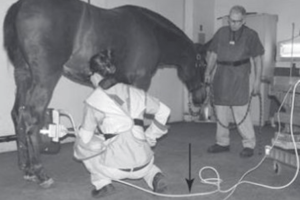
Dynamic Imaging
- Dynamic imaging is also called fluoroscopic imaging.
- Static imaging = an image
- Dynamic imaging - a video or continuous series of images
Fluoroscopy Introduction
- Thomas Edison invented the first fluoroscope shortly after x-rays were discovered
- Fluoroscopy allows real timer visualization of biological functions.
- The main function of modern fluoroscopy equipment is to provide dynamic video imaging, using very low mAs together with methods to increase the brightness of the resultant images, so that they are useful even with lower exposure parameters
- Without low mAs values, the patient dose will be very high due to consistent exposures during the procedure.
- Continuous or pulsed beams of radiation allow visualization of internal structures and is used with contrast media to allow real time body monitoring.
- Contrast administration may be done via catheterisation into the vascular, digestive or urinary system
Fluoroscopy Terms to Research
- Accelerating anode
- Brightness gain
- Camera tube
- Magnification
- Flat panel detector
- Fluoro-loop save
- Continuous versus pulsed fluoroscopy
- Pulse rate
- Image intensifier versus digital units
Fluoroscopy Introductions cont.
- Example procedures include stent insertion, angiography, angioplasty, cystography, and catheter insertion
- Involves cardiac, respiratory, digestive, hepato-biliary, and urinary body systems.
Fluoroscopy: Radiographer's Role
- Patient ID
- Operation of the fluoroscopy equipment including collimation, exposure factor control, positioning, image saving/storing, and the recording of dose
- Minimise patient dose with the accurate use of equipment
- Radiation protection of the patient and staff
- Maintenance of PPE
- Quality assurance of equipment
- Monitoring patient dose
- Wearing TLD (thermoluminescent dosimeter) badges
- Checking pregnancy status of staff in the department.
Fluoroscopy: Department Organization
- Interventional radiology suites
- Fluoroscopy department
- Cath labs (cardiac procedures are performed in catheterization laboratory)
- Staff includes interventional radiologists (specialist areas), radiographers (often dedicated interventional radiographers), radiology nurses, healthcare assistants, radiographer assistants, operating department practitioners, administrative staff and booking teams.
- The basic fluoroscopy controls are Collimation, Magnification, Image brightness control, Image flip, Adjust pulse rate, Automatic exposure control, "Screen grab" , Tube, detector movement controls, Exposure factor controls, Lock and key
- Examples of Exposure buttons are a Hand-held Switch and a Foot Pedal.
Studying That Suits You
Use AI to generate personalized quizzes and flashcards to suit your learning preferences.