Podcast
Questions and Answers
What is the FIRST step in the process of digital radiographic imaging?
What is the FIRST step in the process of digital radiographic imaging?
- Viewing the digital file on a computer monitor.
- Electronic measurement of the pattern of x-ray transmission through the patient. (correct)
- Photographing a radiographic film image with a digital camera.
- Conversion of the electronic measurement into a digital computer file.
Digital radiographic imaging involves photographing a radiographic film image with a digital camera.
Digital radiographic imaging involves photographing a radiographic film image with a digital camera.
False (B)
What type of x-ray machines are typically used for digital radiography?
What type of x-ray machines are typically used for digital radiography?
Conventional x-ray machines
In veterinary medicine, the transition from analog to digital radiographic imaging is nearly ______.
In veterinary medicine, the transition from analog to digital radiographic imaging is nearly ______.
Even after transitioning to digital radiography, which aspects remain as important as they were in analog imaging?
Even after transitioning to digital radiography, which aspects remain as important as they were in analog imaging?
What is the fundamental component of a digital image in digital radiography?
What is the fundamental component of a digital image in digital radiography?
What factor is MOST important to consider when selecting a digital radiography system for a veterinary practice?
What factor is MOST important to consider when selecting a digital radiography system for a veterinary practice?
Explain the analogy between digital radiographic imaging and acquiring a photograph with a digital camera.
Explain the analogy between digital radiographic imaging and acquiring a photograph with a digital camera.
What is the term for individual picture elements that make up a digital image?
What is the term for individual picture elements that make up a digital image?
DICOM images can only be transfered using local area network technology.
DICOM images can only be transfered using local area network technology.
What notation system do computer files use to assign a gray shade to a pixel?
What notation system do computer files use to assign a gray shade to a pixel?
The size of each pixel in a digital image determines the ________ of the image.
The size of each pixel in a digital image determines the ________ of the image.
Match the file size with the corresponding type of digital media:
Match the file size with the corresponding type of digital media:
In binary notation, what gray shade is assigned to '0'?
In binary notation, what gray shade is assigned to '0'?
Which of the following factors has the LEAST impact on the 'clinically useful' spatial resolution of a digital image?
Which of the following factors has the LEAST impact on the 'clinically useful' spatial resolution of a digital image?
Insanely difficult: Assuming a digital image uses 10-bit grayscale, how many shades of gray are possible?
Insanely difficult: Assuming a digital image uses 10-bit grayscale, how many shades of gray are possible?
What is a significant limitation of CCD detectors due to the distance required for light collection and focusing?
What is a significant limitation of CCD detectors due to the distance required for light collection and focusing?
CCD technology can be easily retrofitted into existing x-ray machines.
CCD technology can be easily retrofitted into existing x-ray machines.
Name two limitations that prevent the use of CCD technology for portable imaging.
Name two limitations that prevent the use of CCD technology for portable imaging.
Before the final image is displayed, electronic data from digital radiographic hardware undergoes processing by a ______.
Before the final image is displayed, electronic data from digital radiographic hardware undergoes processing by a ______.
What is one of the primary aims of preprocessing steps in digital radiography?
What is one of the primary aims of preprocessing steps in digital radiography?
In the context of digital radiography, at which stage does assessing image quality at the QC workstation belong?
In the context of digital radiography, at which stage does assessing image quality at the QC workstation belong?
Image processing customization by the user is never possible on digital radiography systems.
Image processing customization by the user is never possible on digital radiography systems.
Name one factor corrected during the preprocessing stage of digital image processing that addresses issues with the detector itself.
Name one factor corrected during the preprocessing stage of digital image processing that addresses issues with the detector itself.
What type of energy is emitted from the PSP plate when it is illuminated by a laser in a CR system?
What type of energy is emitted from the PSP plate when it is illuminated by a laser in a CR system?
In CR systems, the PSP plate is exposed to bright white light after being read to ensure all electrons return to ground state.
In CR systems, the PSP plate is exposed to bright white light after being read to ensure all electrons return to ground state.
What component in the CR reader converts visible light emitted from the PSP plate into an electronic signal?
What component in the CR reader converts visible light emitted from the PSP plate into an electronic signal?
One advantage of CR is that it provides more flexibility in obtaining nonstandard views because the cassette is not ______ to a computer by a cable.
One advantage of CR is that it provides more flexibility in obtaining nonstandard views because the cassette is not ______ to a computer by a cable.
Match the following Direct Digital Radiography (DDR) types with their descriptions:
Match the following Direct Digital Radiography (DDR) types with their descriptions:
In an ambulatory veterinary practice, what is a significant limitation of using CR systems compared to DDR systems?
In an ambulatory veterinary practice, what is a significant limitation of using CR systems compared to DDR systems?
CR systems inherently increase workflow compared to analog imaging due to the immediate availability of digital images.
CR systems inherently increase workflow compared to analog imaging due to the immediate availability of digital images.
An equine veterinarian working in a rural area is considering upgrading from traditional film-based radiography. They often need to take radiographs of horses in barns without immediate access to a clinic. Considering the limitations and advantages of CR and DDR, which of the following factors would be the MOST critical in their decision-making process?
An equine veterinarian working in a rural area is considering upgrading from traditional film-based radiography. They often need to take radiographs of horses in barns without immediate access to a clinic. Considering the limitations and advantages of CR and DDR, which of the following factors would be the MOST critical in their decision-making process?
How does increased ambient light typically affect image interpretation on a monitor?
How does increased ambient light typically affect image interpretation on a monitor?
Viewing digital radiographs in complete darkness enhances interpretation accuracy.
Viewing digital radiographs in complete darkness enhances interpretation accuracy.
What are the three key characteristics that define a medical-grade monochrome LCD monitor?
What are the three key characteristics that define a medical-grade monochrome LCD monitor?
__________ relates to how bright a white screen is on a monitor.
__________ relates to how bright a white screen is on a monitor.
Why is displaying true black challenging for LCD monitors?
Why is displaying true black challenging for LCD monitors?
A monitor with a low contrast ratio is ideal for viewing radiographic images.
A monitor with a low contrast ratio is ideal for viewing radiographic images.
A medical-grade monochrome LCD monitor has a display of 2048 pixels × 1536 pixels. What is the megapixel resolution of this monitor?
A medical-grade monochrome LCD monitor has a display of 2048 pixels × 1536 pixels. What is the megapixel resolution of this monitor?
What is the primary reason given why medical-grade monochrome LCD monitors are not routinely used throughout the veterinary imaging environment, despite their superior quality?
What is the primary reason given why medical-grade monochrome LCD monitors are not routinely used throughout the veterinary imaging environment, despite their superior quality?
What is the primary standard used to calibrate medical-grade monochrome LCD monitors?
What is the primary standard used to calibrate medical-grade monochrome LCD monitors?
Consumer-grade LCD monitors maintain their brightness longer than medical-grade monitors.
Consumer-grade LCD monitors maintain their brightness longer than medical-grade monitors.
Name two advantages of digital radiography related to cost and maintenance.
Name two advantages of digital radiography related to cost and maintenance.
Digital radiography enables adjustments to image blackness and contrast after exposure, a process known as image ______.
Digital radiography enables adjustments to image blackness and contrast after exposure, a process known as image ______.
Match each advantage with its description:
Match each advantage with its description:
Which factor significantly compromises analog image quality due to darkroom errors?
Which factor significantly compromises analog image quality due to darkroom errors?
What makes the use of artificial intelligence (AI) in digital radiography a paradigm shift in imaging?
What makes the use of artificial intelligence (AI) in digital radiography a paradigm shift in imaging?
Explain why local governments are becoming more stringent regarding darkroom chemicals.
Explain why local governments are becoming more stringent regarding darkroom chemicals.
Flashcards
Digital Radiographic Imaging
Digital Radiographic Imaging
Electronic measurement of x-ray transmission, conversion to a digital file, and viewing on a monitor.
Digital Image File
Digital Image File
A computer file containing information from a measured signal.
Digital Radiography Equipment
Digital Radiography Equipment
Using conventional x-ray machines.
Digital Radiography System Quality
Digital Radiography System Quality
Signup and view all the flashcards
Radiation Protection and Patient Positioning
Radiation Protection and Patient Positioning
Signup and view all the flashcards
Digital Transition Benefit
Digital Transition Benefit
Signup and view all the flashcards
Digital Image File Basis
Digital Image File Basis
Signup and view all the flashcards
Digital Image File Information
Digital Image File Information
Signup and view all the flashcards
Browser-Based DICOM Transfer
Browser-Based DICOM Transfer
Signup and view all the flashcards
Pixel
Pixel
Signup and view all the flashcards
Pixel Matrix
Pixel Matrix
Signup and view all the flashcards
Spatial Resolution
Spatial Resolution
Signup and view all the flashcards
Pixel Size (Pixel Pitch)
Pixel Size (Pixel Pitch)
Signup and view all the flashcards
Gray Shades per Pixel
Gray Shades per Pixel
Signup and view all the flashcards
Binary Notation
Binary Notation
Signup and view all the flashcards
Grayscale Assignment
Grayscale Assignment
Signup and view all the flashcards
PSP Plate Illumination
PSP Plate Illumination
Signup and view all the flashcards
Visible Light Emission
Visible Light Emission
Signup and view all the flashcards
Photomultiplier Tube
Photomultiplier Tube
Signup and view all the flashcards
Signal Digitization
Signal Digitization
Signup and view all the flashcards
PSP Plate Reset
PSP Plate Reset
Signup and view all the flashcards
CR vs. Workflow
CR vs. Workflow
Signup and view all the flashcards
CR Replacement Cost
CR Replacement Cost
Signup and view all the flashcards
CR Cassette Flexibility
CR Cassette Flexibility
Signup and view all the flashcards
CCD Detector Size
CCD Detector Size
Signup and view all the flashcards
CCD Retrofitting
CCD Retrofitting
Signup and view all the flashcards
CCD Orientation
CCD Orientation
Signup and view all the flashcards
CCD Cost
CCD Cost
Signup and view all the flashcards
Digital Image Processing
Digital Image Processing
Signup and view all the flashcards
Image Preprocessing
Image Preprocessing
Signup and view all the flashcards
Image Processing Step
Image Processing Step
Signup and view all the flashcards
Image Presets
Image Presets
Signup and view all the flashcards
Medical-grade monochrome monitor
Medical-grade monochrome monitor
Signup and view all the flashcards
DICOM grayscale display function
DICOM grayscale display function
Signup and view all the flashcards
Reduced expendable supply cost
Reduced expendable supply cost
Signup and view all the flashcards
Image postprocessing
Image postprocessing
Signup and view all the flashcards
Improved image accessibility
Improved image accessibility
Signup and view all the flashcards
Enhanced portability
Enhanced portability
Signup and view all the flashcards
Use of artificial intelligence
Use of artificial intelligence
Signup and view all the flashcards
Darkroom errors
Darkroom errors
Signup and view all the flashcards
Ambient Lighting
Ambient Lighting
Signup and view all the flashcards
Viewing radiographs in a dark room
Viewing radiographs in a dark room
Signup and view all the flashcards
Monitor Quality
Monitor Quality
Signup and view all the flashcards
Medical-Grade LCD Monitor
Medical-Grade LCD Monitor
Signup and view all the flashcards
Monitor Brightness
Monitor Brightness
Signup and view all the flashcards
Contrast Ratio
Contrast Ratio
Signup and view all the flashcards
Monitor Resolution
Monitor Resolution
Signup and view all the flashcards
High Resolution Monitor Benefit
High Resolution Monitor Benefit
Signup and view all the flashcards
Study Notes
- Digital radiographic imaging includes electronically measuring x-ray transmission patterns through a patient, converting this data into a digital file, and viewing it on a computer.
- This process resembles taking a photo with a digital camera: both record patterns, transmit the data and can be viewed on a screen.
- It's not about photographing a radiographic film or scanning an analog radiograph.
- Analog x-ray machines are usually compatible with new digital imaging systems.
- Veterinary practices have largely shifted from analog to digital radiography.
- The cost of digital radiography systems has decreased, but substantial investment is still needed.
- Selecting the right system requires careful planning.
- Not all digital systems offer equal quality, and cheaper options might not fully deliver the benefits of digital radiography.
- Getting expert advice is helpful unless the purchaser has knowledge of digital technology.
- Radiation safety and patient positioning remain crucial in digital radiography, as in analog imaging.
- The transition to digital has enhanced the quality of veterinary radiographic studies.
- A comprehensive understanding of digital radiography is vital for those using or considering these systems.
Digital Image Files
- The basis of a digital image is a computer file holding information about a measured signal.
- In medical imaging, signals include x-ray emission patterns (radiography), x-ray attenuation (CT), sound waves (ultrasound), or radiofrequency emission (MRI).
- In medical imaging, the computer file is a DICOM format.
- DICOM files differ from .jpeg and .tiff files, and use .dcm extension.
- Besides image data, DICOM files contain ancillary information called metadata tags:
- Metadata tags detail:
- Device manufacturer.
- Image acquisition date and time.
- Patient demographics.
- Acquisition parameters.
- Identifiers for the referrer, practitioner, and operator.
- Other image parameters.
- The DICOM format mainly seeks to ensure consistency for imaging device interconnectivity.
- Without the DICOM standard, digital images from one vendor might only be viewable with that vendor's software.
- The DICOM standard enables images from any vendor to be viewed on various platforms.
- Compliance with DICOM formatting is voluntary.
- Most vendors acknowledge the benefits of DICOM compliance, and DICOM has emerged to be universal.
- Verifying DICOM adherence from a vendor is key before purchasing a system to maintain viewing and file transmission flexibility.
- The DICOM standard handles medical images with network protocols that run on top of the Internet protocol.
- DICOM images can also be transferred using browser-based technology for enhanced portability.
Components of a Digital Image
- When opened with dedicated software, a digital image has individual picture elements - pixels.
- Pixels form a row-by-column matrix.
- In radiographic, ultrasound, CT, and MRI scans, each pixel has a specific gray shade.
- More pixels yield a larger file size, and a greater array size.
- Uncompressed digital radiographic DICOM files are typically 4 to 12 MB per image.
- A file size of approximately 0.1 MB would be expected for a 30-page document.doc file.
- Pixel size determines spatial resolution, or the smallest detectable object.
- The vendor-supplied hardware dictates the pixel array size in a digital image.
- More pixels is generally better, but benefits plateau past a certain point.
- Pixel size (pixel pitch) may be measured in microns or line pairs per mm.
- Guidelines exist for minimum spatial resolution standards.
- Besides pixel size, other technical factors affect spatial resolution.
- Software optimization of the raw image file can be more important than pixel density.
- In medical digital images, pixels are assigned gray shades.
- The number of shades per pixel influences image contrast.
- Few gray shades result in high contrast with only black, white, and a few intermediate shades.
- Many gray shades allow for a longer contrast scale.
- Computer files apply binary notation to assign pixel shades with 0 as black and 1 as white.
- If "n" equals the quantity of zeroes or ones per pixel, then 2" is the number of possible gray shades.
- The number of possible gray shades are referred to as the bit depth of the image.
- Image file size and bit depth are directly correlated.
- Assigning varying gray shades to each pixel is necessary for diagnostic value.
Digital Radiography Acquisition Hardware
- Two primary types of digital radiography acquisition hardware are available including:
- Computed radiography (CR).
- Direct digital radiography (DDR).
- Though conventional x-ray tubes and tables are used for both CR and DDR, film cassettes are only used with the older analog imaging.
- In CR systems, digital recording is done with a cassette containing a flexible imaging plate.
- With DDR systems, the digital recording device is a rigid imaging plate or chip.
- Special DDR charge-coupled device (CCD) systems might require a new x-ray table purchase.
- This would become the case if hardware components were housed in the x-ray table, and the equipment cannot be retrofitted into an existing x-ray table.
Computed Radiography
- CR was the first digital radiographic system introduced.
- CR cassettes resemble film cassettes. CR cassettes have a flexible imaging plate coated with photostimulable phosphor (PSP) instead of x-ray film.
- With CR an attenuation distribution temporarily stores as what is known as a latent image:
- Latent images are a product of changes in electron energy band.
- The changes are produced by a patient’s x-rays striking the PSP
- Upon exposure, the CR is processed in a plate reader. Key steps when processing CR radiography includes:
- CR cassette removal from the x-ray table before reader insertion. Reader automatically removes the PSP plate from the CR cassette.
- PSP plate illumination via reader laser.
- Laser illumination causes PSP plate emission of visible light.
- Emitted visible light strikes a photomultiplier tube where it is converted into electronic signal.
- The electronic signal is digitized and stored as a digital file.
- The PSP plate is exposed to bright white light to make sure all electrons are at ground state in preparation for the next exposure.
- The PSP plate is automatically returned to the cassette in the reader.
- The cassette is ejected from the reader for use on the next patient. CR isn’t very popular in veterinary imaging.
- With CR, plates are handled like film cassettes, the plate takes 1- 2 minutes to be processed.
- Workflow does not improve via CR compared to analog imaging. CR is slower than film processing.
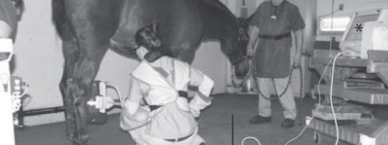
- CR readers are not mobile, while the cassettes themselves tend be portable CR cassettes must be returned to the veterinary practice for processing, preventing rapid evalutation of radiographs in the field.
- Damaged CR cassettes are inexpensive relative to the cost of replacing DDR plates but good-quality CR systems are adequate replacements.
Direct Digital Radiography
- Three types of DDR are identified, including:
- Indirect flat-panel detector system.
- Direct flat-panel detector system.
- CCD system.
- In DDR the plate is swapped out with the detector allowing digital radiographic images for quality-control evaluation.
Indirect Flat-Panel Detectors
- So named due to visible light production as a preliminary step in image making.
- X-ray intensifying screens are used to convert energy into visible light.
- May be composed of gadolinium oxysulfide or cesium iodide.
- Intrinsic capabilities for x-ray detection are had by Cesium Iodide, thinner flat-panel device, allowing lower radiation.
- The intensifying screen is placed on a photodiede panel which is a signal read-out from the TFT.
- 43 x 43 cm. imaging plates may have a photodiode matrix equaling to 2600 x 2600.
- Good resolution is determined by small detector size. Flat-panel detectors may have 6-7 million photodiodes.
- Electronics must spatially localize a signal at each photodiode-this increased expense is contrasted with film-screen cassette systems .
- Bit depth of indirect flat-panel detectors is capable of grayscale resolution of 16,384 shades per pixels.
Direct Flat-Panel Detectors
- Operate without a visible light mediator, as the x-rays strike the photoconductor which is typically amorphous selenium.
- Electrons liberated in the selenium layer by the oncoming x-ray beam are collected to form a charge.
- Charge is read out by the film transistor array the pixel matrix consists of 14 in bit depth.
- Differs from Indirect, whereby light is produced by intensifying effects.
- Diffusion can potentially lead to blurring due to light spread leading to direct and indirect disadvantages and engineering for crystal structures.
- Systems are frequently replacing CR as the preferred digital means of taking diagnostic images in veterinary medicine:
- Higher throughput.
- Recent decrease in price.
- Provide lower doses decreasing radiation for personnel; DDR has tethered and nonwireless attributes
- Batteries and portability help the equine practices to thrive.
Charged-Coupled Device
- Found in camcorders and digital cameras.
- This makes it less common than film / CR / flat-panel for radiography.
- Vendors often advertise the use of CCD chips.
- DDR doesn’t involve equipment between exposure / imaging; No cassette is required and CCD are smaller than flat panels only in area.
- They may contain pixel elements 43 x43 cm for a flat panel which are light sensitive. a light intermediary via intensifying screens standard.
- Collection efficiency and quality of the focusing lenses are key.
- Image quality and lenses help determine quality.
- The lens suffers loss of light and potential distortion, Humans must be reserved;
- If the part is small CCD and veterinary light is needed for parts the physical device and the constant vertical must be met the size is limited by horizontal.
- New and old have limited the scope of the current situation.
Image Processing and Viewing
- Electronic data, post-hardware choice, are processed by computers before displaying information, processing occurs during preprocessing.
- Refers to modifications being applied correctively to data for image distortion.
- Eliminates the need for functional or dead pixels. Processing turns corrected data to a visual in the controller. Aims to optimize aspects; A number of the process happens when a vendor makes presets custom; Data does gets overwritten the processing step occurs the the most effective means which in great vendor /product improvement algorithms limit noise and improve process this is usually not the case and can reduce dose as necessary.
- Algorithm and processing are techniques applied that can vary and should improve contrast resolution;
- Processing creates image files ready to be viewed Appropriate header information is attached through the local network
- Software licenses allow the file to be viewed.
- Functionally, viewing software Is a major asset in digital radiography; Post-processing means enhancement and controls are 100% on an end-user basis.
- A vast means of adjustment exists, over normal manipulation.
- Lighting and monitor impact image reviewing; light makes a huge difference so viewing in darkness won’t improve imagery.
- Quality improves accuracy; radiology is best improved with a dedicated monitor; better is brighter, better whites help users resolve more shades of Gray.
Studying That Suits You
Use AI to generate personalized quizzes and flashcards to suit your learning preferences.
Related Documents
Description
Explore the initial steps in digital radiographic imaging. Understand X-ray machine types, the shift from analog, and crucial imaging aspects. Learn about digital image components and system selection.


