Podcast
Questions and Answers
What process primarily facilitates the incorporation of endoderm into the developing embryonic disc to form the primitive gut tube?
What process primarily facilitates the incorporation of endoderm into the developing embryonic disc to form the primitive gut tube?
- Cephalocaudal and lateral folding of the embryo. (correct)
- Gastrulation.
- Neurulation.
- Somite formation.
From which germ layer is the mucosal epithelium of the digestive tract primarily derived?
From which germ layer is the mucosal epithelium of the digestive tract primarily derived?
- Endoderm. (correct)
- Neural crest cells.
- Ectoderm.
- Mesoderm.
The muscular layers of the digestive tract (lamina propria, muscularis mucosae, submucosa, muscularis externa, adventitia/serosa) are derived from which germ layer?
The muscular layers of the digestive tract (lamina propria, muscularis mucosae, submucosa, muscularis externa, adventitia/serosa) are derived from which germ layer?
- Splanchnic/visceral mesoderm. (correct)
- Neural crest.
- Somatic mesoderm.
- Endoderm.
What structure divides the foregut into the esophagus and trachea?
What structure divides the foregut into the esophagus and trachea?
In the development of the stomach, the dorsal border grows more than the ventral border. What anatomical structure does the dorsal border eventually form?
In the development of the stomach, the dorsal border grows more than the ventral border. What anatomical structure does the dorsal border eventually form?
What is the consequence of the 90-degree rotation of the stomach along its longitudinal axis during development?
What is the consequence of the 90-degree rotation of the stomach along its longitudinal axis during development?
From which part of the primitive gut does the segment of the duodenum distal to the opening of the bile duct primarily develop?
From which part of the primitive gut does the segment of the duodenum distal to the opening of the bile duct primarily develop?
During the development of the midgut, what structure temporarily connects the midgut to the yolk sac?
During the development of the midgut, what structure temporarily connects the midgut to the yolk sac?
What anatomical feature suspends the midgut from the midline during its development?
What anatomical feature suspends the midgut from the midline during its development?
During midgut development, herniation of the midgut loop occurs in the 6th week of IUL. What is the primary reason the midgut herniates outside the abdominal cavity?
During midgut development, herniation of the midgut loop occurs in the 6th week of IUL. What is the primary reason the midgut herniates outside the abdominal cavity?
A critical step in normal midgut development involves the return of the herniated intestinal loops back into the abdominal cavity. By what gestational age should this process be complete?
A critical step in normal midgut development involves the return of the herniated intestinal loops back into the abdominal cavity. By what gestational age should this process be complete?
What is the direction of rotation that the midgut loop undergoes as it returns to the abdominal cavity?
What is the direction of rotation that the midgut loop undergoes as it returns to the abdominal cavity?
What is the embryological origin of the upper portion of the anal canal?
What is the embryological origin of the upper portion of the anal canal?
What two openings result from the division of the cloaca?
What two openings result from the division of the cloaca?
Which of the following structures is derived from the ventral part of the cloaca?
Which of the following structures is derived from the ventral part of the cloaca?
From which blood vessel does the midgut primarily receive its blood supply?
From which blood vessel does the midgut primarily receive its blood supply?
What is the term used to describe a congenital anomaly where the oesophagus ends in a blind pouch?
What is the term used to describe a congenital anomaly where the oesophagus ends in a blind pouch?
A newborn presents with bilious vomiting shortly after birth. Imaging reveals a 'double bubble' sign, indicating a blockage in the duodenum. This is most likely a case of what?
A newborn presents with bilious vomiting shortly after birth. Imaging reveals a 'double bubble' sign, indicating a blockage in the duodenum. This is most likely a case of what?
What condition results from the failure or incomplete obliteration of the vitelline duct?
What condition results from the failure or incomplete obliteration of the vitelline duct?
Which of the following congenital anomalies is characterized by the herniation of abdominal contents through an umbilical opening that fails to close properly, with the herniated contents covered by a membrane?
Which of the following congenital anomalies is characterized by the herniation of abdominal contents through an umbilical opening that fails to close properly, with the herniated contents covered by a membrane?
Flashcards
What is the primitive gut?
What is the primitive gut?
Digestive tract develops from this structure, derived from endodermal yolk sac.
What is the endoderm?
What is the endoderm?
The germ layer that contributes to the mucosal epithelium, mucosal glands and submucosal glands.
What is the splanchnic/visceral mesoderm?
What is the splanchnic/visceral mesoderm?
The germ layer that forms the muscular walls of the digestive tract.
What is the neural crest?
What is the neural crest?
Signup and view all the flashcards
What is the tracheoesophageal septum?
What is the tracheoesophageal septum?
Signup and view all the flashcards
What is the oesophagus?
What is the oesophagus?
Signup and view all the flashcards
What is the stomach?
What is the stomach?
Signup and view all the flashcards
What is the duodenum's origin?
What is the duodenum's origin?
Signup and view all the flashcards
What is the U-shaped primary intestinal loop?
What is the U-shaped primary intestinal loop?
Signup and view all the flashcards
When does physiological hernia occur?
When does physiological hernia occur?
Signup and view all the flashcards
What is Omphalocele?
What is Omphalocele?
Signup and view all the flashcards
What is Congenital megacolon?
What is Congenital megacolon?
Signup and view all the flashcards
What is Rectal Fistula?
What is Rectal Fistula?
Signup and view all the flashcards
What is the perineal body?
What is the perineal body?
Signup and view all the flashcards
What is the cloaca?
What is the cloaca?
Signup and view all the flashcards
What is the urogenital sinus?
What is the urogenital sinus?
Signup and view all the flashcards
What is, 90 degree anticlockwise direction?
What is, 90 degree anticlockwise direction?
Signup and view all the flashcards
What is the upper half of anal canal?
What is the upper half of anal canal?
Signup and view all the flashcards
What is Gastroschisis?
What is Gastroschisis?
Signup and view all the flashcards
What is Atresia?
What is Atresia?
Signup and view all the flashcards
Study Notes
Development of Digestive Tract
- The digestive tract forms from the primitive gut
- The primitive gut is derived from the endodermal yolk sac
- Digestive tract development starts at the 4th week of Intrauterine Life (IUL)
- The cephalocaudal and lateral folding of the embryo incorporates endoderm into the embryonic disc, forming the primitive gut tube
- The gut tube extends from the buccopharyngeal membrane (cranial) to the cloacal membrane (caudal)
- The midgut is temporarily connected to the yolk sac via the vitello-intestinal duct/yolk stalk
- The foregut is cranial to this connection and the hindgut is caudal
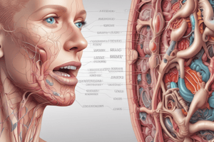
Germ Layer Contribution
- The endoderm becomes the mucosal epithelium, mucosal glands, and submucosal glands
- The splanchnic/visceral mesoderm forms the muscular walls of the digestive tract including the lamina propria, muscularis mucosae, submucosa, muscularis externa, and adventitia/serosa, also forms the mesenteries
- The somatic mesoderm gives rise to the parietal peritoneum
- The neural crest forms neurons and nerves of submucosal and myenteric plexuses
Foregut Derivatives
- The foregut derivatives are the primitive pharynx and respiratory system
- The tracheoesophageal septum divides the foregut into the esophagus and trachea
- Other derivatives:
- Esophagus
- Stomach
- 1st part of the duodenum
- Derivatives of pharyngeal pouches
- Liver
- Pancreas
- Gallbladder
Esophagus Development
- Develops between the pharynx and stomach
- The foregut develops a median laryngotracheal groove
- The groove enlarges to form a diverticulum
- The tracheoesophageal fold develops and divides the tube: ventrally into the trachea, and dorsally into the esophagus
- Initially, the esophageal lumen is occluded by endodermal cells
- Later, these cells break down, and the esophagus recanalizes
Stomach Development
- Fusiform dilatation of the foregut distal to the esophagus occurs at the 4th week of IUL
- This dilatation presents 2 borders: ventral and dorsal
- It also presents 2 surfaces: right and left
- The dilatation has 2 ends: upper (cardiac) and lower (pyloric)
- The dorsal border grows more, forming the greater curvature, while the ventral border forms the lesser curvature
Stomach Rotation
- Rotation occurs along the longitudinal axis
- It initially rotates 90 degrees so that:
- The left surface becomes the anterior surface
- The right surface becomes the posterior surface
- During the anteroposterior axis rotation, the cephalic and caudal ends of the stomach lie in the midline
- After rotation:
- The cardiac end moves from the midline to the left and slightly downward
- The pyloric end moves from the midline to the right and slightly upward
Duodenum Development
- Develops from two sources:
- Proximal half (1st & 2nd part up to opening of bile duct) from the foregut
- Distal half (2nd part below the opening of the bile duct with 3rd & 4th part) from the midgut
Midgut Derivatives
- Lower half of the duodenum
- Small intestine (jejunum, ileum, caecum, appendix)
- Ascending colon
- Right 2/3rd of the transverse colon
- The midgut elongates to form a U-shaped primary intestinal loop
- It is suspended dorsally in the midline by a double-layered dorsal mesentery
- The midgut communicates ventrally with the yolk sac via the vitelline duct/yolk stalk
Physiological Hernia
- The midgut loop elongates in the 6th week of IUL
- The midgut loop and liver grow simultaneously and cannot be accommodated by the abdominal cavity
- The midgut loop herniates through the umbilical opening into the extraembryonic celom
- This herniation of the loop through the umbilical opening is physiological hernia
Reduction of Hernia
- By the end of the 10th week, the abdominal cavity increases in size, and the loops re-enter the abdominal cavity
- By the 12th week, all loops settle inside the abdominal cavity
Midgut Rotation
- After formation, the loop lies outside the abdominal cavity
- The loop has: prearterial/proximal/cranial and postarterial/distal/caudal segments
- Before returning to the abdominal cavity, it undergoes 90 degree anticlockwise direction
- The cranial segment moves to the right, and the caudal segment moves to the left
- The cranial segment elongates and forms the jejunum and ileum, which lie on the right side and outside the abdominal cavity
Cecum and Appendix
- The caudal segment gives rise to the cecal bud (cecum and appendix)
- Coils of the jejunum and ileum take another 2nd 90 degree rotation when going back into the abdominal cavity
- The caudal segment returns to the abdominal cavity after the 3rd 90 degree anti-clockwise rotation
- Now, the cecum & appendix lie on the right side of the abdominal just below the liver
- The descent of the cecum takes place later
- This rotation is important for the definitive relations of the intestines
Hindgut Derivatives
- Left 1/3rd of the transverse colon
- Descending colon
- Sigmoid colon
- Rectum
- Upper part of the anal canal
- The cloaca is the dilated part of the hindgut, which is closed by the cloacal membrane
- During the 7th week, the cloacal membrane disappears, giving rise to two openings with the urorectal septum:
- Urogenital sinus
- Anal opening
- The tip of the urorectal septum later forms the perineal body
Urinary Bladder, Rectum & Anal Canal Development
- The urogenital sinus; the broad ventral part of cloaca, which gives rise to the urinary bladder and urethra
- The anal opening; the narrow dorsal part of the cloaca, called the primitive rectum, which gives rise to the rectum and the upper part of the anal canal
Blood Supply
- Foregut: Celiac artery
- Midgut: Superior mesenteric artery
- Hindgut: Inferior mesenteric artery
Anal Canal Development
- It develops from 2 sources:
- Hindgut develops the upper half from the primitive rectum
- Proctodeum develops the lower half from the anal pit (proctodeum)
- The proctodeum is an ectodermal depression below the anal membrane
- Initially, this anal membrane separates the hindgut and proctodeum
- The later membrane rupture allows the two parts to communicate
- The site of the anal membrane termed as the Pectinate line
Congenital Umbilical Hernia
- Omphalocele is a herniation of bowel loops through the umbilical opening that fails to return to the abdominal cavity
- Gastroschisis is a herniation of bowel loops through the umbilical opening when the umbilicus fails to close properly (weak anterior abdominal wall)
- Meckel’s diverticulum is a failure or incomplete obliteration of the vitello-intestinal duct
Rectal Fistula
- An abnormal connection between two hollow viscera
Congenital Megacolon (Hirschsprung’s Disease)
- A segment of the colon is dilated
Studying That Suits You
Use AI to generate personalized quizzes and flashcards to suit your learning preferences.