Podcast
Questions and Answers
Which of the following is part of the digestive tract?
Which of the following is part of the digestive tract?
- Pancreas
- Salivary glands
- Esophagus (correct)
- Liver
Which of the following is considered an associated gland of the digestive system?
Which of the following is considered an associated gland of the digestive system?
- Liver (correct)
- Small intestine
- Esophagus
- Stomach
What is the primary tissue type of the lamina propria?
What is the primary tissue type of the lamina propria?
- Stratified epithelium
- Simple squamous epithelium
- Loose connective tissue (correct)
- Dense connective tissue
Which layer separates the mucosa from the submucosa?
Which layer separates the mucosa from the submucosa?
Which of the following is found in the submucosa?
Which of the following is found in the submucosa?
The submucosal plexus is composed of what type of nerves?
The submucosal plexus is composed of what type of nerves?
Which nerve plexus generates and coordinates movement of the muscle layer?
Which nerve plexus generates and coordinates movement of the muscle layer?
What is the main function of peristaltic contraction?
What is the main function of peristaltic contraction?
What replaces the serosa in the esophagus?
What replaces the serosa in the esophagus?
What type of epithelium is found in the esophagus?
What type of epithelium is found in the esophagus?
What is the function of esophageal cardiac glands?
What is the function of esophageal cardiac glands?
Which part of the muscularis of the esophagus is exclusively skeletal muscle?
Which part of the muscularis of the esophagus is exclusively skeletal muscle?
What replaces the adventitia in intra-abdominal parts of the esophagus?
What replaces the adventitia in intra-abdominal parts of the esophagus?
Which region of the stomach is responsible for mucus production?
Which region of the stomach is responsible for mucus production?
Which region of the stomach secretes acidic juice?
Which region of the stomach secretes acidic juice?
What are rugae?
What are rugae?
Which cells cover the surface and line the gastric pits?
Which cells cover the surface and line the gastric pits?
Which cells secrete intrinsic factor?
Which cells secrete intrinsic factor?
What do chief cells secrete?
What do chief cells secrete?
What is the function of enteroendocrine cells?
What is the function of enteroendocrine cells?
What is the function of stem cells in gastric glands?
What is the function of stem cells in gastric glands?
What is the function of parietal cells?
What is the function of parietal cells?
What is the primary function of surface mucous cells in the stomach?
What is the primary function of surface mucous cells in the stomach?
What is a key feature of parietal cells?
What is a key feature of parietal cells?
How is the surface area of the small intestine increased?
How is the surface area of the small intestine increased?
What are the permanent transverse folds in the small intestine called?
What are the permanent transverse folds in the small intestine called?
What is the main function of absorptive cells (enterocytes) in the small intestine?
What is the main function of absorptive cells (enterocytes) in the small intestine?
What is the function of goblet cells in the small intestine?
What is the function of goblet cells in the small intestine?
What type of immunity are Paneth cells involved in?
What type of immunity are Paneth cells involved in?
What do duodenal (Brunner's) glands secrete?
What do duodenal (Brunner's) glands secrete?
Flashcards
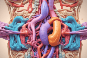
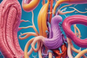
Digestive Tract
Digestive Tract
The GI or alimentary canal encompasses the oral cavity, esophagus, stomach, and small & large intestine, ending at the anus.
Associated Glands
Associated Glands
Glands associated with the digestive tract include salivary glands, the liver, and the pancreas.
Mucosa
Mucosa
The mucosa, a layer of the alimentary canal wall, consists of the epithelium, lamina propria, and muscularis mucosa.
Lamina Propria
Lamina Propria
Signup and view all the flashcards
Muscularis Mucosa
Muscularis Mucosa
Signup and view all the flashcards
Submucosa
Submucosa
Signup and view all the flashcards
Submucosal (Meissner) plexus
Submucosal (Meissner) plexus
Signup and view all the flashcards
Muscularis externa
Muscularis externa
Signup and view all the flashcards
Myenteric (Auerbach) nerve plexus
Myenteric (Auerbach) nerve plexus
Signup and view all the flashcards
Serosa
Serosa
Signup and view all the flashcards
Adventitia
Adventitia
Signup and view all the flashcards
Esophagus Mucosa
Esophagus Mucosa
Signup and view all the flashcards
Esophageal Cardiac Glands
Esophageal Cardiac Glands
Signup and view all the flashcards
Esophageal glands
Esophageal glands
Signup and view all the flashcards
Muscularis of the Esophagus
Muscularis of the Esophagus
Signup and view all the flashcards
Stomach Regions
Stomach Regions
Signup and view all the flashcards
Fundus & Body
Fundus & Body
Signup and view all the flashcards
Gastric Rugae
Gastric Rugae
Signup and view all the flashcards
Surface Mucus Cells
Surface Mucus Cells
Signup and view all the flashcards
Gastric Glands (SBT)
Gastric Glands (SBT)
Signup and view all the flashcards
Mucus neck cells
Mucus neck cells
Signup and view all the flashcards
Parietal (oxyntic) cells
Parietal (oxyntic) cells
Signup and view all the flashcards
HCL production
HCL production
Signup and view all the flashcards
Peptic (chief) Cells
Peptic (chief) Cells
Signup and view all the flashcards
Entero-endocrine EE Cells
Entero-endocrine EE Cells
Signup and view all the flashcards
Stem Cells
Stem Cells
Signup and view all the flashcards
Muscularis Layers of Fundus
Muscularis Layers of Fundus
Signup and view all the flashcards
Increase in surface area
Increase in surface area
Signup and view all the flashcards
Intestinal crypts
Intestinal crypts
Signup and view all the flashcards
Absorptive cells
Absorptive cells
Signup and view all the flashcards
Study Notes
- Digestive tract refers to the GI or alimentary canal
Digestive Tract
- Consists of the oral cavity, esophagus, stomach, and small & large intestines with the anus
Associated Glands
- Salivary glands, liver, and pancreas are associated with the digestive tract
General Structure of the Alimentary Canal Wall
- Mucosa is a layer with epithelium and lamina propria rich in blood, lymph vessels, and small glands
- Muscularis mucosa is a thin layer of smooth muscles that separates mucosa from submucosa and allows local movement
- Submucosa is a layer of denser connective tissue containing large blood and lymph vessels
- Submucosal (Meissner) plexus of autonomic nerves is also found in the submucosa, which may contain glands
Muscularis Externa
- Also known as muscularis propria
- It has two layers of smooth muscles, an inner circular layer and an outer longitudinal layer
- Myenteric (Auerbach) nerve plexus generates and coordinates muscle layer movement
- Connective tissue between muscle layers contains blood and lymph vessels
- Contraction, known as peristaltic contraction, mixes and propels luminal contents forward
Serosa
- It is a thin layer of loose connective tissue covered by simple squamous epithelium
- In the esophagus, the serosa gets replaced by adventitia: a layer of connective tissues without mesothelium
Esophagus
- The mucosa consists of non-keratinized stratified squamous epithelium
- Its lamina propria contains esophageal cardiac glands that secrete mucus and its muscularis mucosa is like other parts of the GI tract
- The submucosa has mucus-secreting esophageal glands
- Upper third of the muscularis is exclusively skeletal
- The middle third has a combination of skeletal and smooth muscles
- The lower third is exclusively smooth which begins with voluntary muscle action and finishes by involuntary peristalsis
- In the intra-abdominal part, the adventitia is replaced by serosa
Stomach Regions
- Cardia and pylorus produce mucus with similar histology
- Fundus and body contain gastric glands that secrete acidic juice
Gastric Rugae
- Large longitudinal folds in the mucosa and submucosa of the empty stomach
- These disappear when the stomach is full
Fundus & Body of Stomach
- The surface of the stomach is covered and lined by surface mucus cells
Epithelium
- Gastric pits lead to gastric glands
- It functions to secrete a thick, adherent, viscous mucous layer high in bicarbonate ions for stomach protection
Lamina Propria
- Contains glands
Gastric Glands
- They are long, branched tubular glands that fills the lamina propria in the mucosa
- Components include gastric pits functioning as ducts, isthmus and neck, and the body/base of the gland
- Secretory cells are mucus neck cells, parietal (oxyntic) cells, peptic (chief) cells, stem cells, and enteroendocrine cells
- Mucus neck cells are in the neck of the gastric glands and are columnar with apical secretory granules
- They secrete less alkaline mucus than surface cells
- Parietal (oxyntic) cells are along the whole gland, especially in the neck and body are large and pyramidal
- They have one, or occasionally two round nuclei; the cytoplasm is strongly acidophilic and rich in mitochondria
Parietal (oxyntic) cells
- They are ion-transporting cells with numerous mitochondria
- Intracellular canaliculi and microvilli lining the lumen of the canaliculi increases surface area
- Cytoplasmic tubules and vesicles are also present
- They are essential for HCL production and rough ER & Golgi apparatus aid synthesis and secretion of intrinsic factor (IF)
HCL Production
- CO2 and Cl- diffuse from the blood into the stomach cell
- CO2 combines with H2O to form H2CO3, which dissociates into bicarbonate (HCO3) and H+
- H+ combines with Cl- in the lumen of the gastric gland, forming HCL
- IF production is glycoprotein material essential for B12 absorption It is stimulated by parasympathetic innervation and gastrin hormone
Damage of oxyntic cells
- Achlorhydria
- Perinicious anemia
Peptic (chief, zymogenic) cells
- Protein- forming cells found in the body and base of gastric glands and are columnar
- The lower third contains abundant rough ER, the middle third has a round euchromatic nucleus & Golgi
- The upper third has numerous secretory granules (supra-nuclear)
- Secretion is Pepsinogen converted into pepsin, which is a proteolytic enzyme and Gastric lipase
Stem cells
- Located in the isthmus, neck, and base
- They renew cells of the fundic glands and migrate to replace surface cells or to form new oxyntic & chief cells
Entero-endocrine Cells
- Members of DNES
- Mainly scattered at the body & base and are stained by silver stain and IHC
- Secrete hormones of paracrine action such as Serotonin, Somatostatin, and Gastrin
Other Layers of the Fundus and Body of Stomach
- The muscularis has 3 layers: inner oblique, middle circular, and outer longitudinal
- In the pylorus, the circular layer is greatly thickened to form the pyloric sphincter
Small Intestine
- The small intestine consists of the duodenum, jejunum and ileum
- Increased surface area is due to plicae circulares (x3), which are permanent transverse mucosal and submucosal folds
- Also due to intestinal villi & crypts (x10) and microvilli (x30)
Intestinal Mucosa
- Villi are finger-like projections covered by epithelium
- Loose connective core of the villus contains a central lymphatic lacteal
- Epithelium has absorptive cells, goblet cells, and few enteroendocrine cells
Intestinal crypts
- Intestinal glands are the crypts of Leiberkühn
- Downward invagination of the mucosa
- Lining has cells covering the villi called Paneth cells, M-cells, and stem cells
Absorptive Cells
- They cover the villi and line the upper parts of the crypts and are tall columnar cells with basal oval nuclei
- They have a brush striated border
Apical Microvilli
- Contains a core of actin filaments
- Covered by a cell coat (glycocalyx), which contains enzymes for absorption
- Terminal web (microfilaments) is under the microvilli
Absorptive Cells (EM)
- Cytoplasm contains few small lipid droplets & well-developed smooth ER mitochondria
Function of Absorptive Cells
- They hydrolyze disaccharides into monosaccharides and dipeptides into amino acids that enter the cell by active transport towards blood capillaries
- They absorb lipids, which are surrounded by proteins forming chylomicrons that are released from the basolateral membrane of the cell to the central lacteal
Goblet Cells
- Covers the villi and lines the upper part of crypts; goblet-like shape and the apical part expanded and unstained because it's filled with mucinogen granules
- The basal part is constricted and contains nucleus and organoids
- Special stain used is PAS which appears a magenta color
- It's function it to synthesize and secrete mucus
Paneth cells
- A typical protein synthesizing cell found at the bottom of the crypts and are columnar
- Lower third is abundant with rough ER, middle third has round euchromatic nucleus and Golgi
- Upper third contains numerous secretory granules
- Functions in innate immunity by synthesizing secreting lysozyme & phospholipase A to break down membranes of micro-organisms and bacterial cell walls
M (microfold) cell
- APC antigen-presenting cell in the crypts of the ileum overlying Peyerʼs patches
- They are dome-shaped with basal pockets containing intraepithelial lymphocytes
Entero-endocrine cells
- Located scattered over the villi lining the crypts
- They secrete peptide hormones like Serotonin, Somatostatin, CCK
Other Layers of Small Intestine
- In the proximal part of the duodenum, the submucosa consists of duodenal or Brunner's glands that secrete alkaline mucus to neutralize chyme.
- In the ileum the lamina propria and submucosa contains Mucosa Associated Lymphoid Tissue consisting of large Peyer's patches
- Muscularis externa has two layers as the other parts of the digestive tract
Studying That Suits You
Use AI to generate personalized quizzes and flashcards to suit your learning preferences.