Podcast
Questions and Answers
What is the primary function of red blood cells?
What is the primary function of red blood cells?
- Carry oxygen (correct)
- Transport carbon dioxide
- Regulate body temperature
- Store nutrients
How many oxygen molecules can one molecule of hemoglobin carry?
How many oxygen molecules can one molecule of hemoglobin carry?
- 6
- 4
- 10
- 8 (correct)
What percentage of carbon dioxide is transported as bicarbonate in the plasma?
What percentage of carbon dioxide is transported as bicarbonate in the plasma?
- 5%
- 90% (correct)
- 50%
- 100%
Which enzyme is crucial for the conversion of carbon dioxide to bicarbonate?
Which enzyme is crucial for the conversion of carbon dioxide to bicarbonate?
What happens to bicarbonate during the chloride shift?
What happens to bicarbonate during the chloride shift?
Where are both red and white blood cells produced?
Where are both red and white blood cells produced?
What occurs in the lungs regarding carbon dioxide and bicarbonate?
What occurs in the lungs regarding carbon dioxide and bicarbonate?
Which red blood cell component primarily carries oxygen?
Which red blood cell component primarily carries oxygen?
What is the primary function of the small intestine?
What is the primary function of the small intestine?
Which of the following structures increases the surface area for absorption in the small intestine?
Which of the following structures increases the surface area for absorption in the small intestine?
What is the main method through which monosaccharides and amino acids are absorbed into the bloodstream?
What is the main method through which monosaccharides and amino acids are absorbed into the bloodstream?
Which component of the small intestine lining facilitates the absorption of lipid breakdown products?
Which component of the small intestine lining facilitates the absorption of lipid breakdown products?
What characteristic of the epithelial lining of the small intestine enhances nutrient absorption?
What characteristic of the epithelial lining of the small intestine enhances nutrient absorption?
Which molecule is NOT typically absorbed in the small intestine?
Which molecule is NOT typically absorbed in the small intestine?
What role do the villi play in the small intestine?
What role do the villi play in the small intestine?
Which characteristic of the small intestine contributes to efficient digestion and absorption?
Which characteristic of the small intestine contributes to efficient digestion and absorption?
What does diastolic pressure represent?
What does diastolic pressure represent?
What structure separates the left and right sides of the heart?
What structure separates the left and right sides of the heart?
Which factor is NOT a risk factor for hypertension?
Which factor is NOT a risk factor for hypertension?
What is atherosclerosis primarily characterized by?
What is atherosclerosis primarily characterized by?
Which valve separates the right atrium from the right ventricle?
Which valve separates the right atrium from the right ventricle?
What condition occurs when blood flow is interrupted to a part of the heart?
What condition occurs when blood flow is interrupted to a part of the heart?
What is the main function of the left side of the heart?
What is the main function of the left side of the heart?
What typically happens during an angina episode?
What typically happens during an angina episode?
Which layer of the heart is primarily responsible for contracting and pumping blood?
Which layer of the heart is primarily responsible for contracting and pumping blood?
Which of the following is a common symptom of myocardial infarction?
Which of the following is a common symptom of myocardial infarction?
What is found within the pericardial space that helps lubricate the heart?
What is found within the pericardial space that helps lubricate the heart?
Which chambers of the heart have thicker walls?
Which chambers of the heart have thicker walls?
Hypertension can lead to which of the following serious conditions?
Hypertension can lead to which of the following serious conditions?
What measurement is used to record blood pressure?
What measurement is used to record blood pressure?
What role do the semilunar valves play in the heart?
What role do the semilunar valves play in the heart?
Which type of muscle composes the heart?
Which type of muscle composes the heart?
What triggers an increase in the rate of breathing during exercise?
What triggers an increase in the rate of breathing during exercise?
Which of the following is a characteristic of chronic bronchitis?
Which of the following is a characteristic of chronic bronchitis?
What is a common feature of emphysema?
What is a common feature of emphysema?
What could potentially trigger an asthma attack?
What could potentially trigger an asthma attack?
Which part of the respiratory system is primarily responsible for the exchange of gases?
Which part of the respiratory system is primarily responsible for the exchange of gases?
What is a common outcome of respiratory tract infections?
What is a common outcome of respiratory tract infections?
Which statement is true regarding COPD?
Which statement is true regarding COPD?
What is a leading cause of death related to respiratory disorders in the US?
What is a leading cause of death related to respiratory disorders in the US?
Flashcards are hidden until you start studying
Study Notes
Anal Canal
- Faeces are expelled from the body through the anal canal.
- Some absorption of water and electrolytes still occurs in the anal canal.
Digestion: Breaking Down Food
- Food must be broken down into smaller molecules for absorption.
- This process involves both mechanical and chemical digestion.
- Mechanical digestion involves the physical breakdown of food, primarily through chewing by teeth.
- Chemical digestion involves enzymes that catalyze reactions to break down food molecules.
- Lipid emulsification is a process where fats are broken down into smaller droplets, increasing their surface area for enzyme action.
- The stomach also contributes to mechanical digestion through churning and mixing of food.
Absorption: Taking in Nutrients
- Absorption is the process of moving digested food molecules into the bloodstream or lymph.
- Monosaccharides, amino acids, fatty acids, and glycerol are the essential components for absorption.
- These molecules are transported across the epithelial cells of the small intestine through diffusion or active transport.
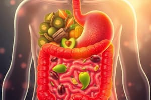
- The small intestine is the primary site of absorption due to its specialized structure.
- The small intestine has a very large surface area for efficient absorption.
- It is long (5-6 meters) to provide adequate time for digestion and absorption.
- The lining is only one cell thick, facilitating easy transport of food molecules.
- The small intestine is characterized by villi and microvilli, further increasing surface area.
- Cell membranes have carrier proteins for active transport of nutrients.
- Monosaccharides, amino acids, minerals, water, and vitamins are absorbed into the blood capillaries.
- Products of lipid breakdown are absorbed into lacteals (lymph vessels).
The Small Intestine: Optimized for Absorption
- The ileum, a part of the small intestine, is the main area for nutrient absorption.
- The epithelial lining has numerous villi, finger-like projections that increase surface area.
- Each villus is accompanied by blood vessels and lymph vessels for nutrient transport.
- The epithelial lining of the villi is only one cell thick for efficient absorption.
- The cells of the villi are further characterized by microvilli, projections of the cell surface membrane, which further increase surface area.
Exercise and Respiration Rate
- Exercise increases the rate of cellular respiration.
- Increased cellular respiration leads to a rise in CO2 concentration in the blood.
- Chemical receptors in blood vessels detect this increase in CO2.
- Impulses are sent to the breathing center in the medulla oblongata.
- The breathing center sends impulses to the intercostal muscles and diaphragm, increasing rate and depth of breathing.
- This increased breathing rate and depth helps to eliminate excess CO2 and restore its normal concentration in the blood.
Respiratory Tract Disorders
- COPD (Chronic Obstructive Pulmonary Disease):
- A group of lung diseases characterized by decreased airflow in the lungs.
- Includes chronic bronchitis and emphysema, primarily caused by cigarette smoking.
- Chronic Bronchitis:
- Inflammation of the airways with excessive mucus production, clogging airways.
- Overproduction and hypersecretion of mucus by goblet cells.
- Results in cough and difficulty breathing.
- Often associated with a "blue bloater" appearance due to cyanosis (blue discoloration of the skin).
- Emphysema:
- Overinflation of the lungs with air trapped in terminal bronchioles and alveolar sacs.
- Alveolar walls are destroyed (elastin destruction by elastases), impairing gas exchange.
- Reduced surface area for gas exchange, leading to decreased oxygen absorption into the bloodstream.
- Patients often have a "barrel chest" appearance.
- Often associated with a "pink puffer" appearance due to ruddy complexion despite low oxygen levels.
- Asthma:
- Episodic, triggered by allergens, exercise, infections, or stress.
- Inflammation of the bronchi, swelling, and constriction of smooth muscles in airways.
- Difficulty breathing, cough, and wheezing.
- Lung Cancer:
- Leading cause of death in the US for both men and women.
- Respiratory Tract Infections:
- Range from the common cold to life-threatening illnesses.
- Pneumonia:
- Inflammation of the alveolar spaces, caused by bacteria or viruses.
Gas Exchange Surfaces
- Human skin is not a primary gas exchange surface.
- The cell membrane of an amoeba is a gas exchange surface.
- The cockroach exoskeleton is not a gas exchange surface.
- Fish lamellae are gas exchange surfaces.
- Air sacs at the end of bronchioles are gas exchange surfaces.
Airflow in the Respiratory Tract - Order of Air Movement
- Nostrils
- Pharynx
- Mouth
- Larynx
- Trachea
- Bronchi
- Bronchioles
- Alveoli
Features of Gas Exchange Surfaces
- Large surface area
- Thin, moist membrane
- Close proximity to a transport system (blood vessels)
- Concentration gradient of gases (higher concentration of oxygen in the air than in the blood, and vice versa for carbon dioxide)
Heart: Structure & Function
- The heart is protected by the sternum (breastbone).
- The heart is primarily composed of cardiac muscle.
- The heart is a double pump:
- Right side pumps deoxygenated blood to the lungs for oxygenation.
- Left side pumps oxygenated blood to the entire body.
- Cardiomyocytes are the specialized cells of cardiac muscle.
- Cardiac muscle is a type of muscle tissue found only in the heart.
Heart Structure
- Septum: Divides the heart into left and right sides, preventing the mixing of oxygenated and deoxygenated blood.
- Chambers: Each side of the heart is divided into two chambers: an atrium and a ventricle. The heart has four chambers: right and left atria, right and left ventricles.
Heart Wall Layers
- Pericardium: Outermost, two-layered sac covering the heart.
- Pericardial space: Space between the two layers containing pericardial fluid, which lubricates the beating heart.
- Fibrous pericardium: Outer layer, strong and fibrous, preventing overfilling of the heart.
- Epicardium: Inner pericardial sac, attached to the heart.
- Myocardium: Muscle layer composed of cardiac muscle. Held in place by elastin and collagen strands.
- Contains coronary arteries that supply oxygen and nutrients to the heart muscle.
- Endocardium: Innermost layer, lining all chambers of the heart.
Heart Wall Thickness
- Atria: Thinner and less muscular walls compared to ventricles. They pump blood to the nearby ventricles.
- Ventricles: Thicker and more muscular walls, especially on the left side.
- Left ventricle: Pumps blood to the entire body at high pressure.
- Right ventricle: Pumps blood to the lungs at lower pressure to protect delicate lung capillaries.
Heart Valves
- Atrioventricular (AV) valves: Located between the atria and ventricles.
- Tricuspid valve: Separates right atrium from right ventricle.
- Bicuspid (mitral) valve: Separates left atrium from left ventricle.
- Semilunar valves: Located at the base of the vessels exiting the heart.
- Aortic valve: At the base of the aorta.
- Pulmonic valve: At the base of the pulmonary artery.
Simplified Blood Flow through the Heart
- Blood enters the left atrium from the lungs through the pulmonary veins.
- Blood flows from the left atrium to the left ventricle through the bicuspid (mitral) valve.
- The left ventricle pumps blood to the aorta through the aortic valve.
- Blood is then pumped from the aorta to the rest of the body.
Blood Pressure (BP)
- Systolic pressure: Highest pressure, occurs as contracting ventricles force blood into arteries.
- Diastolic pressure: Lowest pressure, occurs when ventricles are most relaxed.
- Measured in millimeters of mercury (mmHg).
- Recorded as SBP/DBP.
- Normal blood pressure is 120/80 mmHg.
Diseases of the Heart and Vessels
- Hypertension: Consistently high blood pressure.
- Risk factors include smoking, overweight, excessive alcohol consumption, sedentary lifestyle, high fat/high salt diet.
- Contributes to atheroma development and increases the risk of angina, stroke, and heart attack.
- Cardiovascular disease: A broad category encompassing diseases of the coronary arteries (supply the heart), carotid arteries (supply the brain), and peripheral arteries (supply the body).
- Leading cause of death worldwide.
- Risk factors: Hypertension, smoking, diabetes, obesity, hyperlipidemia, physical inactivity, age over 50, men more than women, family history, ethnicity, and alcohol.
- Atherosclerosis: Hardening of the arteries.
- Lipids build up in the lumen, narrowing the artery.
- Fibrous connective tissue forms over the lipids, creating an atherosclerotic plaque.
- Plaque rupture can cause a clot to form, potentially leading to cardiovascular diseases.
- Plaques breaking off and lodging in smaller arteries can cause stroke or heart attack.
- Myocardial infarction: Heart attack.
- Myocardial - referring to the heart muscle, Infarction - refers to tissue death due to lack of oxygen.
- Occurs when blood flow to a part of the heart is interrupted, causing damage and death to myocardial cells.
- Often caused by a blood clot or ruptured plaque blocking a coronary artery.
- Symptoms include crushing, substernal chest pain radiating to neck, jaw, and arms (can be silent).
- Can lead to sudden death or permanent heart damage.
Angina: A Warning Sign
- Partial blockage of a cardiac artery.
- Muscle exercised without adequate blood supply causes pain, in the heart, it is known as angina.
- Light exercise can cause pain similar to a heart attack.
Combatting Cardiovascular Disease (CVD)
- Lifestyle modifications are key to preventing and managing CVD. These include:
- Quitting smoking
- Maintaining a healthy weight
- Regular exercise
- Eating a healthy diet low in saturated fat and cholesterol
- Managing blood pressure and cholesterol levels
Red Blood Cells (RBCs)
- Red blood cells (erythrocytes) are biconcave disk-shaped cells, allowing them to squeeze through membrane capillaries.
- They originate from bone marrow.
- One drop of blood contains about 5 million RBCs.
- Hemoglobin: Protein within RBCs that carries oxygen.
- Hemoglobin combines with oxygen in the lungs (where oxygen concentration is high) to form oxyhemoglobin.
- One hemoglobin molecule can carry 8 oxygen molecules.
- One red blood cell can carry approximately 250 million hemoglobin molecules.
Oxygen Transport
- In the lungs, hemoglobin (Hb) binds with oxygen to form oxyhemoglobin.
- Oxygenated red blood cells travel to the tissues where oxygen concentration is lower.
- Oxygen is offloaded to the tissues as the concentration gradient favors oxygen movement.
- Hemoglobin then picks up carbon dioxide (CO2) in the tissues, forming carboxyhemoglobin.
- Carboxyhemoglobin travels back to the lungs for gas exchange.
Carbon Dioxide Transport
- Three main ways CO2 is transported in the blood:
- 5%: Attached to proteins as carboxyhemoglobin.
- 5%: Dissolved in blood plasma.
- 90%: Carried in plasma as bicarbonate ions (bicarbonate buffer system).
- Formation of bicarbonate from CO2 requires the enzyme carbonic anhydrase (CA), which is found in RBCs, not the plasma.
Bicarbonate Buffer System
-
In the tissues:
- CO2 diffuses into RBCs.
- Carbonic anhydrase (CA) quickly converts CO2 to carbonic acid (H2CO3).
- Carbonic acid dissociates into H+ ions and bicarbonate ions (HCO3-).
- This conversion drives CO2 uptake down its concentration gradient.
- Bicarbonate ions (HCO3-) are exchanged for chloride ions (Cl-) (chloride shift) and exit into the plasma.
- Cl- enters the RBC.
-
In the lungs:
- Chloride shift reverses: HCO3- enters the RBCs and Cl- leaves the RBCs.
- HCO3- and H+ ions recombine to form carbonic acid.
- Carbonic acid is converted back to CO2 and H2O by carbonic anhydrase.
- CO2 diffuses across membranes and is exhaled.
Blood Cells Origin: Bone Marrow
- Blood cells originate from bone marrow, specifically stem cells.
- Hematopoiesis: Process of blood cell production.
- White blood cells (leukocytes): Different types of WBCs, also produced in the bone marrow.
Studying That Suits You
Use AI to generate personalized quizzes and flashcards to suit your learning preferences.


