Podcast
Questions and Answers
Which substance is non-toxic and relatively cheap?
Which substance is non-toxic and relatively cheap?
- An iodinated contrast media
- An unspecified absorbent (correct)
- Water
- Sodium chloride
What is the first structure in the alimentary canal?
What is the first structure in the alimentary canal?
- Esophagus
- Pharynx
- Small intestine
- Oral cavity (correct)
Which of the following is NOT an accessory organ of digestion?
Which of the following is NOT an accessory organ of digestion?
- Gallbladder
- Spleen (correct)
- Pancreas
- Liver
What is one function of the digestive system?
What is one function of the digestive system?
Which examination focuses on the salivary glands and ducts?
Which examination focuses on the salivary glands and ducts?
Which of these could indicate a need for sialography?
Which of these could indicate a need for sialography?
What contrast media is used in sialography to promote salivation?
What contrast media is used in sialography to promote salivation?
Which salivary gland is demonstrated for stone formation in imaging?
Which salivary gland is demonstrated for stone formation in imaging?
What is the purpose of cholangiography?
What is the purpose of cholangiography?
During which procedure is contrast medium introduced directly to the CBD?
During which procedure is contrast medium introduced directly to the CBD?
What is the concentration of contrast medium used in post-operative (T-Tube) cholangiography?
What is the concentration of contrast medium used in post-operative (T-Tube) cholangiography?
What is the role of kidneys in the body?
What is the role of kidneys in the body?
Which special procedure is done to visualize the biliary tract via a needle through the abdominal wall?
Which special procedure is done to visualize the biliary tract via a needle through the abdominal wall?
What is administered intravenously in intravenous cholecystangiography?
What is administered intravenously in intravenous cholecystangiography?
Which factor is NOT controlled by kidney function?
Which factor is NOT controlled by kidney function?
What type of contrast medium is used during post-operative cholangiography?
What type of contrast medium is used during post-operative cholangiography?
What purpose does the administration of contrast agents serve in special procedures?
What purpose does the administration of contrast agents serve in special procedures?
Which condition is NOT an indication for special procedures using contrast?
Which condition is NOT an indication for special procedures using contrast?
Which of the following conditions is a contraindication for the use of contrast agents?
Which of the following conditions is a contraindication for the use of contrast agents?
What is the significance of conducting the AP view during a special procedure?
What is the significance of conducting the AP view during a special procedure?
During which view is the presence of floating gallstones specifically evaluated?
During which view is the presence of floating gallstones specifically evaluated?
What does the term 'biliary stenosis' refer to?
What does the term 'biliary stenosis' refer to?
Which contrast agent is specifically associated with the procedure using sodium iodide?
Which contrast agent is specifically associated with the procedure using sodium iodide?
What is the expected patient condition regarding food intake before special procedures?
What is the expected patient condition regarding food intake before special procedures?
What is the primary purpose of the Wolf Method in special procedures with contrast?
What is the primary purpose of the Wolf Method in special procedures with contrast?
Which part of the small intestine is the longest?
Which part of the small intestine is the longest?
Which special procedure with contrast is described as a gastrointestinal examination wherein single and double contrast studies are performed on the same day?
Which special procedure with contrast is described as a gastrointestinal examination wherein single and double contrast studies are performed on the same day?
What is the average length of the small intestine as described in special procedures with contrast?
What is the average length of the small intestine as described in special procedures with contrast?
For which condition is a biphasic examination indicated?
For which condition is a biphasic examination indicated?
Which of the following is a contraindication for special procedures with contrast?
Which of the following is a contraindication for special procedures with contrast?
Which method is used to evaluate pancreatic mass by demonstrating retrogastric structures?
Which method is used to evaluate pancreatic mass by demonstrating retrogastric structures?
Which position is typically required for the Hamptons procedure?
Which position is typically required for the Hamptons procedure?
What is the primary purpose of compression applied during an AP urogram?
What is the primary purpose of compression applied during an AP urogram?
When should a compression band not be applied during a urogram procedure?
When should a compression band not be applied during a urogram procedure?
What is typically observed at the 10-minute mark during a urogram?
What is typically observed at the 10-minute mark during a urogram?
Which factor can increase the excretion rate during a Hypertensive IVP?
Which factor can increase the excretion rate during a Hypertensive IVP?
What is the dosage protocol for children aged 2-8 years during contrast procedures?
What is the dosage protocol for children aged 2-8 years during contrast procedures?
What is a common indication for performing a urethrogram?
What is a common indication for performing a urethrogram?
What does a post-micturition examination assess?
What does a post-micturition examination assess?
What value is commonly checked at the 15-minute mark of a urogram procedure?
What value is commonly checked at the 15-minute mark of a urogram procedure?
What is one of the purposes of special procedures with contrast?
What is one of the purposes of special procedures with contrast?
Which of the following is a condition that indicates the need for special procedures with contrast?
Which of the following is a condition that indicates the need for special procedures with contrast?
What is a contraindication for performing special procedures with contrast?
What is a contraindication for performing special procedures with contrast?
During which position is the presence of floating gallstones evaluated?
During which position is the presence of floating gallstones evaluated?
Which of the following substances is used as a contrast medium in special procedures?
Which of the following substances is used as a contrast medium in special procedures?
What is generally discouraged for patients before undergoing special procedures with contrast?
What is generally discouraged for patients before undergoing special procedures with contrast?
What does biliary stenosis refer to?
What does biliary stenosis refer to?
What is a common imaging procedure to confirm the presence of biliary calculi?
What is a common imaging procedure to confirm the presence of biliary calculi?
Which of the following best describes the primary function of the salivary glands?
Which of the following best describes the primary function of the salivary glands?
What is the primary indication for performing a sialography procedure?
What is the primary indication for performing a sialography procedure?
What is the correct sequence in which the alimentary canal begins?
What is the correct sequence in which the alimentary canal begins?
Which salivary gland is associated with Wharton's duct?
Which salivary gland is associated with Wharton's duct?
What procedure is necessary to demonstrate any obstructed lesions in the salivary glands?
What procedure is necessary to demonstrate any obstructed lesions in the salivary glands?
Which of the following is classified as an accessory organ of digestion?
Which of the following is classified as an accessory organ of digestion?
Which symptom is considered a possible indication for a sialography procedure?
Which symptom is considered a possible indication for a sialography procedure?
How is the angle typically positioned for an AP view of the parotid gland during imaging?
How is the angle typically positioned for an AP view of the parotid gland during imaging?
What position is recommended for Poppel's method to evaluate pancreatic mass?
What position is recommended for Poppel's method to evaluate pancreatic mass?
Which method is used to demonstrate small lesions and sliding gastroesophageal herniations?
Which method is used to demonstrate small lesions and sliding gastroesophageal herniations?
Which part of the small intestine is considered the widest?
Which part of the small intestine is considered the widest?
What is a common indication for performing a small intestine follow-through study?
What is a common indication for performing a small intestine follow-through study?
What is the typical length of the small intestine?
What is the typical length of the small intestine?
Which method utilizes a biphasic examination in special procedures with contrast?
Which method utilizes a biphasic examination in special procedures with contrast?
What contraindication involves a condition where no movement of contents occurs within the intestines?
What contraindication involves a condition where no movement of contents occurs within the intestines?
Which of the following is NOT a method for performing special procedures with contrast?
Which of the following is NOT a method for performing special procedures with contrast?
What is the primary purpose of vesiculography?
What is the primary purpose of vesiculography?
What does hysterosalpingography primarily investigate?
What does hysterosalpingography primarily investigate?
What condition does vaginography aim to investigate?
What condition does vaginography aim to investigate?
What is the role of fetography in prenatal care?
What is the role of fetography in prenatal care?
What is the main purpose of pelvimetry?
What is the main purpose of pelvimetry?
What is injected during discography to visualize lumbar intervertebral disks?
What is injected during discography to visualize lumbar intervertebral disks?
Which procedure involves injecting gas or air to demonstrate the brain's ventricular system?
Which procedure involves injecting gas or air to demonstrate the brain's ventricular system?
What type of imaging does fetal imaging primarily utilize to assess developmental abnormalities?
What type of imaging does fetal imaging primarily utilize to assess developmental abnormalities?
What is one of the primary purposes of using contrast media in special procedures?
What is one of the primary purposes of using contrast media in special procedures?
Which type of reaction is characterized by a sudden drop in blood pressure after the injection of contrast media?
Which type of reaction is characterized by a sudden drop in blood pressure after the injection of contrast media?
What is one of the local reactions to contrast media that patients may experience?
What is one of the local reactions to contrast media that patients may experience?
Which of the following is a factor to consider when selecting appropriate contrast media?
Which of the following is a factor to consider when selecting appropriate contrast media?
What is a potential major reaction to contrast media that can occur during special procedures?
What is a potential major reaction to contrast media that can occur during special procedures?
Which method is NOT commonly used to introduce contrast media?
Which method is NOT commonly used to introduce contrast media?
What type of contrast media is used to enhance the visibility of internal structures during imaging?
What type of contrast media is used to enhance the visibility of internal structures during imaging?
An idiosyncratic reaction to contrast media is influenced by which factor?
An idiosyncratic reaction to contrast media is influenced by which factor?
Which structure does the alimentary canal begin with?
Which structure does the alimentary canal begin with?
What is a primary function of the digestive system?
What is a primary function of the digestive system?
Which procedure involves the radiographic examination of salivary glands and ducts?
Which procedure involves the radiographic examination of salivary glands and ducts?
Which of the following is NOT an accessory organ of digestion?
Which of the following is NOT an accessory organ of digestion?
Which condition is an indication for performing sialography?
Which condition is an indication for performing sialography?
Which of the following salivary glands is demonstrated for stone formation?
Which of the following salivary glands is demonstrated for stone formation?
What is a common method used to promote salivation during sialography?
What is a common method used to promote salivation during sialography?
Which of these is a common indication for performing special procedures with contrast?
Which of these is a common indication for performing special procedures with contrast?
What is one of the primary purposes of using contrast media during special procedures?
What is one of the primary purposes of using contrast media during special procedures?
Which procedure is specifically used to evaluate the gallbladder's contracting and emptying power?
Which procedure is specifically used to evaluate the gallbladder's contracting and emptying power?
What condition involves abnormal calcifications or stones in the gallbladder?
What condition involves abnormal calcifications or stones in the gallbladder?
Which of the following is a contraindication for using contrast agents during special procedures?
Which of the following is a contraindication for using contrast agents during special procedures?
Which view is used to visualize floating gallstones specifically?
Which view is used to visualize floating gallstones specifically?
What does non-visualization during a procedure typically indicate?
What does non-visualization during a procedure typically indicate?
Which imaging technique helps delineate trapped gas in the bowel from radiolucent stones in the gallbladder?
Which imaging technique helps delineate trapped gas in the bowel from radiolucent stones in the gallbladder?
Which of the following contrasts is used for enhancement during special procedures involving the biliary system?
Which of the following contrasts is used for enhancement during special procedures involving the biliary system?
What is the primary purpose of the esophagography procedure?
What is the primary purpose of the esophagography procedure?
Which esophageal segment is located posterior to the trachea and pharynx?
Which esophageal segment is located posterior to the trachea and pharynx?
What is the filling phase ratio used during esophagography?
What is the filling phase ratio used during esophagography?
Which condition is associated with retrosternal pain and regurgitation?
Which condition is associated with retrosternal pain and regurgitation?
Which of the following is a contraindication for special procedures with contrast?
Which of the following is a contraindication for special procedures with contrast?
What is the indicator for performing the Modified Valsalva maneuver?
What is the indicator for performing the Modified Valsalva maneuver?
Which procedure is NOT utilized to detect esophageal reflux?
Which procedure is NOT utilized to detect esophageal reflux?
Which of the following indicates a peptic ulcer in the distal esophagus?
Which of the following indicates a peptic ulcer in the distal esophagus?
What is the main purpose of percutaneous cholangiography?
What is the main purpose of percutaneous cholangiography?
In which scenario would intravenous cholecystangiography be performed?
In which scenario would intravenous cholecystangiography be performed?
What is the recommended concentration of contrast medium used during post-operative (T-Tube) cholangiography?
What is the recommended concentration of contrast medium used during post-operative (T-Tube) cholangiography?
Which of the following is NOT a function of the kidneys?
Which of the following is NOT a function of the kidneys?
What is the primary contrast medium method used during operative cholangiography?
What is the primary contrast medium method used during operative cholangiography?
Why might a patient undergo percutaneous cholangiography?
Why might a patient undergo percutaneous cholangiography?
What does the term 'cholangiography' refer to?
What does the term 'cholangiography' refer to?
What is introduced during the post-operative (T-Tube) cholangiography procedure?
What is introduced during the post-operative (T-Tube) cholangiography procedure?
What should be evaluated at the 5-minute mark during a urogram procedure?
What should be evaluated at the 5-minute mark during a urogram procedure?
Why is compression applied during a urogram procedure?
Why is compression applied during a urogram procedure?
What time interval is required to see the ureters during a urogram procedure?
What time interval is required to see the ureters during a urogram procedure?
At what point is the renal parenchyma visible after contrast injection?
At what point is the renal parenchyma visible after contrast injection?
Which of the following is NOT a condition that contraindicates the use of a compression band?
Which of the following is NOT a condition that contraindicates the use of a compression band?
What is the recommended dosage protocol for children aged 1-2 years during contrast procedures?
What is the recommended dosage protocol for children aged 1-2 years during contrast procedures?
What is primarily assessed during a post-micturition examination?
What is primarily assessed during a post-micturition examination?
What should be done during a delayed urogram procedure for obstructive uropathy?
What should be done during a delayed urogram procedure for obstructive uropathy?
What are the two phases employed in esophagography?
What are the two phases employed in esophagography?
Which condition is characterized by retrosternal pain and regurgitation?
Which condition is characterized by retrosternal pain and regurgitation?
What is the correct length of the esophagus?
What is the correct length of the esophagus?
What is NOT an indication for esophagography?
What is NOT an indication for esophagography?
Which segment of the esophagus is NOT one of the three main segments?
Which segment of the esophagus is NOT one of the three main segments?
What condition involves the protrusion of the stomach through the diaphragm?
What condition involves the protrusion of the stomach through the diaphragm?
What is a major contraindication for performing special procedures with contrast?
What is a major contraindication for performing special procedures with contrast?
Which maneuver requires a patient to exhale and try to inhale against a closed glottis?
Which maneuver requires a patient to exhale and try to inhale against a closed glottis?
What is the primary purpose of retrograde pyelography?
What is the primary purpose of retrograde pyelography?
Which of the following is a contraindication for performing Retrograde Urethrography?
Which of the following is a contraindication for performing Retrograde Urethrography?
What is involved in the cystoscopy procedure?
What is involved in the cystoscopy procedure?
What is a key indication for performing a voiding cystourethrogram (VCUG)?
What is a key indication for performing a voiding cystourethrogram (VCUG)?
What type of examination does retrograde pyelography provide?
What type of examination does retrograde pyelography provide?
What is the purpose of using double contrast during bladder examinations?
What is the purpose of using double contrast during bladder examinations?
In performing VCUG for female patients, what additional solution is typically used?
In performing VCUG for female patients, what additional solution is typically used?
During which view is the bladder mechanism evaluated in conjunction with micturition?
During which view is the bladder mechanism evaluated in conjunction with micturition?
What is the primary position of the fundus of the stomach when a person is in the supine position during a Barium Meal examination?
What is the primary position of the fundus of the stomach when a person is in the supine position during a Barium Meal examination?
How long does it typically take for solids or semisolid food to travel from the mouth to the stomach?
How long does it typically take for solids or semisolid food to travel from the mouth to the stomach?
What term describes the semifluid mass that results from the mixing of food with stomach fluids?
What term describes the semifluid mass that results from the mixing of food with stomach fluids?
Which condition is a common indicator for performing special procedures with contrast?
Which condition is a common indicator for performing special procedures with contrast?
What is the primary indication for performing a Barium Meal or Upper Gastrointestinal Series (UGIS)?
What is the primary indication for performing a Barium Meal or Upper Gastrointestinal Series (UGIS)?
What is the position that helps relax abdominal muscles to ease the procedure?
What is the position that helps relax abdominal muscles to ease the procedure?
In pediatric preparation for an UGI, how long should an infant under 1 year old remain NPO before the procedure?
In pediatric preparation for an UGI, how long should an infant under 1 year old remain NPO before the procedure?
Which indication is associated with colitis during special procedures with contrast?
Which indication is associated with colitis during special procedures with contrast?
What is the significance of the container height during the procedure?
What is the significance of the container height during the procedure?
What level is the duodenal bulb typically found in relation to the lumbar vertebrae?
What level is the duodenal bulb typically found in relation to the lumbar vertebrae?
What is the average gastric emptying rate after a meal?
What is the average gastric emptying rate after a meal?
Which type of lesion commonly presents with an 'apple-core' appearance?
Which type of lesion commonly presents with an 'apple-core' appearance?
What common complication might arise in patients undergoing special procedures with contrast?
What common complication might arise in patients undergoing special procedures with contrast?
What form of bezoar is specifically related to the ingestion of hair?
What form of bezoar is specifically related to the ingestion of hair?
Which form of contrast study typically uses a volume range of 75% - 95% WV?
Which form of contrast study typically uses a volume range of 75% - 95% WV?
What sign is associated with a twisting of a portion of the intestine, specifically in males aged 20-50?
What sign is associated with a twisting of a portion of the intestine, specifically in males aged 20-50?
What is the purpose of the Poppel's method in special procedures with contrast?
What is the purpose of the Poppel's method in special procedures with contrast?
What body position is primarily used in the Wolf Method?
What body position is primarily used in the Wolf Method?
Which portion of the small intestine is the shortest?
Which portion of the small intestine is the shortest?
Which condition is indicated for performing special procedures with contrast?
Which condition is indicated for performing special procedures with contrast?
What is the average length of the small intestine described in the special procedures?
What is the average length of the small intestine described in the special procedures?
What does the term 'biphasic exam' refer to in gastrointestinal examinations?
What does the term 'biphasic exam' refer to in gastrointestinal examinations?
What is a defining characteristic of the ileum in the context of the small intestine?
What is a defining characteristic of the ileum in the context of the small intestine?
What is one of the contraindications for performing special procedures with contrast?
What is one of the contraindications for performing special procedures with contrast?
What is the primary function of the pharynx?
What is the primary function of the pharynx?
Which part of the pharynx lies above the soft palate?
Which part of the pharynx lies above the soft palate?
What is the correct sequence of the parts of the pharynx from top to bottom?
What is the correct sequence of the parts of the pharynx from top to bottom?
Which maneuver tests the elasticity and functional integrity of the glottis?
Which maneuver tests the elasticity and functional integrity of the glottis?
Which condition is typically not a reason for performing laryngography?
Which condition is typically not a reason for performing laryngography?
What does the inspiratory phonation maneuver primarily demonstrate?
What does the inspiratory phonation maneuver primarily demonstrate?
Which preparation guideline is typically required before procedures involving contrast media?
Which preparation guideline is typically required before procedures involving contrast media?
Which of the following is NOT an indication for special procedures using contrast?
Which of the following is NOT an indication for special procedures using contrast?
What are the three segments of the esophagus?
What are the three segments of the esophagus?
Which phase of esophagography is primarily focused on distending the lumen of the esophagus?
Which phase of esophagography is primarily focused on distending the lumen of the esophagus?
What condition is associated with abnormal relaxation of the gastro-esophageal junction?
What condition is associated with abnormal relaxation of the gastro-esophageal junction?
Which indication is commonly associated with performing an esophagography?
Which indication is commonly associated with performing an esophagography?
What is the suggested ratio for the filling phase during esophagography?
What is the suggested ratio for the filling phase during esophagography?
Which of the following conditions is NOT a contraindication for special procedures with contrast?
Which of the following conditions is NOT a contraindication for special procedures with contrast?
Which maneuver requires a patient to exhale and then try to inhale against a closed glottis?
Which maneuver requires a patient to exhale and then try to inhale against a closed glottis?
What is the primary function of esophagography?
What is the primary function of esophagography?
What is the position of the duodenal bulb in a hyposthenic individual?
What is the position of the duodenal bulb in a hyposthenic individual?
What occurs to the air and barium distribution in the stomach in the erect position?
What occurs to the air and barium distribution in the stomach in the erect position?
What is the average time it takes for liquids to pass from the mouth to the stomach?
What is the average time it takes for liquids to pass from the mouth to the stomach?
What is the typical gastric emptying time after an average meal?
What is the typical gastric emptying time after an average meal?
For pediatric patients under 1 year old, what is the required NPO duration before a UGI procedure?
For pediatric patients under 1 year old, what is the required NPO duration before a UGI procedure?
What type of mass is a trichobezoar?
What type of mass is a trichobezoar?
What is the primary function of the stomach within the digestive system?
What is the primary function of the stomach within the digestive system?
How long does it typically take for chyme to pass through the entire small intestine?
How long does it typically take for chyme to pass through the entire small intestine?
What is the primary purpose of vesiculography in male reproductive assessments?
What is the primary purpose of vesiculography in male reproductive assessments?
Which condition is NOT typically investigated using hysterosalpingography?
Which condition is NOT typically investigated using hysterosalpingography?
What is the main use of fetography during pregnancy?
What is the main use of fetography during pregnancy?
What information does pelvimetry provide regarding childbirth?
What information does pelvimetry provide regarding childbirth?
Which of the following corrects a misconception about vaginography?
Which of the following corrects a misconception about vaginography?
Encephalography is conducted for which of the following reasons?
Encephalography is conducted for which of the following reasons?
What is the main objective of discography during an imaging procedure?
What is the main objective of discography during an imaging procedure?
Which of the following techniques is used to illustrate conditions in the female reproductive tract?
Which of the following techniques is used to illustrate conditions in the female reproductive tract?
What is the purpose of glucagon administration during contrast filling procedures?
What is the purpose of glucagon administration during contrast filling procedures?
What is the significance of the Enteroclysis procedure in gastrointestinal examinations?
What is the significance of the Enteroclysis procedure in gastrointestinal examinations?
Which of the following best describes the Intubation Method during special procedures?
Which of the following best describes the Intubation Method during special procedures?
What is the primary use of the Hypotonic Duodenography procedure?
What is the primary use of the Hypotonic Duodenography procedure?
What characterizes the Single Contrast Method for examining the large intestine?
What characterizes the Single Contrast Method for examining the large intestine?
What factor is commonly evaluated during the Double Contrast Method of the colon?
What factor is commonly evaluated during the Double Contrast Method of the colon?
Which statement is true regarding the administration of barium during the Enteroclysis procedure?
Which statement is true regarding the administration of barium during the Enteroclysis procedure?
What is the effect of post-operative distention addressed by the Intubation Method?
What is the effect of post-operative distention addressed by the Intubation Method?
What is the primary function of the lateral oblique view in imaging?
What is the primary function of the lateral oblique view in imaging?
Which method is used to evaluate abnormalities during swallowing?
Which method is used to evaluate abnormalities during swallowing?
What is the purpose of administering atropine before nasopharyngography?
What is the purpose of administering atropine before nasopharyngography?
During nasopharyngography, what is one method of introducing contrast media?
During nasopharyngography, what is one method of introducing contrast media?
What is the significance of the Gunson method in pharyngography?
What is the significance of the Gunson method in pharyngography?
What does the infero-superior view primarily assess?
What does the infero-superior view primarily assess?
Which of the following salivary glands is least likely affected by sialography?
Which of the following salivary glands is least likely affected by sialography?
What is a common indication for nasopharyngography?
What is a common indication for nasopharyngography?
What is the primary aim of operative cholangiography?
What is the primary aim of operative cholangiography?
What is the primary cause of peptic ulcers?
What is the primary cause of peptic ulcers?
Which procedure is performed to visualize the biliary tract via a needle puncture in the abdominal wall?
Which procedure is performed to visualize the biliary tract via a needle puncture in the abdominal wall?
What characterizes a partial obstruction in the gastrointestinal tract?
What characterizes a partial obstruction in the gastrointestinal tract?
What type of contrast medium is used in post-operative (T-Tube) cholangiography?
What type of contrast medium is used in post-operative (T-Tube) cholangiography?
Which type of hernia is characterized by a congenital weakening of muscle?
Which type of hernia is characterized by a congenital weakening of muscle?
What is a recommended timeframe for post-operative cholangiography to be performed?
What is a recommended timeframe for post-operative cholangiography to be performed?
What is a common symptom of gastritis?
What is a common symptom of gastritis?
Which of the following is NOT a type of ulcer mentioned?
Which of the following is NOT a type of ulcer mentioned?
In intravenous cholecystangiography, how much contrast medium is administered if the gallbladder is absent?
In intravenous cholecystangiography, how much contrast medium is administered if the gallbladder is absent?
Which of the following is NOT a function of the kidneys?
Which of the following is NOT a function of the kidneys?
What is Schatzke’s ring?
What is Schatzke’s ring?
Which condition is indicated by the presence of diverticula?
Which condition is indicated by the presence of diverticula?
Why is intravenous cholecystangiography conducted in patients whose gallbladder has been removed?
Why is intravenous cholecystangiography conducted in patients whose gallbladder has been removed?
Which of the following is a contraindication for the use of contrast in special procedures?
Which of the following is a contraindication for the use of contrast in special procedures?
What is a primary reason for conducting percutaneous cholangiography in a patient?
What is a primary reason for conducting percutaneous cholangiography in a patient?
What is the average capacity of the bladder?
What is the average capacity of the bladder?
Which of the following terms describes the urinary tract structures that lie behind the peritoneum?
Which of the following terms describes the urinary tract structures that lie behind the peritoneum?
What is a common indication for performing intravenous pyelography (IVP)?
What is a common indication for performing intravenous pyelography (IVP)?
Which of the following is NOT a contraindication for special procedures with contrast?
Which of the following is NOT a contraindication for special procedures with contrast?
How long should a patient typically be NPO before a special procedure with contrast?
How long should a patient typically be NPO before a special procedure with contrast?
What is the average length of the male urethra?
What is the average length of the male urethra?
Which of the following procedures is primarily used to visualize the bladder?
Which of the following procedures is primarily used to visualize the bladder?
Which area of the male urethra is NOT included in the three divisions?
Which area of the male urethra is NOT included in the three divisions?
What is a primary indication for performing Retrograde Pyelography?
What is a primary indication for performing Retrograde Pyelography?
Which of the following is a contraindication for performing any special procedures with contrast?
Which of the following is a contraindication for performing any special procedures with contrast?
During which procedure is the urethra and bladder neck primarily demonstrated?
During which procedure is the urethra and bladder neck primarily demonstrated?
What is the primary purpose of a routine examination in special procedures with contrast?
What is the primary purpose of a routine examination in special procedures with contrast?
What volume of contrast medium is typically administered during male VCUG?
What volume of contrast medium is typically administered during male VCUG?
During Retrograde Urethrography, what is the first step in the procedure?
During Retrograde Urethrography, what is the first step in the procedure?
What is typically demonstrated during Cystoscopy?
What is typically demonstrated during Cystoscopy?
Which position is generally utilized during projection for special procedures?
Which position is generally utilized during projection for special procedures?
Which part of the pharynx is located behind the nose and above the soft palate?
Which part of the pharynx is located behind the nose and above the soft palate?
What is the primary purpose of laryngography?
What is the primary purpose of laryngography?
Which maneuver tests the elasticity and functional integrity of the glottis?
Which maneuver tests the elasticity and functional integrity of the glottis?
Which part of the pharynx connects the oropharynx to the esophagus?
Which part of the pharynx connects the oropharynx to the esophagus?
What is typically required in preparation for procedures involving laryngography?
What is typically required in preparation for procedures involving laryngography?
Which vocal cord function is tested during normal phonation?
Which vocal cord function is tested during normal phonation?
Which indicator is used for demonstrating the presence of an abscess within the pharynx?
Which indicator is used for demonstrating the presence of an abscess within the pharynx?
Which contrast media is used in laryngography?
Which contrast media is used in laryngography?
What is the range of the position of the stomach in hyposthenic individuals?
What is the range of the position of the stomach in hyposthenic individuals?
In which position does the fundus of the stomach hold the highest position during a barium swallow?
In which position does the fundus of the stomach hold the highest position during a barium swallow?
What is the typical time frame for the passage of solid food from the mouth to the stomach?
What is the typical time frame for the passage of solid food from the mouth to the stomach?
What does chyme refer to in the digestive process?
What does chyme refer to in the digestive process?
For how long should infants under 1 year old be NPO before a UGI examination?
For how long should infants under 1 year old be NPO before a UGI examination?
Which of the following conditions is an indication for a barium meal or UGIS?
Which of the following conditions is an indication for a barium meal or UGIS?
What is the expected time for chyme to pass through the entire small intestine?
What is the expected time for chyme to pass through the entire small intestine?
What is a trichobezoar specifically composed of?
What is a trichobezoar specifically composed of?
What is the main objective of intravenous cholecystangiography?
What is the main objective of intravenous cholecystangiography?
Which procedure involves inserting contrast medium directly through the T-tube?
Which procedure involves inserting contrast medium directly through the T-tube?
What is the recommended concentration of contrast medium used in post-operative cholangiography?
What is the recommended concentration of contrast medium used in post-operative cholangiography?
Which of the following is a function of the kidneys?
Which of the following is a function of the kidneys?
In which scenario is percutaneous cholangiography typically performed?
In which scenario is percutaneous cholangiography typically performed?
Which method is used to visualize the biliary tract by introducing contrast medium into the intra-hepatic bile ducts?
Which method is used to visualize the biliary tract by introducing contrast medium into the intra-hepatic bile ducts?
What is the initial step in performing post-operative cholangiography?
What is the initial step in performing post-operative cholangiography?
What anatomical feature are the kidneys primarily responsible for eliminating waste products from?
What anatomical feature are the kidneys primarily responsible for eliminating waste products from?
What is a primary indication for retrograde pyelography?
What is a primary indication for retrograde pyelography?
Which of the following is a contraindication for performing retrograde urethrography?
Which of the following is a contraindication for performing retrograde urethrography?
What is the purpose of using a double contrast with air or gas insufflation during bladder examinations?
What is the purpose of using a double contrast with air or gas insufflation during bladder examinations?
What is the primary role of VCUG in urinary examinations?
What is the primary role of VCUG in urinary examinations?
What is an indication for performing cystoscopy?
What is an indication for performing cystoscopy?
Which statement correctly describes retrograde urethrography?
Which statement correctly describes retrograde urethrography?
During retrograde pyelography, what is the purpose of introducing a ureteric catheter?
During retrograde pyelography, what is the purpose of introducing a ureteric catheter?
What defines the diagnostic approach of a voiding cystourethrogram (VCUG)?
What defines the diagnostic approach of a voiding cystourethrogram (VCUG)?
What is the main purpose of vesiculography in medical imaging?
What is the main purpose of vesiculography in medical imaging?
Which condition is NOT typically investigated through hysterosalpingography?
Which condition is NOT typically investigated through hysterosalpingography?
What does vaginography assess in the female reproductive system?
What does vaginography assess in the female reproductive system?
What is the significance of fetography in prenatal care?
What is the significance of fetography in prenatal care?
Which procedure is specifically designed to examine lumbar intervertebral disks?
Which procedure is specifically designed to examine lumbar intervertebral disks?
What does pelvimetry analyze in obstetrics?
What does pelvimetry analyze in obstetrics?
What type of abnormalities can encephalography detect?
What type of abnormalities can encephalography detect?
Hysterosalpingography is notably useful in evaluating which aspect of female reproductive health?
Hysterosalpingography is notably useful in evaluating which aspect of female reproductive health?
What is the primary purpose of using contrast media in special procedures?
What is the primary purpose of using contrast media in special procedures?
Which of the following is a type of contrast media?
Which of the following is a type of contrast media?
What kind of reaction can occur when a patient cannot tolerate contrast media?
What kind of reaction can occur when a patient cannot tolerate contrast media?
What is one effect of a hemodynamic reaction to contrast media?
What is one effect of a hemodynamic reaction to contrast media?
Which reaction type can be caused by the speed of injection or the amount of the contrast medium injected?
Which reaction type can be caused by the speed of injection or the amount of the contrast medium injected?
Which property must contrast media have to be effective?
Which property must contrast media have to be effective?
What method of administering contrast media involves injecting it directly into the bloodstream?
What method of administering contrast media involves injecting it directly into the bloodstream?
Which of the following is NOT a local reaction to contrast media?
Which of the following is NOT a local reaction to contrast media?
Which organ begins the alimentary canal?
Which organ begins the alimentary canal?
What is one of the primary functions of the digestive system?
What is one of the primary functions of the digestive system?
Which of the following is NOT an accessory organ of digestion?
Which of the following is NOT an accessory organ of digestion?
What is a common indication for a sialography procedure?
What is a common indication for a sialography procedure?
Which salivary gland is primarily evaluated for stone formation during imaging?
Which salivary gland is primarily evaluated for stone formation during imaging?
Which condition would typically not necessitate a sialography?
Which condition would typically not necessitate a sialography?
What is the characteristic of a suitable contrast media used in sialography?
What is the characteristic of a suitable contrast media used in sialography?
How many pairs of salivary glands are present in the human body?
How many pairs of salivary glands are present in the human body?
What procedure is used to assess the extent of nasopharyngeal tumors?
What procedure is used to assess the extent of nasopharyngeal tumors?
Which position is typically used for the Lateral Oblique view in imaging the mandibular ramus?
Which position is typically used for the Lateral Oblique view in imaging the mandibular ramus?
What is a primary function of the pharynx during swallowing?
What is a primary function of the pharynx during swallowing?
What premedication is commonly given to suppress nasopharyngeal secretions before a nasopharyngography exam?
What premedication is commonly given to suppress nasopharyngeal secretions before a nasopharyngography exam?
Which special procedure focuses on abnormalities during deglutition?
Which special procedure focuses on abnormalities during deglutition?
Which method involves synchronizing exposure with the act of swallowing?
Which method involves synchronizing exposure with the act of swallowing?
What is the purpose of instilling contrast media through the nostrils during nasopharyngography?
What is the purpose of instilling contrast media through the nostrils during nasopharyngography?
When performing a Submandibular imaging study, which view is NOT typically utilized?
When performing a Submandibular imaging study, which view is NOT typically utilized?
What is the primary purpose of glucagon during a special procedure with contrast?
What is the primary purpose of glucagon during a special procedure with contrast?
What is administered in an Enteroclysis procedure?
What is administered in an Enteroclysis procedure?
During the Intubation Method, which tube is utilized?
During the Intubation Method, which tube is utilized?
The Hypotonic Duodenography procedure is primarily used for evaluating which condition?
The Hypotonic Duodenography procedure is primarily used for evaluating which condition?
Which is NOT a method used in the examination of the large intestine?
Which is NOT a method used in the examination of the large intestine?
In the Single contrast method for large intestine examination, what is primarily used?
In the Single contrast method for large intestine examination, what is primarily used?
What is a feature of the Double contrast method in studying the colon?
What is a feature of the Double contrast method in studying the colon?
Which condition is commonly indicated for special procedures involving contrast medium?
Which condition is commonly indicated for special procedures involving contrast medium?
What is the primary purpose of using contrast media in special procedures?
What is the primary purpose of using contrast media in special procedures?
Which of the following conditions is characterized by narrowing of the biliary ducts?
Which of the following conditions is characterized by narrowing of the biliary ducts?
Which procedure position is used to demonstrate the presence of floating gallstones?
Which procedure position is used to demonstrate the presence of floating gallstones?
What is the recommended action regarding food intake prior to special procedures with contrast?
What is the recommended action regarding food intake prior to special procedures with contrast?
What is one of the indications for performing special procedures with contrast?
What is one of the indications for performing special procedures with contrast?
Which of the following is a contraindication for using contrast agents during special procedures?
Which of the following is a contraindication for using contrast agents during special procedures?
What is the significance of conducting an oblique (RAO) view during a special procedure?
What is the significance of conducting an oblique (RAO) view during a special procedure?
Which contrast medium is used to evaluate the functionality of the gallbladder?
Which contrast medium is used to evaluate the functionality of the gallbladder?
What is the primary function of the digestive system?
What is the primary function of the digestive system?
Which pair of salivary glands share a common duct system?
Which pair of salivary glands share a common duct system?
What is a common indication for performing sialography?
What is a common indication for performing sialography?
Which structure begins the alimentary canal?
Which structure begins the alimentary canal?
What is the function of the parotid gland during imaging procedures?
What is the function of the parotid gland during imaging procedures?
During which procedure is the water-soluble iodinated contrast media used?
During which procedure is the water-soluble iodinated contrast media used?
What role does lemon juice play in the examination of salivary glands?
What role does lemon juice play in the examination of salivary glands?
Which of the following is NOT an accessory organ of digestion?
Which of the following is NOT an accessory organ of digestion?
What position causes the fundus of the stomach to be filled with air during a barium swallow procedure?
What position causes the fundus of the stomach to be filled with air during a barium swallow procedure?
What is the average time for gastric emptying after an average meal?
What is the average time for gastric emptying after an average meal?
In what way does the anatomy of a hyposthenic individual differ from an asthenic individual?
In what way does the anatomy of a hyposthenic individual differ from an asthenic individual?
How long should an infant under one year old be NPO before a UGI procedure?
How long should an infant under one year old be NPO before a UGI procedure?
What happens to the barium and air distribution in the stomach when a person is in an erect position?
What happens to the barium and air distribution in the stomach when a person is in an erect position?
What is the term for a mass of undigested material trapped in the stomach?
What is the term for a mass of undigested material trapped in the stomach?
What duration does liquid food take to pass from the mouth to the stomach?
What duration does liquid food take to pass from the mouth to the stomach?
What describes the mixing of food with stomach fluids into a semifluid mass?
What describes the mixing of food with stomach fluids into a semifluid mass?
What is the primary anatomical position for performing Poppel's method?
What is the primary anatomical position for performing Poppel's method?
Which part of the small intestine has the smallest diameter?
Which part of the small intestine has the smallest diameter?
Which examination method is specifically used to evaluate small lesions in the esophagus?
Which examination method is specifically used to evaluate small lesions in the esophagus?
What is the average length of the small intestine?
What is the average length of the small intestine?
Which special procedure involves both single and double contrast studies performed on the same day?
Which special procedure involves both single and double contrast studies performed on the same day?
In which region of the abdomen is the duodenum primarily located?
In which region of the abdomen is the duodenum primarily located?
What is a common indication for performing special procedures with contrast?
What is a common indication for performing special procedures with contrast?
What is a contraindication for performing special procedures using contrast?
What is a contraindication for performing special procedures using contrast?
What is the primary purpose of using double contrast in special procedures?
What is the primary purpose of using double contrast in special procedures?
What dosage of BaSO4 preparation is typically administered to a child aged 2 to 4 years?
What dosage of BaSO4 preparation is typically administered to a child aged 2 to 4 years?
Which specific view is utilized to visualize the pyloric canal and duodenal bulb?
Which specific view is utilized to visualize the pyloric canal and duodenal bulb?
What should be the patient's status in preparation for special procedures with contrast?
What should be the patient's status in preparation for special procedures with contrast?
In which special procedure is the Gordons Maneuver typically applied?
In which special procedure is the Gordons Maneuver typically applied?
Which condition would warrant a 24-hour delayed assessment in small intestines?
Which condition would warrant a 24-hour delayed assessment in small intestines?
What is the role of gas-producing tablets during double contrast procedures?
What is the role of gas-producing tablets during double contrast procedures?
What is being visualized in an LPO view during special procedures?
What is being visualized in an LPO view during special procedures?
What procedure involves the introduction of a special tube to examine the small bowel?
What procedure involves the introduction of a special tube to examine the small bowel?
Which method is used to relieve post-operative distention in patients?
Which method is used to relieve post-operative distention in patients?
What is administered to relax the intestines during the complete reflux procedure?
What is administered to relax the intestines during the complete reflux procedure?
Which of the following is NOT an indication for Hypotonic Duodenography?
Which of the following is NOT an indication for Hypotonic Duodenography?
What type of diet is typically required before enteroclysis?
What type of diet is typically required before enteroclysis?
In what type of study is the colon examined using only barium sulfate?
In what type of study is the colon examined using only barium sulfate?
What is the purpose of using a double contrast method in colon studies?
What is the purpose of using a double contrast method in colon studies?
Which contrast medium is typically used in intubation procedures?
Which contrast medium is typically used in intubation procedures?
What positioning technique is used for the Lateral Oblique view?
What positioning technique is used for the Lateral Oblique view?
Which of the following terms describes a radiographic examination of the nasopharynx with contrast media?
Which of the following terms describes a radiographic examination of the nasopharynx with contrast media?
What is a primary function of the pharynx as indicated in the content?
What is a primary function of the pharynx as indicated in the content?
What premedication is suggested to suppress secretions before nasopharyngography?
What premedication is suggested to suppress secretions before nasopharyngography?
Which contrast media is specifically mentioned for use in nasopharyngography?
Which contrast media is specifically mentioned for use in nasopharyngography?
During pharyngography, what method is used to synchronize exposure with swallowing studies?
During pharyngography, what method is used to synchronize exposure with swallowing studies?
What type of imaging view helps demonstrate any calcific deposits around the parotid gland?
What type of imaging view helps demonstrate any calcific deposits around the parotid gland?
What procedure is indicated to assess the extent of a nasopharyngeal tumor?
What procedure is indicated to assess the extent of a nasopharyngeal tumor?
Which part of the pharynx is located behind the nose and above the soft palate?
Which part of the pharynx is located behind the nose and above the soft palate?
What is a primary indication for performing laryngography?
What is a primary indication for performing laryngography?
What technique is used during the Valsalva maneuver in laryngeal examination?
What technique is used during the Valsalva maneuver in laryngeal examination?
Which procedure tests the adduction of the vocal cords?
Which procedure tests the adduction of the vocal cords?
What structure does the laryngopharynx connect with above and below?
What structure does the laryngopharynx connect with above and below?
What is the purpose of the aspirant maneuver during laryngeal assessment?
What is the purpose of the aspirant maneuver during laryngeal assessment?
Prior to laryngography, how long should a patient remain NPO?
Prior to laryngography, how long should a patient remain NPO?
Which part of the pharynx is common to both respiratory and alimentary tracts?
Which part of the pharynx is common to both respiratory and alimentary tracts?
What is the primary purpose of retrograde pyelography?
What is the primary purpose of retrograde pyelography?
Which condition is contraindicated for performing retrograde urethrography?
Which condition is contraindicated for performing retrograde urethrography?
What indicates the need for a voiding cystourethrogram (VCUG)?
What indicates the need for a voiding cystourethrogram (VCUG)?
What material is used during cystoscopy to examine the bladder?
What material is used during cystoscopy to examine the bladder?
What is an indication for conducting a routine examination in special procedures with contrast?
What is an indication for conducting a routine examination in special procedures with contrast?
In the context of VCUG, what is the primary difference between male and female procedures?
In the context of VCUG, what is the primary difference between male and female procedures?
What is the primary aim of retrograde urethrography?
What is the primary aim of retrograde urethrography?
Which special procedure is utilized to evaluate ureteric reflux?
Which special procedure is utilized to evaluate ureteric reflux?
What is the recommended preparation time for a patient undergoing special procedures with contrast in cases of suspected pyloric stenosis?
What is the recommended preparation time for a patient undergoing special procedures with contrast in cases of suspected pyloric stenosis?
For which age group is a dosage of 4 to 6 oz. of BaSo4 preparation indicated?
For which age group is a dosage of 4 to 6 oz. of BaSo4 preparation indicated?
What is the purpose of administering gas-producing tablets during double contrast studies?
What is the purpose of administering gas-producing tablets during double contrast studies?
In which position is the patient typically placed to demonstrate a hiatal hernia during special procedures with contrast?
In which position is the patient typically placed to demonstrate a hiatal hernia during special procedures with contrast?
What radiographic view is used to demonstrate the pyloric canal and duodenal bulb?
What radiographic view is used to demonstrate the pyloric canal and duodenal bulb?
What is the main focus of a single contrast study during special procedures?
What is the main focus of a single contrast study during special procedures?
What is the objective of performing a double contrast study in special procedures?
What is the objective of performing a double contrast study in special procedures?
Which procedural modification is used to demonstrate the pylorus and the bulb in a prone position?
Which procedural modification is used to demonstrate the pylorus and the bulb in a prone position?
What is the main purpose of the toe-touch maneuver in special procedures?
What is the main purpose of the toe-touch maneuver in special procedures?
In which body habitus classification is the stomach positioned at the level of T9 to T12?
In which body habitus classification is the stomach positioned at the level of T9 to T12?
Which imaging position is preferred to clearly visualize the esophagus away from the spine?
Which imaging position is preferred to clearly visualize the esophagus away from the spine?
What is the approximate location of the duodenal bulb in a sthenic body habitus?
What is the approximate location of the duodenal bulb in a sthenic body habitus?
Which type of body habitus is most commonly observed, accounting for about 50% of patients?
Which type of body habitus is most commonly observed, accounting for about 50% of patients?
What is the significance of having a patient exhale fully during procedures to demonstrate esophageal varices?
What is the significance of having a patient exhale fully during procedures to demonstrate esophageal varices?
What characteristic defines an asthenic body habitus?
What characteristic defines an asthenic body habitus?
During which type of procedure is contrast used to visualize the entire esophagus?
During which type of procedure is contrast used to visualize the entire esophagus?
Flashcards are hidden until you start studying
Study Notes
Digestive System - Contrast Procedures
- Absorbent material in the gastrointestinal system should have the following characteristics:
- Absorbs water
- High atomic number
- Insoluble in water and stable
- Non-toxic
- Relatively cheap
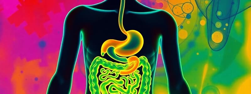
Alimentary Canal
- Starts at the oral cavity
- Includes the pharynx, esophagus, small intestine, and large intestine
- Terminates at the anus
Accessory Organs of Digestion
- Salivary glands
- Pancreas
- Liver
- Gallbladder
Functions of Digestive System
- Intake and digestion of food.
- Absorption of digested food particles.
- Elimination of undigested waste products.
Sialography
- Radiographic examination of salivary glands and ducts
- Uses water-soluble iodinated contrast media.
- Visualizes salivary glands and calculi in their ducts
Sialography Indications
- Inflammatory lesions (obstructed or not obstructed)
- Pain or swelling in the area
- Palpable mass
- Calculi
- Strictures
- Tumor
- Infection
- Dryness of mouth and eyes
Sialography Procedures
-
Plain films can determine calculi and calcified cervical glands.
-
A 2ml syringe and lemon juice are used to promote salivation and identify the orifice of the duct.
-
1-2ml of contrast media is injected.
Salivary Glands
-
Parotid (Stensen's duct)
-
Submandibular/submaxillary (Wharton's duct)
-
Sublingual (Bartholins duct)
Parotid Gland Radiographic Views
- AP
- Lateral
- Lateral Oblique
Parotid Gland - AP View
- Patient supine, head rotated away 50 degrees
- Central ray level of the lower lip
Gugliantini Method
- For infants
- Patient prone
- Central ray 20-250 cephalad
Hamptons Method
- Demonstrates the leaf-like pattern of the pylorus and bulb
- LPO position
Poppel's Method
- Demonstrates retrogastric space and evaluates for pancreatic mass
- Right lateral recumbent position
Wolf Method
- Applies greater intra-abdominal pressure.
- Demonstrates small lesions and sliding gastroesophageal herniations.
- RAO position
- Rotated body 40-45 degrees
- Right posterior, T6/T7
- Central ray 10-200 caudally
Biphasic Exam
- Gastrointestinal examination with single and double contrast study performed on the same day.
Small Intestine (SIS/Ba. Meal Follow Through)
- Extends from pyloric sphincter to ileocecal valve
- Averages 22 feet (6 and 1/2 meters) long
- Proximal 1 and 1/2 inch (4 cm)
- Distal part 1 inch (2-5 cm)
- Duodenum is 8-10 inches (20-25 cm)
Parts of the Small Intestine
- Duodenum
- Jejunum
- Ileum
Duodenum
- First part, shortest and widest.
- 8-10 inches (20-25 cm)
- Located in the right upper quadrant and left upper quadrant.
Jejunum
- Located in left upper quadrant and left lower quadrant.
- 3-3.5 cm diameter.
Ileum
- Right lower quadrant and left lower quadrant.
- Longest portion of the small intestine.
- 2.5 cm diameter.
Small Intestine Procedures
- Oral administration (by mouth)
- Complete reflux examination
- Intubation examination
- Enteroclysis
Small Intestine Indications
- Pain
- Diarrhea
- Bleeding
- Partial obstruction
- Abdominal mass
- Failed small bowel enema
- Enteritis (inflammation of the small intestine)
- Giardiasis (infection of the small intestine)
- Ileus
- Meckel's diverticulum
Meckel's Diverticulum
- Saclike outpouching of the intestinal wall.
- Birth defect.
Small Intestine Contraindications
- Complete obstruction
- Suspected perforation
Oral Contrast for Small Intestine
- Telepaque (Ioponoic acid) - 6 tablets
- Cholebrin (Iocetanic acid) - 6 tablets
- Biloptin (Sodium iopadate) - 6 tablets
- Solu-biloptin (Calcium iopadate) - 3 grams/sachet
Oral Contrast Administration
-
5-6 tablets given 10-12 hours before exam for normal function
-
No food 10-12 hours before exam
-
Encourage water intake.
Cholecystography
-
Evaluates the liver and its ability to remove contrast medium from the bloodstream and excrete it into the bile.
-
Determines the patency of the biliary ducts.
-
Evaluates the contracting and emptying power of the gallbladder.
-
Detects biliary calculi.
Cholecystography Indications
- Nausea
- Heartburn
- Vomiting
- Cholelithiasis
Cholelithiasis
- Abnormal calcifications or stones in the gallbladder.
Cholecystography Indications (Continued)
- Milk calcium bile
- Non-visualization
- Cholecystitis
- Acute or chronic inflammation of the gallbladder.
- Blockage of the cystic duct due to a stone lodge in the neck of the gallbladder.
- Biliary stenosis
- Congenital anomalies
Cholecystography Contraindications
- Hepatorenal disease with renal impairment
- Active GI disease (vomiting, severe diarrhea)
- Pyloric obstruction
- Severe Jaundice
- Liver dysfunction
- Hypersensitivity to iodinated contrast media
- Pregnancy
Cholecystography Procedures
- AP
- Oblique (RAO)
- Post motor meal (evaluate contracting power of the gallbladder)
- Erect LAO
- Right Lateral Decubitus
Cholesystography - AP View
- Demonstrates the presence and location of the gallbladder
- Demonstrates choleliths.
Cholecystography - RAO View
- Delineates trapped gas in the bowel from radiolucent stones in the gallbladder.
- 15-30 degree body rotation.
Cholecystography - Fatty Meal Post Motor Meal
Cholecystography - Erect LAO View
- Demonstrates floating gallstones
- 20 degree body rotation.
Cholecystography - Right Lateral Decubitus View
- Demonstrates stones heavier than the bile.
- Stones lighter than the bile can be visualized only by stratification (dropping).
Cholangiography (Choledochography)
- Demonstrates the hepatic and common bile ducts during an operation, especially patency and retained calculi.
Cholangiography Procedures
- Operative cholangiography
- Post-operative (T-tube) cholangiography
- Percutaneous cholangiography
- Intravenous cholangiography
Operative Cholangiography
- Contrast medium is introduced directly into the common bile duct (CBD).
Post-Operative Cholangiography (T-Tube Method)
-
Contrast medium is introduced through the T-tube.
-
Done 10 days after surgery.
-
RPO position.
-
Water-soluble contrast medium
-
25-30% concentration
Percutaneous Cholangiography
-
Visualizes the biliary tract where the contrast medium is injected directly through the lateral abdominal wall, into the intrahepatic bile ducts via a thin flexible needle.
-
Performed in patients with jaundice of unknown cause.
-
Pre-operative radiographic exploration of the biliary tract.
Intravenous Cholangiography
- Investigates the biliary duct system, especially in a patient whose gallbladder has been removed.
- 30 ml contrast medium intravenously.
- If the gallbladder is present, a fatty meal may be given.
Urinary System
- Kidneys are paired, bean-shaped organs.
- Located in the retroperitoneal space, posteriorly on either side of the vertebral column.
- Upper portion of the abdomen.
- Remove waste materials from the blood and eliminate them as urine.
Kidney Functions
- Control of water balance.
- Control of blood pH.
- Control of electrolyte balance.
- Excretion of waste products and drugs.
- Control of blood pressure.
Intravenous Pyelogram (IVP) Views
- AP urogram
- RAO and LAO
- Prone
- Postvoiding
Intravenous Pyelogram (IVP) Procedures
- Immediate (10-14 seconds after injection) - to show the nephrogram (renal parenchyma, renal tubules).
- 1 minute - to see nephrogram (enables visualization of filling defects).
- 5 minutes - to see early calyceal filling or filling defects.
- 10 minutes -to show the filled calyces
- 15 minutes - to demonstrate the ureters.
- 20 minutes - to show drainage of the kidneys, ureters, and bladder filling.
- 30-45 minutes - to open out the calyces or remove overlying opacities.
- Post micturation to assess bladder emptying.
Compression
- Compression should be applied to the bladder.
- Slows drainage of urine from the calyceal system.
- Fills out the calyces for clearer images.
- Midline, upper margin of the sacrum.
Compression Contraindications
- Renal failure
- Obstruction
- Children
- Recent abdominal surgery
Intravenous Pyelogram (IVP) Additional Views
- Tomography - to help assess filling defects by removing overlying gas shadows.
Intravenous Pyelogram (IVP) Dosage Protocol for Children
- 0-1 year - 3ml/kg
- 1-2 years - 30 ml/kg
- 2-8 years - 40 ml/kg
- 8-18 years - 50 ml/kg
Hypertensive IVP
- Increased blood pressure causes an increase in excretion rate.
Urethrograms
- Examines the urethra to determine the caliber, or width, of the urethra.
Urethrogram Indications
- Strictures
- Tears
- Congenital abnormalities
- Fistulas
- After surgery
- Diverticula
- Urethral valves
Urethrogram Contraindications
- Not mentioned in text.
Contrast Media
- Purpose: To make structures visible, increase contrast in images
- Types:
- Positive: Increase density, appear white on images
- Negative: Decrease density, appear darker on images
Reactions to Contrast Media
- Histamine Imbalance: Occurs immediately after injecting contrast, common in patients with allergies to contrast media
- Hemodynamic: Can cause systemic shock (sudden blood pressure drop), myocardial infarction, renal shutdown, hypertension, and urticaria
- Psychosomatic: Transient effect linked to patient anxiety, fatigue, or dehydration
- Technique: Extravasation (injection outside the vessel) causes burning pain, hematoma, numbness, and vascular constriction
- Pyrogenic: Reaction without a known cause that leads to a rise in body temperature
Effects of Hemodynamic Reaction
- Systemic Shock: Sudden drop in blood pressure after contrast injection
- Myocardial Infarction: Heart attack
- Renal Shut-down: Kidney failure
Effects of Contrast Media on Organs
- Chemotoxic: Toxic to living organisms causing cell damage
- Idiosyncratic: Reactions caused by the speed or amount of contrast injected
Major Reactions
- Cardiac Arrest: Heart stops beating
- Severe Bronchial Spasms: Prolonged contraction of the muscle in the wall of the bronchi
- Severe Hypertension: Extremely high blood pressure
Local Reactions
- Pain
- Burning Sensation
- Itching
- Vomiting
- Rashes
- Increased Salivation
- Increase of Tears
- Choking
Selecting the Appropriate Contrast Media
- Non-toxic when administered
- Produces adequate contrast
- Suitable viscosity - The quality of a sticky fluid
- Suitable persistence - It stays visible in the body long enough to obtain images
Methods of Introducing Contrast Media
- Ingestion: By mouth
- Retrograde Administration: Through a natural opening, but backwards
- Intrathecal: Injection of contrast into a sheath
- Parenteral: Injection into the bloodstream
- IV (intravenous): Most common route
- Intra-arterial: Directly into an artery
Properties of BaSo4 (Barium Sulfate)
- Absorbs water
- High atomic number - This makes it highly radiopaque, meaning it absorbs x-rays well
- Insoluble in water and stable - It doesn't dissolve in the body liquids
- Cannot be absorbed in the GIT (Gastrointestinal Tract): It passes through undigested
- Non-toxic: Generally safe for consumption
- Relatively cheap
Upper Gastrointestinal (UGI) System
- Alimentary Canal: The digestive system
- Oral cavity
- Pharynx
- Esophagus
- Small intestine
- Large intestine
- Anus
- Accessory Organs of Digestion
- Salivary Glands
- Pancreas
- Liver
- Gallbladder
Functions of the Digestive System
- Intake and/or digestion of food
- Absorption of digested food particles
- Elimination of unused material in the form of semi-solid waste
Sialography
- Radiographic examination of the salivary glands and ducts using water-soluble iodinated contrast media
- Purpose:
- Demonstrate the salivary glands and ducts
- Show calculi (stones) in the ducts
Indications for Sialography
- Inflammatory lesions (obstructed or not obstructed)
- Pain or swelling
- Palpable mass
- Calculi
- Strictures: Narrowing of a duct
- Tumor
- Infection
- Dryness of mouth & eyes
Sialography Procedure
- Plain films: To determine the presence of calculi and calcified cervical glands.
- 2 ml syringe: Used to inject the contrast
- Lemon juice: Stimulates salivation for better image quality
- 1-2 ml contrast media
Three Pairs of Salivary Glands
- Parotid gland (Stensen’s duct)
- Submandibular / submaxillary gland (Wharton’s duct)
- Sublingual gland (Bartholins duct)
Parotid Gland Projections:
- AP (Anteroposterior)
- Lateral
- Lateral Oblique
AP Projection:
- Position: Supine, head rotated away from the side being examined
- Central Ray (CR): Level of the lower lip
UGI Series (Upper Gastrointestinal Series)
- Biphasic Exam: Combines single and double contrast studies in a single day
Small Intestine
- Small bowel (SI) Series / Barium Meal Follow-Through
- Extends from the pyloric sphincter (stomach outlet) to the ileocecal valve (junction of the small and large intestines)
- Average Length: 22 feet (6.5 meters)
- Proximal part: 1.5 inches (4 cm) in diameter
- Distal part: 1inch (2-5 cm) in diameter
- Duodenum: 8-10 inches (20-25 cm) long
Parts of the Small Intestine
- Duodenum: Shortest and widest part
- 8-10 inches (20-25 cm) long
- Lies in the RUQ & LUQ (right and left upper quadrants)
- Jejunum: 3-3.5 cm in diameter
- Lies in the LUQ & LLQ ( left upper and left lower quadrants)
- Ileum : Longest part of the Small Intestine
- 2.5 cm in diameter
- RLQ & LLQ (right and left lower quadrants)
Small Bowel Series Procedures
- Oral: Ingestion of barium contrast
- Complete reflux exam: Contrast is passed both ways through the Small Intestine
- Intubation exam: Using a tube to introduce contrast
- Enteroclysis: Use of a tube to introduce a large volume of diluted barium into the small bowel
Small Bowel Series Indications
- Pain
- Diarrhea
- Bleeding
- Partial obstruction
- Abdominal Mass
- Failed small bowel enema
- Enteritis: Inflammation of small bowel
- Giardiasis: Infection of the small intestine
- Ileus: Obstruction of the small intestine
- Meckel’s diverticulum: Congenital outpouching of the intestinal wall
Small Bowel Series Contraindications
- Complete Obstruction: Total blockage
- Suspected Perforation: Hole in the intestinal wall
Small Bowel Series Contrast Media
- Telepaque: Ioponoic acid (6 tablets)
- Cholebrin: Iocetanic acid (6 tablets)
- Biloptin: Sodium iopadate (6 tablets)
- Solu-biloptin: Calcium iopadate (3 grams per sachet)
Cholecystography (Gallbladder Study)
- Purpose:
- Determine the function of the liver's ability to remove contrast media from the bloodstream and excrete it into bile
- Determine the patency (openness) of the biliary ducts
- Evaluate the contracting and emptying power of the gallbladder
- Detect gallstones
Indications for Cholecystography
- Nausea
- Heartburn
- Vomiting
- Cholelithiasis: Existence of gallstones
- Milk Calcium Bile: Presence of gallstones
- Non-visualization: Inability to visualize the gallbladder
- Cholecystitis: Inflammation of the gallbladder, can be acute or chronic
- Biliary stenosis: Narrowing of one of the biliary ducts
- Congenital anomalies: Abnormalities present at birth
Cholecystography Contraindications:
- Hepatorenal disease with renal impairment
- Active GI disease: Vomitting, severe diarrhea
- Pyloric obstruction
- Severe jaundice
- Liver dysfunction
- Hypersensitivity to iodinated contrast media
- Pregnancy
Cholecystography Procedure:
- AP (Anteroposterior)
- Oblique (RAO - Right Anterior Oblique)
- Post motor meal: To evaluate the contracting power of the gallbladder
- Erect LAO (Left Anterior Oblique)
- Right Lateral Decubitus
AP Projection:
- Demonstrates the presence and location of the gallbladder and any gallstones
RAO Projection:
- Delineates the difference between gas trapped in the bowel and a radiolucent (dark) gallstone
- 15-30 degrees body rotation is required
Fatty Meal/ Post Motor Meal:
- Evaluates the functioning of the gallbladder and the expulsion of bile after a fatty meal
Erect LAO Projection:
- Demonstrates floating gallstones
- 20 degrees body rotation is required
Right Lateral Decubitus Projection:
- Demonstrates gallstones in the dependent position
Male Reproductive System:
- Vesiculography
- Demonstrates the seminal ducts (tubes that carry sperm)
- Investigates abnormalities, such as:
- Cysts
- Abscesses
- Tumors
- Inflammation
- Sterility: Inability to have children
- Demonstrates the vas deferens (tubes that carry sperm from the testes to the seminal vesicles) and seminal vesicles using contrast media
Female Reproductive System:
- Hysterosalpingography
- Investigates the uterus (womb) and the uterine tubes (fallopian tubes)
- Delineates abnormalities of the uterus, such as:
- Polyps
- Submucous tumors (tumors in the lining of the uterus)
- Masses
- Fistula tracts (abnormal connections between organs)
- Evaluates the patency (openness) of the uterine tubes - Demonstrates the uterus and fallopian tubes, aiding in infertility investigations
Other Female Reproductive Imaging
- Vaginography - Investigates congenital abnormalities, vaginal fistulas (abnormal openings), and other pathological conditions of the vagina
- Fetography - Demonstrates the fetus in the uterus - Taken 18 weeks after gestation - Detects suspected abnormalities - Confirms suspected fetal death - Determines the presentation and position of the fetus - Determines whether the pregnancy is single or multiple
Pelvimetry
- Demonstrates the architecture of the maternal pelvis: Shape and bony structure of the pelvis
- Compares the size of the fetal head with the size of the maternal bony outlet: To assess if the baby will be able to pass through the birth canal
Discogram
- Demonstrates lumbar intervertebral discs by injection of 0.5 ml. of contrast media through a lumbar puncture needle.
Encephalography
- Demonstrates the ventricular and cisternal system (fluid-filled spaces) of the brain by injecting gas or air into the lumbar subarachnoid space (the space that surrounds the spinal cord and brain) or directly into the cisterna magna. The contrast media is used to visualize the ventricles(fluid-filled chambers in the brain).
Contrast Media Properties
- Absorbs water
- High atomic number
- Insoluble in water and chemically stable
- Cannot be absorbed in the gastrointestinal tract
- Non-toxic
- Relatively cheap
Alimentary Canal
- Starts at the oral cavity
- Continues with the pharynx, esophagus, small intestine, and large intestine
- Ends at the anus
Accessory Organs of Digestion
- Salivary glands
- Pancreas
- Liver
- Gallbladder
Functions of the Digestive System
- Intake and digestion of food
- Absorption of digested food particles
- Elimination of unused material as semi-solid waste
Sialography
- Radiographic examination of the salivary glands and ducts using water-soluble iodinated contrast media
- Used to visualize salivary glands and calculi in their ducts
Sialography Indications
- Inflammatory lesions (obstructed or not obstructed)
- Pain or swelling in the area
- Palpable mass
- Calculi
- Strictures
- Tumors
- Infections
- Dryness of mouth and eyes
Sialography Procedure
- Plain films assess for calculi and calcified cervical glands
- 2 ml syringe used for contrast administration
- Lemon juice used to stimulate salivation and identify the orifice of the duct
- 1-2 ml of contrast media administered
Salivary Glands
- Three pairs:
- Parotid (Stensen's duct)
- Submandibular/submaxillary (Wharton's duct)
- Sublingual (Bartholins duct)
Parotid Gland Projections
- AP: Patient supine, head rotated away 50 degrees, CR levels with the lower lip
- Lateral
- Lateral Oblique
Modified Valsalva Maneuver
- Tests the elasticity of the hypopharynx and piriform sinuses
Esophagography/Barium Swallow
- Demonstrates the pharynx and esophagus
- Indirectly investigates suspected lesions of the heart and great vessels
Esophagus
- Length: 10 inches / 25-30 cm
- Located:
- Posterior to the trachea and pharynx
- Anterior to the vertebral column
- Passes through the diaphragm in front of the aorta to enter the stomach
Esophagus Segments
- Cervical
- Thoracic
- Intra-abdominal
Esophagography Phases
- Filling Phase: Distends the lumen of the esophagus using a 2:1 or 3:1 ratio of barium to water
- Mucosal Phase: Demonstrates the mucosal pattern of the esophagus using a 4:1 ratio of barium to water
Esophagography Indications
- Dysphagia
- Barrett's esophagus/syndrome
- Thyroid gland enlargement
- Esophageal carcinoma
- Mediastinal mass
- Chalasia
- Achalasia
- Hiatal hernia
- Cardiomegaly
- Foreign bodies
Esophagography Contraindications
- Suspected leakage from the esophagus into the mediastenum, pleural, or peritoneal cavities
- Aspiration into the bronchial tree
Esophageal Reflux Tests
- Breathing exercises:
- Valsalva maneuver
- Mueller maneuver
- Water test: Swallowing a mouthful of water through a straw
- Telepaque (6-12 tablets)
- Food forbidden, water encouraged
Esophagography Contrast Media
- Telepaque (Ioponoic - 6 tabs)
- Cholebrin/iocetanic acid (6 tabs)
- Biloptin/Sodium iopadate (6 tabs)
- Solu-biloptin/Calcium iopadate (3 g/sachet)
Cholecystography Purpose
- To determine the function of the liver and its ability to remove contrast media from the bloodstream and excrete it in the bile
- To determine patency of the biliary ducts
- To evaluate the contracting and emptying power of the gallbladder
- To detect biliary calculi
Cholecystography Indications
- Nausea
- Heartburn
- Vomiting
- Cholelithiasis (condition with abnormal calcifications or stones in the gallbladder)
- Milk calcium bile (biliary stones in the gallbladder)
- Non-visualization
- Cholecystitis (acute or chronic inflammation of the gallbladder)
- Biliary stenosis
- Congenital anomalies
Cholecystography Contraindications
- Hepatorenal disease with renal impairment
- Active GI disease (vomiting, severe diarrhea)
- Pyloric obstruction
- Severe jaundice
- Liver dysfunction
- Hypersensitivity to iodinated contrast media
- Pregnancy
Cholecystography Procedure
- AP
- Oblique (RAO)
- Post motor meal (to evaluate the contracting power of the gallbladder)
- Erect LAO
- Right Lateral Decubitus
Cholecystography AP Projection
- Demonstrates the presence and location of the gallbladder
- Shows the presence of choleliths (gallstones)
Cholecystography RAO Projection
- Delineates between gas trapped in the bowel and radiolucent stones in the gallbladder
- 15-30 degrees body rotation
Cholecystography Fatty Meal (Post Motor Meal) Projections
- To evaluate the contracting power of the gallbladder
Cholecystography Erect LAO Projection
- Demonstrates floating gallstones
- Body rotation 20 degrees
Cholecystography Lateral Decubitus Projection
- Demonstrates gallstones heavier than bile
- Stones lighter than bile can be visualized only by stratification or layering
Cholangiography (Choledocography)
- Demonstrates the hepatic and common bile ducts during surgery, particularly focusing on patency and retained calculi
Cholangiography Types
- Operative Cholangiography: Contrast media is introduced directly into the common bile duct (CBD)
- Post-operative (T-tube): Contrast media is introduced through the T-tube (done 10 days after surgery)
- Percutaneous Cholangiography: Contrast media is introduced through the lateral abdominal wall into the intra-hepatic bile ducts (done when the patient has jaundice of unknown cause)
- Intravenous Cholangiography: Investigates the biliary duct system in patients who have had their gallbladder removed
Cholangiography Post-operative T-tube Procedure
- RPO position
- Water-soluble contrast media
- 25-30% concentration
Percutaneous Cholangiography
- Used to visualize the biliary tract by directing the contrast media through the lateral abdominal wall into the intra-hepatic bile ducts via a thin flexible needle.
- Performed when the patient has jaundice of unknown cause.
- Used for pre-operative radiographic exploration of the biliary tract.
Intravenous Cholangiography
- Investigates the biliary duct system, particularly in a patient who has had their gallbladder removed.
- 30 ml of contrast media is administered intravenously.
- A fatty meal may be given if the gallbladder is present.
Kidneys
- Pair of grand-like organs
- Located in the retroperitoneal space
- Posteriorly on either side of the vertebral column
- Upper portion of the abdomen
- Remove waste materials from the blood and excrete them as urine
- Bean-shaped organs
Kidney Functions
- Control water balance
- Control blood pH
- Control electrolyte balance
- Excretion of waste products and drugs
- Control of blood pressure
Intravenous Pyelogram (IVP) Projections
- AP
- RAO and LAO
- Prone
- Postvoiding
IVP Procedure
- Immediate: 10-14 seconds after injection to show the nephrogram (renal parenchyma and renal tubules)
- 1 minute: Demonstrates the nephrogram (enables visualization of filling defects in the renal parenchyma)
- 5 minutes: Shows early calyceal filling or filling defects
- Compression: Slows the drainage of urine from the calyceal system and fills out the calyces for clearer images
- 10 minutes: Shows the filled calyces
- 15 minutes: Demonstrates the ureters
- 20 minutes: Demonstrates the drainage of the kidneys, ureters, and bladder filling
- 30 and 45 minutes: Used to open out the calyces or remove overlying opacities
- Post micturation: Assess the quality of bladder emptying, any abdominal bladder shapes, and any reflux
- Delayed 24 hours: Demonstrates obstructive uropathy
IVP Compression Contraindications
- Renal failure
- Obstruction
- Children
- Recent abdominal surgery
IVP Additional Views
- Tomography: Used to remove overlying gas shadows or identify the extent of filling defects
IVP Dosage Protocol for Children
- 0-1 year: 3 ml/kg
- 1-2 years: 30 ml/kg
- 2-8 years: 40 ml/kg
- 8-18 years: 50 ml/kg
Hypertensive IVP
- Increased blood pressure causes an increased excretion rate
Urethrograms
- Examination of the urethra to assess the caliber of the urethra
Urethrogram Indications
- Strictures
- Tears
- Congenital abnormalities
- Fistulas
- Post-surgery
- Diverticulae
- Urethral valves
Urethrogram Contraindications
- Not mentioned in the text
Special Procedures with Contrast
- Modified Valsalva tests the elasticity of the hypopharynx and piriform sinuses.
Esophagography / Ba.Swallow
- Demonstrates the pharynx and esophagus.
- Indirectly investigates suspected lesions of the heart and great vessels.
Esophagus
- It is approximately 10 inches or 25-30 cm long.
- Located posterior to the trachea and pharynx.
- Anterior to the vertebral column.
- Passes through the diaphragm in front of the aorta to enter the stomach.
Segments of the Esophagus
- Cervical segment
- Thoracic segment
- Intra-abnormal segment
Esophagography Phases:
- Filling Phase
- Distends the lumen of the esophagus.
- 2:1 or 3:1 ratio of contrast agent to air.
- Mucosal Phase
- Demonstrates the mucosal pattern of the esophagus.
- 4:1 ratio of contrast agent to air.
Esophagography Indications:
- Dysphagia.
- Barret's esophagus/syndrome:
- Stricture in the distal esophagus.
- Developed peptic ulcer in the distal esophagus.
- Thyroid gland enlargement.
- Esophageal carcinoma (Ca of the esophagus).
- Mediastenal mass.
- Chalasia: Abnormal relaxation of the gastro-esophageal junction.
- Heartburn, retrosternal pain, regurgitation, and eructation.
- Achalasia: Abnormal constriction of the gastroesophageal junction.
- Hiatal hernia: Protrusion of the stomach through the wall of the diaphragm.
- Cardiomegally.
- Foreign body (FB).
Esophagography Contraindications:
- Suspected leakage from the esophagus into the mediastinum, pleural, or peritoneal cavities.
- Aspiration into the bronchial tree.
Esophageal Reflux Procedures:
- Breathing Exercises
- Valsalva maneuver: Forced exhalation against a closed glottis.
- Mueller maneuver: The patient exhales and tries to inhale against a closed glottis.
- Water Test: Swallow a mouthful of water through a straw.
- Hyposthenic: Modification of extreme asthenic stature (35% of the population).
- The stomach is approximately at T11 to L5 or even lower.
- Duodenal bulb is at the level of L3 or L4.
Upper Gastrointestinal Series (UGIS) - Air Barium Distribution
- Supine Position: The fundus of the stomach is in the lowest part, where the heavy barium settles.
- Prone Position: The fundus is in the highest position, causing the air to fill the fundus of the stomach.
- Erect Position: Air raises to fill the fundus, whereas barium descends by gravity to fill the pyloric portion of the stomach.
Peristaltic Activity of the GI Tract
- The passage of solid or semi-solid food from the mouth to the stomach takes 4 to 8 seconds, liquids pass in about 1 second.
- Waves of muscular contraction in the esophagus take 1 to 8 seconds.
- Food is churned or mixed with stomach fluid into a semi-fluid mass called chyme within 2 to 6 hours.
- Gastric emptying rate after an average meal is 2 to 6 hours.
- Chyme normally takes 3 to 4 hours to pass through the entire small intestine.
Pediatric UGI Preparation
- Infants under 1 year old: NPO for 4 hours.
- Children older than 1 years old: NPO for 6 hours.
Barium Meal / UGIS
- Radiographic examination of the gastrointestinal tract.
- The stomach is the dilated sac-like portion of the digestive tract extending from the esophagus to the small intestine.
- From the Greek word "GASTER".
- Dilated portion of the alimentary canal.
- Reservoir for swallowed foods and fluids.
Barium Meal/UGIS Examination
- Examination of the gastrointestinal tract function:
- Distal esophagus.
- Stomach.
- Duodenum.
Barium Meal/UGIS Indications
- Bezoar: A mass of undigested material trapped in the stomach.
- Trichobezoar: Ingested Hair.
- Gugliantini: Infant, prone.
- Demonstrates the stomach, CR 20-250 cephalad.
- Hamptons: Demonstrates the leaf-like pattern of the pylorus and bulb, LPO position.
- Poppel's Method: Demonstrates retrogastric space and evaluates pancreatic mass, right lateral recumbent position.
- Wolf Method: Applies greater intra-abdominal pressure to demonstrate small lesions and sliding gastroesophageal herniations.
- RAO position, body rotated 40-45 degrees.
- RPT 6/7, CR 10-200 caudally.
Biphasic Exam
- Gastrointestinal examination wherein single and double contrast study performed on the same day.
Small Intestine Series (SIS) / Ba. Meal Follow Through:
- Extends from the pyloric sphincter of the stomach to the ileocecal valve.
- Length averages about 22 feet (6 1/2 meters).
- Proximal 1 1/2 inches (4 cm) in diameter.
- Distal part 1 inch (2-5 cm) in diameter.
- The duodenum is 8-10 inches (20-25 cm) long.
Parts of the Small Intestine:
- Duodenum: First, shortest, and widest part, 8-10 inches (20-25 cm) long, located in the RUQ and LUQ.
- Jejunum: 3-3.5 cm diameter, located in the LUQ and LLQ.
- Ileum: Longest portion of the small intestine, 2.5 cm diameter, located in the RLQ and LLQ.
SIS Procedure:
- Oral: By mouth.
- Complete reflux exam: Complete examination of the esophagus.
- Intubation exam: Uses a tube to administer barium.
- Enteroclysis: Flush with barium mixed with saline.
SIS Indications:
- Pain.
- Diarrhea.
- Bleeding.
- Partial obstruction.
- Abdominal Mass.
- Failed small bowel enema.
- Enteritis: Inflammation of the small intestine.
- Giardiasis: Infection of the lumen of the small intestine.
- Ileus: Paralysis of the intestinal muscles.
- Meckel's diverticulum: Sac-like outpouching of the intestinal wall, birth defect.
SIS Contraindications:
- Complete obstruction.
- Suspected perforation.
- Intraluminal lesions such as polyps.
- Cleansing enemas shall be prohibited to patients having:
- Diarrhea
- Gross bleeding
- Symptoms of obstruction
Barium Sulfate
- 500-1200 ml barium sulfate.
- Warm: 85-90 degrees Fahrenheit (29-30 degrees Celsius).
- Cold: 41 degrees Fahrenheit (5 degrees Celsius).
- 15%-25% WV single contrast.
- 75%-95% WV double contrast.
Barium Enema (BE)
- Performed to evaluate the large intestine (colon).
- Often used as a diagnostic tool for various conditions of the colon.
- The patient typically receives a cleansing enema before the procedure.
- The enema is administered rectally, using a flexible tube.
- Barium sulfate is a contrast agent, which helps to highlight the colon on X-rays.
Reasons for using Warm Barium Sulfate:
- Produces less irritation.
- Stimulates tonic contraction of the anal sphincter.
- The container is typically placed 90 cm (36 inches) above the table to avoid undue discomfort to the patient.
- Barium solution should be 18 to 24 inches (45-60 cm) above the level of the anus.
- Use 90 cc of air to inflate the rectum for better visualization.
Sim's Position
- Used for BE procedures as it relaxes abdominal muscles.
- Reduces intra-abdominal pressure on the rectum.
- Promotes relaxation of the anal sphincter.
BE Indications
- Changes in bowel habits.
- Pain in the abdomen.
- Abdominal mass.
- Obstruction in the colon.
- Colitis: Inflammation of the large intestine; saw-tooth or "jagged" appearance.
- Polyps: Sac-like projections growing inward from the colon's lining.
- Volvulus: Twisting of a portion of the intestine, most common in males 20-50 years old, "beak" appearance.
- Intussusception: Telescoping or invagination of one part of the intestine into another, common in infants under 2 years old, "mushroom-shaped" appearance.
- Ulcerative colitis: Severe form of colitis, cobblestone appearance, can develop "stovepipe colon".
- Diverticulum: Common in adults over 40 years old.
- Neoplasm: Benign tumor, "apple-core or napkin ring" appearance.
BE Contraindications:
- Toxic megacolon: A rare but serious condition where the colon becomes dangerously enlarged.
- Pseudomembranous colitis: A serious infection of the colon caused by bacteria.
- Recent rectal biopsy.
- Incomplete bowel preparation.
- Recent barium meal.
BE Positions:
- AP/PA.
- LAO.
- RAO.
- Left lateral.
- UTI (not a BE position, likely a typo).
Retrograde Urethrography:
- Examination of the ureters and pelvicalyceal system.
Retrograde Urethrography Indications:
- Identify if an opacity is in the ureter.
- When previous examinations show an obstruction, but the cause cannot be identified.
Retrograde Urethrography Contraindications:
- Current urinary tract infection (UTI).
- Strictures (narrowing of the urethra).
Retrograde Urethrography Procedure:
- Control: Locates the position of a calculus to identify the tip of any catheters.
- 35-degree Oblique (RPO): Full length.
- A cystoscope is passed per urethra and bladder.
Retrograde Pyelography
- Non-functional examination of the urinary system (US).
- Directly injected into the pelvicalyceal system.
- Demonstrates the kidneys and ureters in cases of doubtful lesions.
Retrograde Pyelography Procedure
- A ureteric catheter is introduced via the urethra and bladder into the ureteric orifice.
- 35 cc of contrast medium is injected.
Retrograde Pyelography Indications:
- Demonstrates the site, length, and lower limit of an obstructive lesion.
- Demonstrates the pelvicalyceal system after unsatisfactory excretion urograms.
Retrograde Pyelography Contraindications:
- Urinary tract infection (UTI),
Cystoscopy:
- A non-functional examination of the urinary system (US).
- Demonstrates the bladder, urethra, and ureteric reflux.
- Contrast medium of 150 ml.
Routine Cystography Exams:
- AP Projection
- RPO and LPO Projections
- Lateral Projection
- Voiding Cystourethrogram (VCUG)
- PA Projection
- Chassard-Lapine Projection
Cystography with Double Contrast:
- Used to detect bladder tumors.
- Air or gas insufflation is used to enhance visualization.
Voiding Cystourethrogram (VCUG):
- Study of the urethra and evaluates the patient's ability to urinate.
VCUG Indications:
- Trauma to the urethra.
- Involuntary loss of urine (incontinence).
Male VCUG:
- Demonstrates the urethra and bladder neck.
- Requires 150 ml of contrast medium plus 20 ml of oily medium.
Female VCUG:
- Requires 150 ml of contrast medium plus 20 ml of a more dense oily medium.
- Outlines the base of the bladder and urethral catheter in situ.
- Demonstrates defects in the bladder mechanism.
AP Projection (VCUG):
- Performed during urination.
- Demonstrates defects in the bladder mechanism.
Urography:
- Radiographic investigation of the renal drainage or collecting system.
Pharynx
- Resonating chamber for sounds produced in the larynx
- Extends from the base of the skull to the cricoid cartilage
- Situated behind the nose, mouth and upper part of the throat
Parts of the Pharynx
- Nasopharynx: Lies behind the nose & above the soft palate
- Oropharynx: Lies behind the mouth and extends from the soft palate to the epiglottis
- Common to both respiratory and alimentary tracts
- Laryngopharynx: Extends from the upper border of the epiglottis to the lower border of the cricoid cartilage
- Connects with the oropharynx above and the esophagus below
Pharynx Imaging Indications
- Demonstrate tumor, abscess, and presence of foreign bodies
- Procedures: AP and lateral views
Laryngography
- Radiographic examination of the larynx
- Indications: Demonstrate muscular weakness due to disease, edema or fibrosis, investigation of malignancy
- Preparation: NPO for 5 hours, contrast media - Dionosil 10-15 ml
- Quiet inspiration: Test abduction of the vocal cords
- Normal Phonation: Test adduction of the vocal cords
- Take a deep breath and exhale slowly, phonate a high or low pitch
- Inspiratory phonation/reverse phonation/aspirant maneuver: Demonstrate the laryngeal ventricle
- Exhale completely and then slowly inhale to make a harsh, stridulous sound
- Valsalva maneuver: Shows complete closure of the glottis
- Test the elasticity and function integrity of the glottis
- Modified Valsalva: Test the elasticity of the hypopharynx and piriform
Esophagography/Ba. Swallow
- Demonstrate pharynx and esophagus
- Indirectly investigate suspected lesions of the heart and greater vessels
- Length: 10” / 25-30 cm
- Posterior to the trachea and pharynx
- Anterior to the vertebral column
- Passes through the diaphragm in front of the aorta to enter the stomach
Segments of the Esophagus
- Cervical Segment
- Thoracic Segment
- Intra-abnormal Segment
Esophagography - Two Phases
- Filling Phase: Distend the lumen of the esophagus - ratio 2:1 or 3:1
- Mucosal Phase: Demonstrate the mucosal pattern of the esophagus - 4:1 ratio
Esophagography Indications
- Dysphagia
- Barrett's esophagus/syndrome
- Stricture in the distal esophagus
- Developed peptic ulcer in the distal
- Thyroid gland enlargement
- Esophageal carcinoma - Ca of the esophagus
- Mediastenal mass
- Chalasia: Abnormal relaxation of the gastro-esophageal junction
- Heartburn, retrosternal pain, regurgitation and eructation
- Achalasia: Abnormal constriction of the gastroesophageal junction
- Hiatal hernia: Protrusion of the stomach through the wall of the diaphragm
- Cardiomegally
- Foreign bodies
Esophagography Contraindications
- Suspected leakage from the esophagus into the mediastenum, pleural or peritoneal cavities
- Aspiration into the bronchial tree
Esophageal Reflux Procedures
- Breathing exercises: Valsalva maneuver
- Mueller maneuver: Exhale and try to inhale against a closed glottis
- Water test: Swallow a mouthful of water through a straw
- Hyposthenic: Modification of extreme asthenic (35%)
- Stomach is approximately T11 to L5 or even lower
- Duodenal bulb is at the level of L3 or L4
Air Barium Distribution in the Stomach
- Supine: The fundus of the stomach is in the lowest part, where the heavy barium settles.
- Prone Position: The fundus is in the highest position, causing the air to fill this part of the stomach.
- Erect Position: Air raises to fill the fundus, while barium descends by gravity to fill the pyloric portion of the stomach.
Peristaltic Activity
- The passage of solid or semisolid food from the mouth to the stomach takes from 4 to 8 seconds, liquids pass in about 1 second
- Waves of muscular contraction in the esophagus: 1 to 8 seconds
- Food is churned or mixed with stomach fluid into a semifluid mass called CHYME in 2 to 6 hours
- Gastric emptying rate after an average meal is 2 to 6 hours
- Chyme normally takes 3 to 4 hours to pass through the entire small intestine
Pediatric UGI Preparation
- Infants under 1 year old: NPO for 4 hours
- Children older than 1 year old: NPO for 6 hours
Barium Meal / UGIS
- Radiographic examination of the GIT
- Stomach: Dilated sac-like portion of the digestive tract extending from the esophagus to the small intestine
- From the Greek word "GASTER"
- Dilated portion of the alimentary canal - reservoir for swallowed foods and fluids
Barium Meal / UGIS Examination
- Distal esophagus
- Stomach
- Duodenum
Barium Meal / UGIS Indications
- Bezoar - mass of undigested material trapped in the stomach.
- Trichobezoar: Ingested hair
- Dehydration
Barium Meal / UGIS Procedures
- AP
- Complete Reflux: Filling of the small bowel about 4500 ml. to fill the colon and small intestines
- Glucagon may be administered to relax the intestines
- Diazepam: Diminish discomfort during initial filling
Enteroclysis
- Procedure in which contrast media is introduced into the duodenum to examine the small bowel
- Special polyethylene tube (Bilbao or Sellink tube) is introduced through the esophagus into the stomach
- 30 ml of barium with 60 ml of water
- 1200 ml
- No cleansing enema
- Low residue diet
Intubation Method
- Procedure in which a tube is inserted through the nose and is passed into the stomach
- To relieve post-operative distention or deflate/decompress an obstructed small bowel
- Miller-Abbot
- Water-soluble iodinated or thin BaSo4
Hypotonic Duodenography
- Evaluation of post-bulbar duodenal lesions and for the detection of pancreatic disease
- Indications: Duodenitis, hepatitis, pancreatitis, abdominal mass affecting the C-Loop
Large Intestine/B-E/Lower GI Tract
- Study the form and function of the LI
- Detect any abnormal conditions
- 5 to 5 1/2 ft. (1 ½ to 3” diameter)
Large Intestine / B-E / Lower GI Tract Methods
- Single Contrast Method: Colon is examined with barium sulfate only
- 40-450F
- Reduction of intussusception
- Common in infancy
- Prolapse of one part of the intestine
- Double Contrast Method: Demonstrates the colon and the cecum
Vesiculography
- Radiographic examination of the seminal ducts
- Used to investigate abnormalities such as: cysts, abscesses, tumors, inflammation, sterility
- Demonstrate the vas deferens and seminal vesicle using contrast media
Hysterosalpingography
- Determine the uterus and uterine tubes
- Delineate lesions such as polyps, submucous tumors, masses, and fistula tract
- Investigate the patency of the uterine tube
- Demonstrate the uterus and fallopian tube, and investigate infertility
Vaginography
- Investigate congenital abnormalities, vaginal fistulas, and other pathologic conditions of the vagina
Fetography
- Demonstrate the fetus in the uterus
- Taken 18 weeks after gestation
- Detect suspected abnormalities of development
- Confirm suspected fetal death
- Determine the presentation and position of the fetus
- Determine whether the pregnancy is single or multiple
Pelvimetry
- Demonstrating the architecture of the maternal pelvis
- Comparing the size of the fetal head with the size of the maternal bony outlet
Discography
- Demonstrate lumbar intervertebral disks by injection of 0.5 ml. / lumbar puncture needle
Encephalography
- Demonstrate the ventricular and cisternal system of the brain by injection of a gas or air into the lumbar subarachnoid space or directly into the cisterna magna.
Lateral Projection
- Semi-prone position
- Extends the neck
- 1 inch (2.5 cm) superior to the mandibular angle
- Demonstrates bony structures and calcific deposits
- Highlights the parotid gland
Special Procedures with Contrast
- Performed to visualize anatomical structures and identify pathologies
Submandibular Gland
- Requires three projections:
- Lateral
- Infero-superior
- Lateral Oblique
Nasopharyngography
- Radiographic examination of the nasopharynx
- Contrast media is used to visualize the area
- Indications:
- Assessing the extent of nasopharyngeal tumors
- Investigating carcinoma
Nasopharyngography Procedure
- Two methods for instillation:
- Continuous breathing modified valsalva
- Contrast media is introduced through the nostrils
- Premedication with atropine 30 minutes prior to suppress secretions
- Contrast media: Dionosil aqueous (8-10 ml)
- Projections:
- SMV (OML 40-45° to horizontal)
- Lateral
Pharyngography
- Identifies abnormalities during swallowing
- Gunson Method:
- Synchronizes exposure with swallowing studies
- Uses a shoestring to monitor swallowing
Pharynx Functions
- Passageway for food
- Air passage from nasal cavity or mouth to larynx
- Identifies phytobezoar (ingested vegetation, fiber, or seeds)
Pharyngography Radiographic Appearance
- Light coating of barium sulfate (BaSo4)
- Visualizes:
- Diverticula (weakening and blind out pouching of the mucosal wall)
- Emesis (act of vomiting) to identify the cause
- Gastritis (inflammation of the stomach lining)
- Acute (severe pain/discomfort)
- Chronic (intermittent, often due to diet or stress)
- Dyspepsia (uncomfortable feeling of fullness, nausea, bloating)
- Gastrointestinal (GI) Hemorrhage
- Upper Abdominal Mass
- Partial Obstruction
- Hiatal Hernia
- Congenital
- Weakening of the muscle
- Sliding of Hiatal Hernia indicates the degree of herniation, visible radiographically
- Schatzke’s Ring (ringlike constriction at the distal esophagus)
- Ulcers
- Erosions in the stomach or duodenal mucosa
- Can cause weight loss
- Types of Ulcers:
- Peptic (ulceration of the mucous membrane due to acid gastric juice)
- Duodenal (type of peptic ulcer, common in the second and third aspect of the duodenum)
- Gastric (ulcer of the gastric mucosa)
- Perforating Ulcer (ulcer that extends through the entire thickness of the stomach or intestinal wall)
- Stomach Sections:
- Cardia
- Fundus (lies above the horizontal junction of the stomach and esophagus)
- Body
- Pyloric Portion
Stomach Contraindications:
- Complete large bowel obstruction
- History of bowel perforation or laceration
Stomach Functions
- Two openings:
- Cardiac Orifice (esophagus and stomach)
- Pyloric Orifice (stomach and small intestine)
Stomach Habitus
- Classified into three types:
- Eutonic or Normotonic (incisura and pylorus are at the same level)
- Hypotonic (pylorus is higher than incisura angularis by 1cm)
- Stone heavier than bile (stones lighter than bile are visible only by stratification)
Cholangiography (Choledocography)
- Examines hepatic and common bile ducts during surgery
- Evaluates patency and retained calculi
- Two types:
- Operative (contrast media is introduced directly into the common bile duct during surgery)
- Postoperative (T-Tube) where contrast media is introduced through the T-tube (10 days after surgery)
- Performed in RPO projection
- Uses water-soluble contrast media (25-30% concentration)
Percutaneous Cholangiography
- Visualizes the biliary tract by introducing contrast media through the lateral abdominal wall into the bile ducts via a thin, flexible needle.
- Performed for patients with jaundice of unknown cause.
- Offers pre-operative exploration of the biliary tract.
Intravenous Cholecystangiography
- Investigates the biliary duct system, especially for patients with a removed gallbladder.
- 30 ml of contrast media is administered intravenously
- If the gallbladder is present, a fatty meal might be given
Urinary System
- Includes: Kidneys, Ureters, Bladder, Urethra
Kidney Functions
- Bean-shaped organs lying retroperitoneally
- Control Water Balance
- Control Blood pH
- Control Electrolyte Balance
- Excretion of Waste Products and Drugs
- Control of Blood Pressure
- Activation of Vitamin D
Ureters
- Retroperitoneal and run along the psoas muscle, 5 cm from the midline
- Lie anterior to the kidney
Bladder
- Hollow muscular organ located behind the symphysis pubis
- Acts as a reservoir for urine and contracts to expel it
- Average capacity is 500 ml
Urethra
- Infraperitoneal structure with a length of 18-20 cm for males and 4 cm for females.
- Divided into three parts:
- Prostatic Part
- Membranous Part
- Spongy Part
Intravenous Pyelography (IVP) / Intravenous Urography (IVU)
- Investigates suspected urinary pathologies
- Demonstrates anatomy and physiology of the urinary tract
- Visualizes the minor and major calyces, renal pelvis, ureters, and bladder
Other Urinary System Procedures
- Percutaneous antegrade Pyelography
- Retrograde urography
- Cystoscopy
- Percutaneous renal puncture
- Angiographic procedures of the kidney and adrenals
Urinary System Indications for Special Procedures
- Calculi
- Infections
- Tumors
- Cysts
- Malformations of the urinary tract
- Obstruction
- Strictures
- Trauma
- Non-functioning kidney
Urinary System Contraindications for Special Procedures
- Renal Failure
- Severe Heart Disease
- Pregnancy
- Hypersensitivity
Urinary System Patient Preparation
- NPO (Nothing by Mouth) for 6-8 hours
- 50-100 ml of contrast media administered
- Restrict fluid intake to dehydrate the patient and prevent dilution of the contrast media
- Exceptions to fluid restriction:
- Diabetic patient
- Renal failure
- Children
- Recommended doses for children:
- Neonates: 4.0 ml/kg
- Babies: 3.0 ml/kg
- Small Children: 1.5 ml/kg
Urinary System Scout Film
- Verifies patient preparation
- Determines exposure technique
- Verifies positioning
- Detects any abnormal calcifications
Routine Urinary System Examinations
-
UTI (Urinary Tract Infection)
-
AP Projection
-
RPO and LPO Projections
-
Lateral Projection
-
Voiding Cystourethrogram (VCUG)
-
PA Projection
-
Chassard-lapine Projection
-
Double contrast using air or gas insufflation is recommended for detecting bladder tumors.
Voiding Cystourethrogram (VCUG)
- Evaluates the urethra and bladder function during urination.
- Indications:
- Trauma
- Involuntary loss of urine
Male VCUG
- Demonstrates the urethra and bladder neck
- 150 ml of contrast media plus 20 ml of oily medium used
Female VCUG
- 150 ml of contrast media plus 20 ml of a more dense oily medium is used
- Outlines the base of the bladder and urethral catheter in place
- Demonstrates defects in bladder mechanism
AP Projection
- Taken during micturation (urination)
- Demonstrates defects in bladder mechanism
- Useful for assessing the urethra
Retrograde Urethrography
- Examination of the ureters and pelvicalyceal system
- Indications:
- Identification of opacity in the ureter
- Obstruction with unclear cause from previous examinations
- Contraindications:
- Current urinary tract infection (UTI)
- Strictures (narrowing of the ureters)
Retrograde Urethrography Procedure:
- Control: To locate the position of a calculus and guide catheter insertion.
- 35° Oblique (RPO) projection: To capture the full length of the urethra.
- Cystoscope: Passed through the urethra and into the bladder.
Retrograde Pyelography
- Non-functional examination of the urinary system where contrast media is injected directly into the pelvicalyceal system.
- Indications:
- Visualization of the site, length, and nature of obstructive lesions.
- Examination of the pelvicalyceal system when excretion urograms are insufficient.
- Contraindications:
- UTI
Retrograde Pyelography Procedures:
- Urethral catheter introduced through urethra and bladder to the ureteric orifice
- 35 cc of contrast media administered
Cystoscopy
- Non-functional examination of the urinary system which demonstrates the bladder, urethra, and ureteric reflux.
- Uses 150 ml of contrast media.
Pharynx
- Resonating chamber for sounds generated in the larynx.
- Extends from the base of the skull to the cricoid cartilage.
- It is situated behind the nose, mouth, and the upper part of the throat.
Parts of the Pharynx
- Nasopharynx: Lies behind the nose and above the soft palate.
- Oropharynx: Lies behind the mouth, extends from the soft palate to the epiglottis. It is common to both the respiratory and alimentary tracts.
- Laryngopharynx: Extends from the upper border of the epiglottis to the lower border of the cricoid cartilage, leading to the esophagus. It connects to the oropharynx above and the esophagus below.
Laryngography
- Radiographic examination of the larynx.
- Indications:
- Demonstrates muscular weakness due to disease.
- Demonstrates edema or fibrosis.
- Investigates malignancy.
- Preparation:
- NPO for 5 hours.
- Contrast Media: Dionosil (10-15 ml).
Laryngography Procedures
- Quiet Inspiration: Tests abduction of the vocal cords.
- Normal Phonation: Tests adduction of the vocal cords. Patient takes a deep breath, then exhales slowly phonating a high or low pitch.
- Inspiratory Phonation/Reverse Phonation/Aspirant Maneuver: Demonstrates the laryngeal ventricle. Patient exhales completely, then inhales slowly creating a harsh, stridulous sound.
- Valsalva Maneuver: Shows complete closure of the glottis. Tests the elasticity and functional integrity of the glottis.
Hyposthenic
- Modification of extreme asthenic body type (35%).
- Stomach is at the level of T11 to L5 or even lower.
- Duodenal bulb is at the level of L3 or L4.
Air Barium Distribution
- In the supine position, the fundus of the stomach is in the lowest part where the heavy barium settles.
- In the prone position, the fundus is in the highest position, causing the air to fill this part of the stomach.
- In the erect position, air raises to fill the fundus, whereas barium descends by gravity to fill the pyloric portion of the stomach.
Peristaltic Activity
- Solid or semisolid food takes 4 to 8 seconds to pass from the mouth to the stomach, while liquids pass in about 1 second.
- Waves of muscular contraction in the esophagus take 1 to 8 seconds.
- Food is churned or mixed with stomach fluid into semifluid mass termed CHYME in 2 to 6 hours.
- Gastric emptying rate after an average meal is 2 to 6 hours.
- Chyme normally takes 3 to 4 hours to pass through the entire small intestine.
Pediatric UGI Preparation
- Infants under 1 year old: NPO for 4 hours
- Children older than 1 year old: NPO for 6 hours
Barium Meal / UGIS
- Radiographic Examination of the GIT.
- Examines the distal esophagus, stomach, and duodenum.
- Indications:
- Bezoar (mass of undigested material trapped in the stomach):
- Trichobezoar: Ingested hair.
- Stone: Stones heavier than bile are visible. Stones lighter than bile may only be visible through layering/dropping.
- Bezoar (mass of undigested material trapped in the stomach):
Cholangiography (Choledocography)
- Demonstrates the hepatic and common bile ducts during operation, specifically patency and retained calculi.
- Types:
- Operative Cholangiography: Contrast media is introduced directly into the CBD.
- Post-operative (T-Tube): Contrast media is introduced through the T-tube, done 10 days after surgery in RPO position using water-soluble contrast media (25-30% concentration).
Percutaneous Cholangiography
- Visualizes the biliary tract when contrast media is directly injected through the lateral abdominal wall into the intra-hepatic bile ducts using a thin flexible needle.
- Done in patients with jaundice of unknown cause. Also used for pre-operative radiographic exploration of the biliary tract/duct.
Intravenous Cholecystangiography
- Investigates the biliary duct system, particularly in patients who have had their gallbladder removed.
- 30 ml of contrast media is administered intravenously.
- A fatty meal may be given if the gallbladder is present.
Kidney
- A pair of bean-shaped organs, lying retroperitoneally in the upper abdomen on either side of the vertebral column.
- Remove waste materials from the blood and eliminate waste in the urine.
Kidney Functions
- Control water balance.
- Control blood pH.
- Control electrolyte balance.
- Excretion of waste products and drugs.
- Control of blood pressure.
- Prevention of UTI.
Retrograde Urethrography
- Examination of the ureters and pelvicalyceal system.
- Indications:
- Identify if an opacity is in the ureter.
- To identify the cause of an obstruction seen in a previous examination.
- Contraindications:
- Current UTI.
- Strictures.
- Procedure:
- Control: Identify the position of a calculus to identify the tip of any catheters.
- 350 Oblique (RPO): Full length.
- Cystoscope passed per urethra and bladder.
Retrograde Pyelography
- Non-functional examination of the urinary system directly into the pelvicalyceal system.
- Demonstrates the kidneys and ureters in cases of doubtful lesions by introducing a ureteric catheter via the urethra, bladder into the ureteric orifice.
- 35 cc of contrast media used.
Retrograde Pyelography Indications
- Demonstrate the site, length and nature of an obstructive lesion.
- Demonstrate the pelvicalyceal system after unsatisfactory excretion urograms.
Retrograde Pyelography Contraindications
- UTI.
Cystoscopy
- Non-functional examination of the urinary system.
- Demonstrates the bladder, urethra and ureteric reflux.
- 150 ml of contrast media used.
Routine Examination
- AP projection.
- RPO and LPO.
- Lateral projection.
- Voiding cystourethrogram.
- PA projection.
- Chassard-lapine.
Cystoscopy Notes
- Double contrast using air or gas insufflation is used to detect bladder tumors.
VCUG (Voiding Cystourethrogram)
- Studies the urethra and the patient's ability to urinate.
VCUG Indications
- Trauma or involuntary loss of urine.
VCUG for Male
- Demonstrates the urethra and bladder neck.
- 150 ml of contrast media plus 20 ml of oily medium is used.
VCUG for Female
- 150 ml of contrast media plus 20 ml of more dense oily medium is used to outline the base of the bladder and the urethral catheter in situ.
- Demonstrates defects in the bladder mechanism.
AP Projection During Micturation
- Demonstrates defects in the bladder mechanism.
Urography
- Radiographic investigation of the renal drainage or collecting system.
Vesiculography
- Radiographic examination of the seminal ducts.
- Used to investigate abnormalities such as:
- Cysts.
- Abscesses.
- Tumors.
- Inflammation.
- Sterility.
- Demonstrates the vas deferens and seminal vesicle using contrast media .
Hysterosalpingography
- Determines the uterus and uterine tubes.
- Delineates lesions like polyps, submucous tumours, masses and fistula tract.
- Investigates the patency of the uterine tube.
- Demonstrates the uterus and fallopian tube and used to investigate infertility.
Vaginography
- Investigates congenital abnormalities, vaginal fistulas and other pathologic conditions of the vagina.
Fetography
- Demonstrates the fetus in the uterus starting from 18 weeks of gestation.
- Detects suspected abnormalities of development.
- Confirms suspected fetal death.
- Determines the presentation and position of the fetus.
- Determines if the pregnancy is single or multiple.
Pelvimetry
- Demonstrating the architecture of the maternal pelvis.
- Comparing the size of the fetal head with the size of the maternal bony outlet.
Discography
- Demonstrates lumbar intervertebral disks by injection of 0.5 ml of contrast media through a lumbar puncture needle.
Encephalography
- Demonstrates the ventricular and cisternal system of the brain by injecting gas or air into the lumbar subarachnoid space or directly into the cisterna magna.
Contrast Media
- Used for opacification, visualization, and contrast studies.
Types of Contrast Media
- Positive: Used to increase the radiographic density of structures.
- Negative: Used to decrease the radiographic density of structures.
Contrast Media Reactions
- Histamine Imbalance: Occurs when the patient cannot tolerate the contrast media, leading to reactions such as hives, itching, and swelling.
- Hemodynamic: Reaction occurs upon injection, causing sudden drops in blood pressure, as well as potential myocardial infarction, renal shutdown, and immediate reactions like hypertension and urticaria.
- Psychosomatic: Mild transient effect associated with patient anxiety, fatigue, and dehydration.
- Technique: Complications include extravasation with burning pain, hematoma numbness, and vascular constriction.
- Pyrogenic: A reaction without apparent cause that tends to cause a rise in body temperature.
Major Reactions to Contrast Media
- Cardiac arrest.
- Severe bronchial spasms (prolonged contraction of muscles in the wall of the bronchi).
- Severe hypertension.
Local Reactions to Contrast Media
- Pain.
- Burning sensation.
- Itching.
- Vomiting.
- Rashes.
- Increased salivation.
- Increase of tears.
- Chocking.
Factors in Selecting the Right Contrast Media
- Must be non-toxic when administered.
- Must produce adequate contrast.
- Must have suitable viscosity (quality of a sticky fluid).
- Must have suitable persistence.
Methods of Introducing Contrast Media
- Ingestion: Taken orally.
- Retrograde Administration: Inserted into a body cavity through a natural opening.
- Intrathecal: Introduced into a sheath (e.g., the spinal canal).
- Parenteral: Injecting into the bloodstream, the most common method, which can be done intravenously or intra-arterially.
Properties of Barium Sulfate (BaSo4)
- Absorbs water.
- High atomic number, meaning it absorbs x-rays more effectively.
- Insoluble in water and stable.
- Cannot be absorbed in the gastrointestinal tract (GIT).
- Non-toxic.
- Relatively inexpensive.
Upper Gastrointestinal System
- Alimentary canal begins at the oral cavity, followed by the pharynx, esophagus, small intestine, large intestine, and terminates at the anus.
Accessory Organs of Digestion
- Salivary glands.
- Pancreas.
- Liver.
- Gallbladder.
Functions of the Digestive System
- Intake and/or digestion of food.
- Absorb digested food particles.
- Eliminate any unused material in the form of semi-solid waste products.
Sialography
- Radiographic examination of the salivary glands and ducts using water-soluble iodinated contrast media.
- Used to demonstrate salivary glands and calculi in their ducts.
Indications for Sialography
- Inflammatory lesions, obstructed or not.
- Pain or swelling in the area.
- Palpable mass.
- Calculi.
- Strictures.
- Tumor.
- Infection.
- Dryness of mouth and eyes.
Sialography Procedure
- Plain Films: Used to determine the presence of calculi and calcified cervical glands.
- 2 mL syringe: Used during the procedure.
- Lemon juice: Used to promote salivation and identify the orifice of the duct.
- 1-2 mL contrast media: Administered to the duct.
Three Pairs of Salivary Glands
- Parotid (Stensen's duct): Located near the ear, emptying into the mouth opposite the upper second molar.
- Submandibular/Submaxillary (Wharton's duct): Located under the jaw, emptying into the mouth under the tongue.
- Sublingual (Bartholin's duct): Located under the tongue, emptying into the floor of the mouth.
Parotid Gland Projections for Sialography
- AP: Patient supine, head rotated away 50 degrees, CR level of the lower lip. Demonstrates stone formation.
- Lateral: Patient semi-prone, extending the neck, RP 1” (2.5 cm) superior to the mandibular angle. Demonstrates bony structure, calcific deposits, swelling, and the three pairs of salivary glands.
- Lateral Oblique (PA Tangential): Patient PA, head rotated 10-15 degrees towards the lateral surface of the mandibular ramus.
Submandibular Gland Projections for Sialography
- Lateral
- Infero-superior
- Lateral Oblique
Nasopharyngography
- Radiographic examination of the nasopharynx following instillation of contrast media.
- Used to assess the extent of nasopharyngeal tumors and investigate carcinoma.
Methods for Nasopharyngography
- Continuous breathing modified valsalva: The patient breathes continuously while performing a modified valsalva maneuver (holding the breath while gently pushing out with the mouth closed).
- Contrast media is introduced/instilled through the nostrils: The contrast media is introduced through the patient's nostrils.
Premedication for Nasopharyngography
- Atropine (30 mins before exam): Used to suppress nasopharyngeal and buccal secretions.
Contrast Media Used for Nasopharyngography
- Dionosil aqueous, 8-10 mL.
Projections for Nasopharyngography
- SMV: OML 40-45 degrees to horizontal.
- Lateral
Pharyngography
- Used to identify abnormalities during the active progress of deglutition (swallowing).
Gunson Method for Pharyngography
- Synchronizes the exposure with the height of the swallowing studies of the pharynx and superior esophagus.
- A dark colored shoestring is tied snugly around the patient's throat above the thyroid cartilage to serve as a visual marker.
Functions of the Pharynx
- Acts as a passageway of food.
- Air passage from the nasal cavity or mouth to the larynx.
- Dehydration.
Procedures for Pharyngography
- AP
- Complete Reflux: Filling of the small bowel with approximately 4500 mL to fill the colon and small intestines. Glucagon may be administered to relax the intestines. Diazepam may be administered to diminish discomfort during initial filling.
Enteroclysis
- Procedure in which contrast media is introduced into the duodenum to examine the small bowel.
- A special polyethylene tube (Bilbao or Sellink tube) is introduced into the esophagus and into the stomach.
- 30 mL of barium is mixed with 60 mL of water, followed by 1200 mL of contrast media.
- No cleansing enema is required, and the patient is given a low residue diet.
Intubation Method
- A procedure in which a tube is inserted through the nose and passed into the stomach.
- Used to relieve post-operative distention or deflate or decompress an obstructed small bowel.
Miller-Abbott Tube
- Frequently used for intubation.
Contrast Media for Intubation Method
- Water-soluble iodinated contrast media or thin Barium sulfate.
Hypotonic Duodenography
- Evaluation of post-bulbar duodenal (the area of the duodenum after the bulb) lesions and for the detection of pancreatic disease.
Indications for Hypotonic Duodenography
- Duodenitis
- Hepatitis
- Pancreatitis
- Abdominal mass affecting the C-loop (the second curve of the duodenum).
Large Intestine/B-e/Lower GI Tract
- Study the form and function of the large intestine.
- Detect any abnormal conditions.
- The large intestine is approximately 5 to 5 1/2 feet long and between 1 ½ to 3 inches in diameter.
Methods for Barium Enema (BE)
- Single Contrast Method: The colon is examined with barium sulfate only.
- Double Contrast Method: Barium sulfate and air are used together to highlight the colon and cecum.
Contrast Media for Double Contrast Barium Enema
- Telepaque (Ioponoic acid): 6 or 12 tablets.
- Cholebrin (Iocetanic acid): 6 tablets.
- Biloptin (Sodium Iopadate): 6 tablets.
- Solu-biloptin (Calcium Iopadate): 3 g/sachet.
Purpose of Cholecystography (GB)
- Determine the liver's function and its ability to remove contrast media from the bloodstream and excrete it through the bile.
- Determine the patency (openness) of the biliary ducts.
- Evaluate the contracting and emptying power of the gallbladder.
- Detect biliary calculi (gallstones).
Indications for Cholecystography (GB)
- Nausea.
- Heartburn.
- Vomiting.
- Cholelithiasis.
- Milk calcium bile, which indicates biliary stones in the gallbladder.
- Non-visualization of the gallbladder.
- Cholecystitis.
- Biliary stenosis (narrowing of one of the biliary ducts).
- Congenital anomalies.
Contraindications for Cholecystography (GB)
- Hepatorenal disease with renal impairment.
- Active GI disease (vomiting, severe diarrhea).
- Pyloric obstruction.
- Severe jaundice.
- Liver dysfunction.
- Hypersensitivity to iodinated contrast media.
- Pregnancy.
Procedure for Cholecystography (GB)
- AP
- Oblique (RAO)
- Post motor meal: Used to evaluate the contracting power of the gallbladder.
- Erect LAO: Demonstrate floating gallstones.
- Right Lateral Decubitus: Demonstrate gallstones.
AP Projection for Cholecystography (GB)
- Demonstrates the presence and location of the gallbladder.
- Demonstrates the presence of choleliths (gallstones).
RAO Projection for Cholecystography (GB)
- Delineates between gas trapped in the bowel and radiolucent stones in the gallbladder.
- Patient is rotated 15-30 degrees.
Erect LAO Projection for Cholecystography (GB)
- Demonstrates floating gallstones.
- Patient is rotated 20 degrees.
Right Lateral Decubitus Projection for Cholecystography (GB)
- Demonstrates gallstones.
Fatty Meal for Post Motor Meal
- A high-fat meal is given to the patient to stimulate the release of bile.
BaSO4 Properties
- Barium Sulfate (BaSO4) is used as a contrast media in many special procedures
- BaSO4 absorbs water
- BaSO4 has a high atomic number
- BaSO4 is insoluble in water
- BaSO4 is stable
- BaSO4 cannot be absorbed in the gastrointestinal tract (GIT)
- BaSO4 is non-toxic
- BaSO4 is relatively cheap
Alimentary Canal
- The alimentary canal begins at the oral cavity
- The alimentary canal moves through the pharynx
- The alimentary canal moves through the esophagus
- The alimentary canal moves through the small intestine
- The alimentary canal moves through the large intestine
- The alimentary canal terminates at the anus
Accessory Organs of Digestion
- The salivary glands are an accessory organ of digestion
- The pancreas an accessory organ of digestion
- The liver an accessory organ of digestion
- The gallbladder is an accessory organ of digestion
Functions of the Digestive System
- The digestive system is responsible for the intake and digestion of food.
- The digestive system is responsible for absorbing digested food particles.
- The digestive system is responsible for eliminating any unused material in the form of semi-solid waste product.
Sialography
- Sialography is a radiographic examination of the salivary glands and ducts using a water-soluble iodinated contrast media.
- Sialography is used to demonstrate the salivary glands and calculi in their ducts.
Sialography Indications
- Sialography is used for inflammatory lesions, obstructed or non-obstructed
- Sialography is used for pain or swelling in the area
- Sialography is used for palpable mass
- Sialography is used for calculi
- Sialography is used for strictures
- Sialography is used for tumour
- Sialography is used for infection
- Sialography is used for dryness of the mouth and eyes
Sialography Plain Films
- Plain Films are used to determine if there are calculi or calcified cervical glands
- Plain Films use a 2 ml syringe
- Lemon juice is used to promote salivation and to identify the orifice of the duct
- 1 to 2 ml of contrast media is used
Pairs of Salivary Glands
- The Parotid (Stensen’s duct) is a pair of salivary glands
- The Submandibular/submaxillary (Wharton’s duct) are a pair of salivary glands
- The Sublingual (Bartholins duct) are a pair of salivary glands
Parotid Gland
- The parotid gland can be used to demonstrate stone formation
- The parotid gland is demonstrated by 1. AP, 2. Lateral, and 3. Lateral Oblique views
Parotid Gland AP View
- The patient should be in the supine position, head rotated away 50 degrees
- The CR (central ray) should be at the level of the lower lip
Body Types
- Asthenic body type has a long and slender body
- Hyposthenic body type - a modification of the asthenic type - 35%
- Sthenic body type - a modification of the asthenic type - 50%
- Hypersthenic body type - a modification of the asthenic type - 75%
- Extreme Asthenic body type - a modification of the asthenic type - 100%
UGI (Upper Gastrointestinal System)
- The UGI system is a system that examines the digestive system from the esophagus to the duodenum.
- An UGI is usually performed via a "barium meal"
- In an UGI, barium absorbs fluid and moves through the body, filling the digestive system
- The position of the body determines the distribution of barium and air in the stomach during an UGI
Air Barium Distribution in the Stomach
- Supine position - the fundus of the stomach is in the lowest part, where the heavy barium settles.
- Prone position - the fundus is in the highest position, causing the air to fill this part of the stomach.
- Erect position - air raises to fill the fundus, whereas barium descends by gravity to fill the pyloric portion of the stomach.
Peristaltic Activity
- The passage of solid or semisolid food from the mouth to the stomach takes from 4 to 8 seconds, whereas liquids pass in about 1 second.
- Waves of muscular contraction in the esophagus take 1 to 8 seconds.
- Churned or mixed with stomach fluid into a semifluid mass termed CHYME in 2 to 6 hours.
- Gastric emptying rate after an average meal is 2 to 6 hours.
- Chyme normally takes 3 to 4 hours to pass through the entire small intestine
Pediatric UGI Preparation
- For infants under 1 year old: NPO (nothing by mouth) for 4 hours
- For children older than 1 year old: NPO (nothing by mouth) for 6 hours
Barium Meal / UGIS
- Upper Gastrointestinal Series (UGIS) is a radiographic examination of the GIT (gastrointestinal tract).
- The stomach is a dilated sac-like portion of the digestive tract extending from the esophagus to the small intestine.
- Stomach is from the Greek word GASTER.
- The stomach is the dilated portion of the alimentary canal
- The stomach is a reservoir for swallowed foods and fluids.
Barium Meal / UGIS
- Barium Meal is the examination of the GIT and its function including the distal esophagus, stomach, and duodenum.
Bezoar
- Bezoar is a mass of undigested material trapped in the stomach.
- Trichobezoar is ingested hair that forms a bezoar.
- Steerhorn bezoar - the incisura is higher by 1 cm than the pylorus.
Barium Meal / UGIS - Indications
-
Barium Meal / UGIS - NPO 6-8 hours before the procedure
-
6 hours delayed*
-
Barium Meal / UGIS - to assess gastric emptying rate
-
24 hours delayed*
-
Barium Meal / UGIS - to assess suspected pathologies in the small intestine, appendix or colon
BaSO4 Preparation
- 1 yr.= 2 to 4 oz.
- 1- 3 yrs.= 4 to 6 oz.
- 3 – 10 yrs.= 6 t0 12 oz.
- Older than 10 yrs.= 12 to 16 oz.
- 30 – 50% Weight Volume.
- 50-80 ml. - esophagus.& mocusal pattern of the stomach.
- 300 ml – stomach & duodenum.
- 100 g/ 8 oz of water.
Single Contrast
- Single Contrast is used to demonsrate gross pathology only
- Single Contrast is used for children
- Single Contrast is used for very ill patients
Double Contrast
- Double Contrast is used to demonstrate mucosal pattern of the stomach and to detect small lesions.
- Gas-producing tablets are given
- The patient is rotated 360 degrees
- This helps coat gastric mucosa
Barium Meal / UGIS - Views
- PA View - demonstrates the relationship of the stomach and retrogastric space
- AP View - demonstrates the antrum/body of the stomach
- RAO (Right Anterior Oblique) - demonstrates the pyloric canal and duodenal bulb
- RLR (Right Lateral Recumbent) - demonstrates the duodenal loop and duodenojejunal junction
- LPO (Left Posterior Oblique) - demonstrates hiatal hernia
Barium Meal / UGIS - Modifications
- Gordon's Manuever - to demonstrate the pylorus and the bulb - prone - CR (central ray) 35 – 450 cephalad.
- Gugliantini - for infants - prone - CR (central ray) 20 – 250 cephalad.
- Hamptons – to demonstrate the leaf-like pattern of the pylorus and the bulb - LPO.
- Poppel’s method- to demonstrate the retrogastric space and evaluate pancreatic masses - right lateral recumbent.
- Wolf Method- to demonstrate small lesions, or sliding gastroesophageal herniations - RAO - rotated body 40-450 - R.P.T6 / T7 - CR (central ray) 10- 200 caudally
Bipasic Exam
- A Gastrointestinal examination where single & double contrast studies are performed on the same day.
SIS/ Ba.Meal Follow Through
- The small intestine (SI) extends from the pyloric sphincter of the stomach to the ileocecal valve.
- The SI averages about 22 ft. (61/2) meters
- The proximal portion of the SI is 1 ½ “ (4 cm.).
- The distal portion of the SI is 1” (2-5 cm.).
- The duodenum is 8-10 (20-25 cm.).
Small Intestine Parts
- Duodenum - The first part, shortest, widest
- Duodenum - located right upper quadrant (RUQ) & left upper quadrant (LUQ)
- Duodenum - 8-10” (20-25 cm.)
- Ileum - located right lower quadrant (RLQ) & left lower quadrant (LLQ)
- Ileum - the longest portion of SI
- Ileum - 2.5 cm diameter
- Jejunum - located LUQ & LLQ
- Jejunum - 3-3.5 cm diameter
SIS/ Ba.Meal Follow Through - Procedures
- Oral/by the mouth
- Complete reflux exam
- Intubation exam
- Enteroclysis
SIS/ Ba.Meal Follow Through - Indications
- Pain
- Diarrhea
- Bleeding
- Partial obstruction
- Abdominal mass
- Failed small bowel enema
- Enteritis - inflammation of the small intestine
- Giardiasis - infection of the lumen of the small intestine
- Ileus
- Meckel’s diverticulum - a saclike outpouching of the intestinal wall - a birth defect
SIS/ Ba.Meal Follow Through - Contraindications
- Complete obstruction
- Suspected perforation
- Dehydration
SIS/ Ba.Meal Follow Through - Procedures
- AP view
- Complete Reflux - filling of the small bowel about 4500 ml to fill the colon and small intestines - glucagon may be administered to relax the intestines - Diazepam may be administered to diminish discomfort during initial filling
- Enteroclysis - a procedure where the contrast media is introduced into the duodenum to examine the small bowel. A special polythene tube (Bilbao or sellink tube) is intro. to the esophagus into the stomach. 30 ml of barium with 60 ml of water is used, as well as 1200 ml in total. No cleansing enema is required and the patient should be on a low-residue diet.
- Intubation Method - A procedure where a tube is inserted through the nose and passed into the stomach. Used to relieve post-operative distention or deflate, or to decompress an obstructed small bowel. Miller-Abbott tubes are used. Water-soluble iodinated or thin BaSO4 is used.
Hypotonic Duodenography
- Evaluation of post bulbar duodenal lesions & for the detection of pancreatic disease.
Hypotonic Duodenography - Indications
- Duodenitis
- Hepatitis
- Pancreatitis
- Abdominal mass affecting the C-Loop
Large Intestine / B-E / Lower GI Tract
- The large intestine is also called the Lower GI Tract (LGI)
- The large intestine is about 5 to 5 1/2 ft. (1 ½ to 3” diameter)
- The purpose of B-E / Lower GI Tract is to study the form and function of the LGI and to detect any abnormal conditions.
- An LGI study is a procedure that uses barium to assess the large intestine
Large Intestine / B-E / Lower GI Tract - Methods
- There are two methods of performing an LGI study
- Single Contrast Method - This procedure examines the colon only and uses barium sulfate as a contrast media
- Single Contrast Method - The tube used for contrast is a 40-450F, with a reduction to make it easier to perform in infants (commonly used in cases of intussusception, which is a prolapse of one part of the intestine.)
- Double Contrast Method - This procedure demonstrates both the colon and the cecum.
- Double Contrast Method- This procedure also helps to demonstrate small lesions.
Intussusception
- Intussusception is a prolapse of one part of the intestine.
- Intussusception is common in infancy.
Lateral Projection
- Semi-prone position, extending the neck
- Radiographic position: 1 inch (2.5 cm) superior to the mandibular angle
- Demonstrates the bony structure and potential calcifications or swelling in the parotid gland area
- Demonstrates the three pairs of salivary glands
Submandibular
- Lateral projection
- Infero-superior projection
- Lateral oblique projection
Nasopharyngography
- Radiographic examination of the nasopharynx following contrast media instillation
- Indicates need to assess the extent of nasopharyngeal tumors and investigate carcinoma
- Two approaches:
- Continuous breathing modified Valsalva
- Contrast media introduced via nostrils
- Premedication with atropine 30 minutes before the exam to suppress nasopharyngeal and buccal secretions
- Contrast media used: Dionosil aqueous, 8-10ml
- Procedure Positions:
- SMV: OML 40-45 degrees to horizontal
- Lateral projection
Pharyngography
- Identifies abnormalities during the active progress of deglutition
- Gunson Method: Synchronize exposure with the height of the swallowing studies of the pharynx and superior esophagus
- Used to tie a dark-colored shoestring snugly around the patient’s throat above the thyroid cartilage
- Function of the Pharynx:
- Passageway for food
- Air passage from the nasal cavity or mouth to the larynx
- Resonating chamber for sounds produced in the larynx
- Extends from the base of the skull to the cricoid cartilage and is situated behind the nose, mouth, and upper part of the throat
Parts of The Pharynx
- Nasopharynx: Lies behind the nose and above the soft palate
- Oropharynx: Lies behind the mouth and extends from the soft palate to the epiglottis. Common to both the respiratory and alimentary tracts
- Laryngopharynx: Extends from the upper border of the epiglottis to the lower border of the cricoid cartilage, where it continues to the esophagus. Connects with the oropharynx above and the esophagus below.
Indications for Pharyngography
- Demonstration of tumors
- Demonstration of abscesses
- Demonstration of foreign bodies
Procedure Positions for Pharyngography
- AP (anteroposterior projection)
- Lateral (side view)
Laryngography
- Radiographic examination of the larynx
- Indicates the need to demonstrate muscular weakness due to disease
- Indicates the need to demonstrate edema or fibrosis and investigate malignancy
Preparation for Laryngography
- NPO for 5 hours
- Contrast media - Dionosil 10-15 ml.
- Procedures:
- Quiet inspiration: Test abduction of the vocal cords
- Normal Phonation: Test adduction of vocal cords. Instruct the patient to take a deep breath and then exhale slowly, phonating a high or low pitch.
- Inspiratory phonation/reverse phonation/aspirant maneuver: Demonstrate of the laryngeal ventricle. Exhale completely and then slowly inhale to make a harsh, stridulous sound
- Valsalva maneuver: Shows complete closure of the glottis, test the elasticity and functional integrity of the glottis.
- Compression technique: Use a compression paddle in prone to provide pressure
- Toe-touch maneuver: Study possible regurgitation into the esophagus from the stomach. Used for esophageal reflux and hiatal hernias
Esophagography
- Procedure Positions:
- AP or PA
- RPO: 35-40 degrees
- LPO
- RAO: To throw the esophagus clear of the spine
Indications for Esophagography
- Demonstrate esophageal varices. Instruct the patient to exhale fully and then follow the barium flow - avoid inhaling until exposure is made.
- Demonstrate entire esophagus
Body Habitus (Classification)
- Hypersthenic
- Sthenic
- Hyposthenic
- Asthenic
Hypersthenic
- Massive build
- Stomach level: T9 to T12
- Duodenal bulb level: T11 to T12
- Stomach & GB occupy high, almost horizontal position, less than 5% of the population fits into this category.
Sthenic
- Modification of Hypersthenic. Predominant type
- 50% of patients fit into this category.
- Stomach level: T10 to T11 to L2
- Duodenal bulb: L2
Asthenic
- Extremely slender build
- Stomach & GB are low, vertical, and near the midline
- Lower extreme, about 10% of the population.
- Stomach Level: T11 to L4
- Duodenal bulb level: L3
Steershorn
- Incisura is higher by 1 cm. than the pylorus.
Preparation for Upper Gastrointestinal Series
- NPO for 6-8 hours
- 6 hours delayed: Used in cases of suspected pyloric stenosis to assess the gastric emptying rate.
- 24 hours delayed: Used if there is suspicion in the small intestine, appendix or colon.
- Barium Sulfate (BaSO4) Preparation:
- 1 year: 2-4 oz.
- 1-3 years: 4-6 oz.
- 3-10 years: 6-12 oz.
- Older than 10 years: 12-16 oz.
- 30-50% weight volume - 50-80 ml for esophagus and mucosa pattern of the stomach
- 300 ml for stomach and duodenum
- 100 g per 8 oz of water
Single Contrast
- Used for gross pathology only
- Applicable for children and very ill patients.
Double Contrast
- Demonstrate mucosal pattern of the stomach and to detect small lesions
- Gas-producing tablets are given
- Rotate the patient 360 degrees to coat the gastric mucosa
Procedure Positions for a Upper Gastrointestinal Series
- PA: Demonstrate the relationship of the stomach and the retrogastric space (AP - Antrum/Body)
- RAO: Demonstrate the pyloric canal and duodenal bulb. (Antrum/Greater curve/peristalsis of the stomach)
- RLR: Demonstrate the duodenal loop, duodenojejunal junction, and the right retrogastric space
Variations for Upper Gastrointestinal Series
- LPO: Demonstrate a hiatal hernia. Head down by 25-30 degrees with 10-15 degrees of body rotation.
Modifications for Upper Gastrointestinal Series
- Gordons Maneuver: Demonstrate the pylorus and bulb. Patient is prone with CR 35-45 degrees cephalad. Demonstrate the greater and lesser curvatures and the antral portion of the stomach.
- UTI: Upper tract investigations
Retrograde Urethrography
- Examination of the urethra and pelvicalyceal system.
- Indications:
- Identify if an opacity is in the ureter.
- Previous examinations show obstruction, but the cause is unidentifiable.
- Contraindications: Current UTI or strictures.
Procedure for Retrograde Urethrography
- Control: Identify the position of a calculus to identify the tip of any catheters.
- 35 degrees Oblique (RPO): Full length.
- Cystoscope is passed per urethra and bladder.
- Positioning: AP
- Positioning: 35 degrees Oblique (RPO)
Retrograde Pyelography
- Non-functional examination of the urinary system, injecting contrast media directly into the pelvicalyceal system.
- Demonstrates the kidneys and ureters in cases of doubtful lesions.
- Urethral catheter is introduced via the urethra and bladder into the ureteric orifice. 35 cc of contrast media are injected
- Indications:
- Demonstrate the site, length, and nature of obstruction lesions
- Demonstrate the pelvicalyceal system after unsatisfactory excretion urograms.
- Contraindications: UTI
Cystoscopy
- Non-functional examination of the urinary system.
- Demonstrates the bladder, urethra, and ureteric reflux
- 150 ml of contrast media.
Routine Examination – Upper Urinary Tract
- AP projection
- *RPO and LPO (right posterior oblique and left posterior oblique)
- Lateral projection
- Voiding cystourethrogram
- PA projection
- Chassard-lapine
Indications for a Double Contrast Cystogram
- To detect bladder tumors.
- Air or gas insufflation is used.
Voiding Cystourethrogram (VCUG) - Indications
- Study of the urethra and evaluate the patient's ability to urinate.
- Evaluate trauma or involuntary loss of urine.
VCUG - Male
- Demonstrate the urethra and bladder neck.
- 150 ml + 20 ml of oily medium.
VCUG - Female
- 150 ml of contrast media + 20 ml of a more dense oily medium.
- Highlight the base of the bladder and the urethral catheter in situ.
- Demonstrate defects in bladder mechanism.
AP Projection - VCUG
- During micturation.
- Demonstrate defects in bladder mechanism.
Urography
- Radiographic investigation of the renal drainage or collecting system.
- Note: The text you provided is a list of procedures, and the detailed description of each procedure is often lacking. It might be helpful to refer to additional resources for a comprehensive study of each procedure
Studying That Suits You
Use AI to generate personalized quizzes and flashcards to suit your learning preferences.


