Podcast
Questions and Answers
Which type of aphasia involves patients who can speak but do not make sense?
Which type of aphasia involves patients who can speak but do not make sense?
- Transcortical sensory aphasia
- Broca's aphasia
- Wernicke's aphasia (correct)
- Mixed aphasia
In which brain region is the lesion typically located for patients with Broca's (expressive) aphasia?
In which brain region is the lesion typically located for patients with Broca's (expressive) aphasia?
- Adjoining parietal cortex
- Inferior frontal gyrus (correct)
- Frontal lobe
- Superior temporal gyrus
What type of aphasia results from a lesion involving the superior temporal gyrus or adjoining parietal cortex?
What type of aphasia results from a lesion involving the superior temporal gyrus or adjoining parietal cortex?
- Broca's aphasia
- Wernicke's aphasia (correct)
- Transcortical motor aphasia
- Global aphasia
Which term is associated with Broca's aphasia as a mnemonic for remembering the lesion location?
Which term is associated with Broca's aphasia as a mnemonic for remembering the lesion location?
When is aphasia likely to occur according to the provided information?
When is aphasia likely to occur according to the provided information?
Patients with Wernicke's aphasia typically have difficulty with:
Patients with Wernicke's aphasia typically have difficulty with:
What is the typical pattern of upper motor neuron deficits from lesions in the cerebral cortex?
What is the typical pattern of upper motor neuron deficits from lesions in the cerebral cortex?
Why does hyperglycemia and fever worsen brain injury due to ischemia?
Why does hyperglycemia and fever worsen brain injury due to ischemia?
In LMNL, what is a characteristic feature related to muscle tone?
In LMNL, what is a characteristic feature related to muscle tone?
What is a key difference between the impact of lesions in the cerebral cortex and those in the internal capsule?
What is a key difference between the impact of lesions in the cerebral cortex and those in the internal capsule?
What does a transient ischemic attack (TIA) involve?
What does a transient ischemic attack (TIA) involve?
What distinguishes an ischemic stroke from a hemorrhagic stroke?
What distinguishes an ischemic stroke from a hemorrhagic stroke?
Which of the following best describes the communication characteristics of patients with Wernicke's aphasia?
Which of the following best describes the communication characteristics of patients with Wernicke's aphasia?
In Broca's aphasia, patients are unable to:
In Broca's aphasia, patients are unable to:
Where is the typical anatomical localization of lesions for patients with Wernicke's aphasia?
Where is the typical anatomical localization of lesions for patients with Wernicke's aphasia?
What distinguishes Broca's aphasia from Wernicke's aphasia in terms of speech ability?
What distinguishes Broca's aphasia from Wernicke's aphasia in terms of speech ability?
Which region of the brain is affected in patients with Broca's aphasia according to the provided information?
Which region of the brain is affected in patients with Broca's aphasia according to the provided information?
What clinical deficit is common between patients with upper motor neuron lesions and those with lower motor neuron lesions?
What clinical deficit is common between patients with upper motor neuron lesions and those with lower motor neuron lesions?
What is a common feature of upper motor neuron lesions in the cerebral cortex?
What is a common feature of upper motor neuron lesions in the cerebral cortex?
What is the significant difference between resistance in biceps and triceps muscles in upper motor neuron lesions?
What is the significant difference between resistance in biceps and triceps muscles in upper motor neuron lesions?
Which characteristic is associated with LMNL rather than UMNL?
Which characteristic is associated with LMNL rather than UMNL?
Why does resistance typically occur in quads but not hamstrings in UMNL?
Why does resistance typically occur in quads but not hamstrings in UMNL?
What is a distinguishing factor between the impact of lesions in the internal capsule versus the cerebral cortex?
What is a distinguishing factor between the impact of lesions in the internal capsule versus the cerebral cortex?
What is a characteristic symptom associated with a transient ischemic attack (TIA)?
What is a characteristic symptom associated with a transient ischemic attack (TIA)?
Which type of aphasia results from a lesion in the superior temporal gyrus or adjoining parietal cortex according to the information provided?
Which type of aphasia results from a lesion in the superior temporal gyrus or adjoining parietal cortex according to the information provided?
What communication characteristics are typically observed in patients with Broca's aphasia?
What communication characteristics are typically observed in patients with Broca's aphasia?
In upper motor neuron lesions, what is a common clinical manifestation related to muscle tone?
In upper motor neuron lesions, what is a common clinical manifestation related to muscle tone?
What is a distinguishing feature between Broca's aphasia and Wernicke's aphasia?
What is a distinguishing feature between Broca's aphasia and Wernicke's aphasia?
What clinical deficit is commonly observed in lower motor neuron lesions?
What clinical deficit is commonly observed in lower motor neuron lesions?
Which brain region is associated with Broca's aphasia based on the provided information?
Which brain region is associated with Broca's aphasia based on the provided information?
What is the characteristic muscle tone change seen in upper motor neuron lesions?
What is the characteristic muscle tone change seen in upper motor neuron lesions?
Which type of stroke involves the death of brain tissues within 4-10 minutes of zero cerebral blood flow?
Which type of stroke involves the death of brain tissues within 4-10 minutes of zero cerebral blood flow?
Why does resistance occur in 'anti-gravity' muscles in upper motor neuron lesions?
Why does resistance occur in 'anti-gravity' muscles in upper motor neuron lesions?
What differentiates transient ischemic attacks (TIAs) from ischemic strokes?
What differentiates transient ischemic attacks (TIAs) from ischemic strokes?
In LMNL, what is a common feature related to muscle atrophy?
In LMNL, what is a common feature related to muscle atrophy?
What role does perfusion imaging play in the diagnosis of ischemic strokes?
What role does perfusion imaging play in the diagnosis of ischemic strokes?
What is the main purpose of using soft tissue treatment techniques before starting DMFR or HVLA for lumbar vertebrae dysfunction?
What is the main purpose of using soft tissue treatment techniques before starting DMFR or HVLA for lumbar vertebrae dysfunction?
When applying Direct Myofascial Release (DMFR) to a lumbar somatic dysfunction, what should be done after moving the planes of motion into restriction?
When applying Direct Myofascial Release (DMFR) to a lumbar somatic dysfunction, what should be done after moving the planes of motion into restriction?
During muscle energy technique for Type I lumbar somatic dysfunction, where should the patient place their hands?
During muscle energy technique for Type I lumbar somatic dysfunction, where should the patient place their hands?
What is a relative contraindication for applying Direct Myofascial Release (DMFR) to lumbar somatic dysfunctions?
What is a relative contraindication for applying Direct Myofascial Release (DMFR) to lumbar somatic dysfunctions?
In muscle energy technique for lumbar somatic dysfunctions, where should the physician stand in relation to the rotational component of the dysfunction?
In muscle energy technique for lumbar somatic dysfunctions, where should the physician stand in relation to the rotational component of the dysfunction?
What is a key step when utilizing post-isometric relaxation in muscle energy technique for Type I lumbar dysfunction?
What is a key step when utilizing post-isometric relaxation in muscle energy technique for Type I lumbar dysfunction?
How long should the isometric contraction be maintained during the osteopathic technique for Type II Dysfunction Example: L2 ERRSR Post-Isometric Relaxation?
How long should the isometric contraction be maintained during the osteopathic technique for Type II Dysfunction Example: L2 ERRSR Post-Isometric Relaxation?
In the Non-specific Supine Lumbar Roll osteopathic technique, what does the physician instruct the patient to do after maintaining isometric contraction?
In the Non-specific Supine Lumbar Roll osteopathic technique, what does the physician instruct the patient to do after maintaining isometric contraction?
What is the role of the physician's left hand during the lateral recumbent lumbar HVLA technique?
What is the role of the physician's left hand during the lateral recumbent lumbar HVLA technique?
What should the patient gently pull backward in the lateral recumbent lumbar HVLA technique?
What should the patient gently pull backward in the lateral recumbent lumbar HVLA technique?
In the Non-specific Supine Lumbar Roll osteopathic technique, what is the purpose of monitoring the transverse processes of L2 and L3?
In the Non-specific Supine Lumbar Roll osteopathic technique, what is the purpose of monitoring the transverse processes of L2 and L3?
During the Type II Dysfunction Example: L2 ERRSR Post-Isometric Relaxation, what should be monitored to localize side bending and rotation?
During the Type II Dysfunction Example: L2 ERRSR Post-Isometric Relaxation, what should be monitored to localize side bending and rotation?
Which type of barrier does the physician reposition the patient's trunk to the edge of in both osteopathic techniques described?
Which type of barrier does the physician reposition the patient's trunk to the edge of in both osteopathic techniques described?
What is instructed to be maximally improved at the dysfunctional segment after repeating steps 6 to 8 in both osteopathic techniques?
What is instructed to be maximally improved at the dysfunctional segment after repeating steps 6 to 8 in both osteopathic techniques?
During both osteopathic techniques, what is the least amount necessary to produce a palpable muscle twitch at the segmental level being monitored?
During both osteopathic techniques, what is the least amount necessary to produce a palpable muscle twitch at the segmental level being monitored?
What distinguishes Type II Dysfunction Example: L2 ERRSR Post-Isometric Relaxation from Non-specific Supine Lumbar Roll in terms of patient position during treatment?
What distinguishes Type II Dysfunction Example: L2 ERRSR Post-Isometric Relaxation from Non-specific Supine Lumbar Roll in terms of patient position during treatment?
What is the purpose of the rotational movement described in the text during the HVLA technique?
What is the purpose of the rotational movement described in the text during the HVLA technique?
In the HVLA technique for L5/S1 radiculitis, what is the significance of flexing the patient's hips and knees until L5 is fully flexed in relation to S1?
In the HVLA technique for L5/S1 radiculitis, what is the significance of flexing the patient's hips and knees until L5 is fully flexed in relation to S1?
Why does the physician place the caudad forearm in a line between the patient's PSIS and greater trochanter during the HVLA technique?
Why does the physician place the caudad forearm in a line between the patient's PSIS and greater trochanter during the HVLA technique?
What does it mean for the physician to deliver an impulse that separates L5 from S1 during the HVLA technique?
What does it mean for the physician to deliver an impulse that separates L5 from S1 during the HVLA technique?
What is the role of reassessing the severity of radicular symptoms after performing the HVLA technique for L5/S1 radiculitis?
What is the role of reassessing the severity of radicular symptoms after performing the HVLA technique for L5/S1 radiculitis?
Which action by the physician helps create joint gapping between L5 and S1 during the HVLA technique?
Which action by the physician helps create joint gapping between L5 and S1 during the HVLA technique?
Where is the urethra located in the penis?
Where is the urethra located in the penis?
What covers the glans in uncircumcised men?
What covers the glans in uncircumcised men?
Which structure forms the ejaculatory duct?
Which structure forms the ejaculatory duct?
What is the function of the epididymis in relation to sperm?
What is the function of the epididymis in relation to sperm?
Where is the external inguinal ring located?
Where is the external inguinal ring located?
Which structure forms the majority of the scrotum's underlying structure?
Which structure forms the majority of the scrotum's underlying structure?
What is the function of the vas deferens in relation to sperm?
What is the function of the vas deferens in relation to sperm?
What is the primary function of the seminal vesicle in male reproduction?
What is the primary function of the seminal vesicle in male reproduction?
Where is an indirect inguinal hernia most likely to develop?
Where is an indirect inguinal hernia most likely to develop?
Which structure is responsible for separating each testis into distinct compartments within the scrotum?
Which structure is responsible for separating each testis into distinct compartments within the scrotum?
For a dysfunction at L1 on L2, what is required in order to treat the dysfunction?
For a dysfunction at L1 on L2, what is required in order to treat the dysfunction?
In the lateral recumbent lumbar technique, what placement and thrust would make the technique not follow the definition of the dysfunction?
In the lateral recumbent lumbar technique, what placement and thrust would make the technique not follow the definition of the dysfunction?
In treating a Type I (Neutral) Dysfunction at L5, which technique is emphasized?
In treating a Type I (Neutral) Dysfunction at L5, which technique is emphasized?
What is one crucial step in determining the effectiveness of a lateral recumbent lumbar technique for Type II (Non-neutral) Dysfunction?
What is one crucial step in determining the effectiveness of a lateral recumbent lumbar technique for Type II (Non-neutral) Dysfunction?
In an HVLA technique for Type I Dysfunction, how is the shoulder usually moved during the impulse thrust?
In an HVLA technique for Type I Dysfunction, how is the shoulder usually moved during the impulse thrust?
What is emphasized in HVLA for Type II (Non-neutral) Dysfunction at L4?
What is emphasized in HVLA for Type II (Non-neutral) Dysfunction at L4?
What is the best position for examining the male genitalia to allow for better assessment of hernias or varicocele?
What is the best position for examining the male genitalia to allow for better assessment of hernias or varicocele?
What is the white cheesy material that may accumulate under the foreskin known as?
What is the white cheesy material that may accumulate under the foreskin known as?
What should a physician do if a patient reports a discharge from the male genitalia that is not visible during examination?
What should a physician do if a patient reports a discharge from the male genitalia that is not visible during examination?
What is the normal location of the urethral meatus during male genitalia examination?
What is the normal location of the urethral meatus during male genitalia examination?
Which structure should be palpated on the superior posterior surface of each testicle during examination?
Which structure should be palpated on the superior posterior surface of each testicle during examination?
How can abnormal swelling of the testicles during examination be further evaluated?
How can abnormal swelling of the testicles during examination be further evaluated?
What is the first priority when stroke is suspected in a patient?
What is the first priority when stroke is suspected in a patient?
What is the blood pressure threshold that should prompt a reduction in patients with suspected stroke?
What is the blood pressure threshold that should prompt a reduction in patients with suspected stroke?
What is the primary reason for lowering blood pressure in stroke patients?
What is the primary reason for lowering blood pressure in stroke patients?
When should supplemental oxygen be recommended in acute stroke cases?
When should supplemental oxygen be recommended in acute stroke cases?
Which vessels are typically involved in large-vessel intracranial occlusion in ischemic stroke?
Which vessels are typically involved in large-vessel intracranial occlusion in ischemic stroke?
What is the primary benefit of endovascular mechanical thrombectomy in acute stroke patients?
What is the primary benefit of endovascular mechanical thrombectomy in acute stroke patients?
In what time range from onset of stroke does IV thrombolytics show benefits to clinical outcomes?
In what time range from onset of stroke does IV thrombolytics show benefits to clinical outcomes?
What is the primary concern associated with giving IV thrombolytics to stroke patients?
What is the primary concern associated with giving IV thrombolytics to stroke patients?
What is the primary factor that can improve outcomes from endovascular therapy?
What is the primary factor that can improve outcomes from endovascular therapy?
What is the recommended range for maintaining blood glucose levels in patients with acute ischemic stroke?
What is the recommended range for maintaining blood glucose levels in patients with acute ischemic stroke?
Flashcards are hidden until you start studying
Study Notes
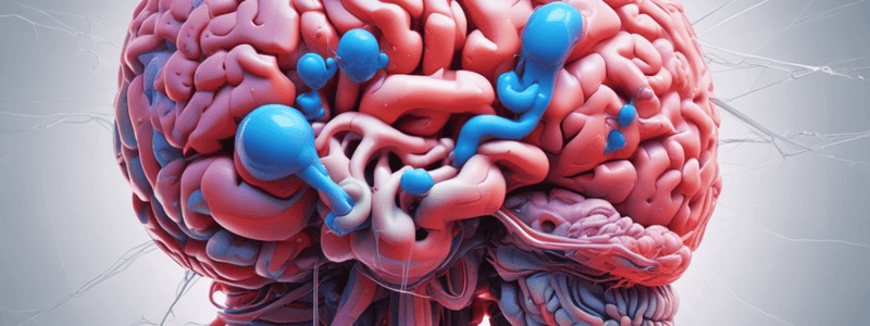
Aphasia
- Broca's (expressive) aphasia:
- Caused by lesions in the frontal lobe, specifically the posterior aspect of the inferior frontal gyrus
- Patients are unable to express spoken words
- "Can't bark out their thoughts"
- Wernicke's (receptive) aphasia:
- Caused by lesions in the temporal-parietal region, specifically the superior temporal gyrus or adjoining parietal cortex
- Patients cannot understand written or verbal language
- Can speak but do not make sense
- Aphasia only occurs when the dominant hemisphere is affected
Upper Motor Neuron (UMNL) vs. Lower Motor Neuron (LMNL) Lesions
- Upper Motor Neuron (UMNL) Lesions:
- Characterized by clumsiness to a greater degree than a loss of power
- Babinski sign is present immediately
- Decreased muscle tone initially, but increased tone (spasticity) develops later
- Spasticity is a unidirectional increase in muscular tone in "anti-gravity" muscles
- Resistance in biceps (not triceps) and quads (not hamstrings)
- Little or no atrophy
- Lower Motor Neuron (LMNL) Lesions:
- Characterized by loss of power to a greater degree than clumsiness
- Hypotonia
- Muscle atrophy
- Muscle fasciculations (involuntary twitching)
Upper Motor Neuron Deficits in Cerebral Cortex vs. Internal Capsule
- Cerebral Cortex:
- Specific fibers destined for one part of the body (e.g., leg and foot, but not face and arm)
- Contralateral effects
- Fibers are spread far apart
- Internal Capsule:
- All fibers on one side of the body
- Contralateral effects
- Fibers travel in a compact bundle
Transient Ischemic Attacks (TIAs) and Strokes
- Transient Ischemic Attacks (TIAs):
- Episodes of stroke symptoms that last less than 24 hours
- Blood flow is restored to ischemic tissue before significant infarction develops
- Ischemic Stroke:
- Caused by a lack of adequate blood flow
- Cerebral blood flow of zero causes death of brain tissues within 4-10 minutes
- Ischemic penumbra: ischemic tissue that can become restored surrounding a central area of infarction
- Treatment of Acute Ischemic Stroke:
- Do ABC's (Airway, Breathing, Circulation)
- Treat hyper/hypoglycemia
- Perform non-contrast head CT
- 6 Treatment Types:
- Medical support
- IV thrombolysis
- Endovascular revascularization
- Antithrombotic treatment
- Neuroprotection
- Stroke centers and rehabilitation
Osteopathic Techniques
- Non-specific Supine Lumbar Roll (5238):
- HVLA technique
- Given a segmental definition
- Lateral Recumbent Lumbar HVLA (5239):
- HVLA technique
- Given a segmental definition
- Direct Myofascial Release (DMFR) (5236):
- Physiology: engage myofascial layer to influence local and surrounding ligaments, articular processes, vasculature, and lymphatics
- Indications: restricted tissue, areas of hypertonicity, prominent tight-loose asymmetry of tissue
- Contraindications: sprain/strain, fracture, malignancy, osteopetrosis or osteopenia, infection
- Steps:
- Find dysfunction
- Move all planes of motion into restriction
- Hold in restriction until release
- Reassess
- Muscle Energy (5237):
- Type I Dysfunction: L2 NSLRR
- Post-Isometric Relaxation:
- Patient is seated
- Physician stands to the side opposite the rotational component of the dysfunction
- Patient places the right hand behind the neck and the left hand on the right elbow
- Soft Tissue Treatment (5235):
- Given a segmental definition of a lumbar vertebra
- Techniques:
- Prone Pressure
- Prone Traction
- B/L Thumb Pressure
- Scissor Technique
- Prone Pressure with Counterleverage
- Lateral Recumbent Position
- Supine Extension
- Lon Lever Counterlateral with Knees### Osteopathic Manipulative Treatment (OMT)
- HVLA (High-Velocity, Low-Amplitude) technique for L4/L5 dysfunction:
- Patient's foot does not touch the floor
- Physician's cephalad hand is in the antecubital fossa of the patient's left arm
- Physician's caudad hand stabilizes L5
- Patient's shoulder and pelvis are axially rotated in opposite directions
- During exhalation, further rotational slack is taken up
- If necessary, physician can grasp the patient's right arm to draw the shoulder forward
- Impulse is delivered with the forearms, moving the shoulder slightly caudad and the pelvis and sacrum cephalad
GYN/GU Demo
- Normal external female genitalia:
- Labeling required
- Normal internal female genitalia:
- Labeling required
- 5 P's of sexual history:
- Describe the 5 P's
- Anatomy of male genitalia:
- Penis:
- Shaft formed by 3 columns of vascular erectile tissue
- Corpus spongiosum contains urethra
- 2 corpora cavernosa
- Urethra located in ventral midline of the shaft
- Testes:
- Paired ovoid glands
- Tunica albuginea (fibrous outer coating)
- Scrotum (loose, wrinkled pouch of skin and underlying dartos muscle)
- Tunica vaginalis (serous membrane covering the testes)
- Epididymis:
- On posterolateral surface of each testis
- Softer, comma-shaped
- Contain tightly coiled tubules
- Vas deferens:
- Firm muscular cord
- Ascends from scrotal sac into the pelvic cavity
- Merges with seminal vesicle to form the ejaculatory duct
- Spermatic cord:
- Vas deferens + blood vessels + nerves + muscle fibers
- Penis:
- Inguinal canal:
- Medial to and parallel to inguinal ligament
- Forms tunnel for vas deferens
- Internal inguinal ring (1 cm above midpoint of inguinal ligament)
- External inguinal ring (triangular slit-like structure palpable just above and lateral to pubic tubercle)
- Inguinal hernias:
- Occur when loops of bowel force their way through the inguinal canal
- Indirect inguinal hernia (develop at internal inguinal ring)
- Direct inguinal hernia (arise more medially due to weakness in the floor of the inguinal canal)
Rectum and Prostate
- Rectum:
- Lies against sacrum and coccyx
- Merges with short segment of anal canal
- Extends from rectosigmoid junction to anorectal junction
- External margin of anal canal is poorly demarcated
- Note direction of anal cavity
- Anorectal junction (pectinate line): boundary between somatic and visceral nerve supply
- Contains 3 inward foldings (valves of Houston)
- Prostate:
- Surrounds urethra
- Lies next to bladder outlet
- Small during childhood, increases 5-fold between puberty and 20 years old
- R/L lateral lobes lie anterior against rectal wall
- Palpable as rounded, heart-shaped structure
- Separated by shallow median sulcus or groove
- Anterior and central areas of prostate cannot be examined
General Notes on Segmental Definitions of the Lumbar Spine
- If the dysfunction is at L1, L1 is restricted on L2, and L2 is not dysfunctional under L1
- If the dysfunction is at L1, L1 is not dysfunctional as it relates to T12
- To treat a dysfunction of L1 on L2, L1 must move through its restrictive barrier (bind) while L2 is either held stable in neutral or carried through the described ease of L1
HVLA for Type I (Neutral) Dysfunction
- Long-lever, rotational/side-bending emphasis
- Step-by-step instructions for example: L5 NSLRR
HVLA for Type II (Non-neutral) Dysfunction
- Long-lever, rotational/side-bending emphasis
- Step-by-step instructions for example: L4 FRRSR
Studying That Suits You
Use AI to generate personalized quizzes and flashcards to suit your learning preferences.


