Podcast
Questions and Answers
What is developmental biology?
What is developmental biology?
Developmental biology is the study of the sequence of events from the fertilization of a secondary oocyte by a sperm cell to the formation of an adult organism.
What period marks fertilization through the 8th week of development?
What period marks fertilization through the 8th week of development?
- Zygotic period
- Neonatal period
- Fetal period
- Embryonic period (correct)
Which period spans from week 9 until birth?
Which period spans from week 9 until birth?
- Morula period
- Fetal period (correct)
- Neonatal period
- Embryonic period
What is the neonatal period?
What is the neonatal period?
Fertilization usually occurs in the uterus.
Fertilization usually occurs in the uterus.
What is capacitation?
What is capacitation?
A clear glycoprotein layer between the corona radiata and oocyte plasma membrane is called the:
A clear glycoprotein layer between the corona radiata and oocyte plasma membrane is called the:
Polyspermy is fertilization by only one sperm.
Polyspermy is fertilization by only one sperm.
In the "fast block to polyspermy", what happens to the oocyte cell membrane?
In the "fast block to polyspermy", what happens to the oocyte cell membrane?
What does the oocyte divide into during meiosis II?
What does the oocyte divide into during meiosis II?
What is syngamy?
What is syngamy?
What is a zygote?
What is a zygote?
What is cleavage?
What is cleavage?
What are blastomeres?
What are blastomeres?
What is a morula?
What is a morula?
What is a blastocyst?
What is a blastocyst?
Which of the following is the outer cell layer of the blastocyst that becomes part of the chorion?
Which of the following is the outer cell layer of the blastocyst that becomes part of the chorion?
What does the inner cell mass (or embryoblast) of the blastocyst become?
What does the inner cell mass (or embryoblast) of the blastocyst become?
Roughly how many days after fertilization does the blastocyst attach to the endometrium of the uterine wall (implantation)?
Roughly how many days after fertilization does the blastocyst attach to the endometrium of the uterine wall (implantation)?
What is the decidua?
What is the decidua?
What does the trophoblast develop into around the 8th day?
What does the trophoblast develop into around the 8th day?
What two layers does the inner cell mass differentiate into?
What two layers does the inner cell mass differentiate into?
What is the bilaminar embryonic disc?
What is the bilaminar embryonic disc?
What does the chorion develop from?
What does the chorion develop from?
What is the role of human chorionic gonadotropin (hCG)?
What is the role of human chorionic gonadotropin (hCG)?
The amnion is a thick, structural membrane.
The amnion is a thick, structural membrane.
What fills the amniotic cavity?
What fills the amniotic cavity?
What are some key functions of the yolk sac?
What are some key functions of the yolk sac?
What are lacunae?
What are lacunae?
What is the connecting stalk?
What is the connecting stalk?
What process transforms the bilaminar embryonic disc into a trilaminar embryonic disc?
What process transforms the bilaminar embryonic disc into a trilaminar embryonic disc?
Name the 3 primary germ layers that make up the trilaminar embryonic disc.
Name the 3 primary germ layers that make up the trilaminar embryonic disc.
What tissues does the ectoderm become?
What tissues does the ectoderm become?
What does mesoderm become?
What does mesoderm become?
What does the endoderm become?
What does the endoderm become?
Name key structures that develop during gastrulation.
Name key structures that develop during gastrulation.
What is the notochord?
What is the notochord?
What is neurulation?
What is neurulation?
What are somites?
What are somites?
What is angiogenesis?
What is angiogenesis?
What is the fetal part of the placenta?
What is the fetal part of the placenta?
Name 3 functions of the placenta.
Name 3 functions of the placenta.
What does embryonic folding convert the embryo into?
What does embryonic folding convert the embryo into?
What are the pharyngeal (branchial) arches?
What are the pharyngeal (branchial) arches?
What is a teratogen?
What is a teratogen?
What is the maternal alphafetoprotein (AFP) test?
What is the maternal alphafetoprotein (AFP) test?
Flashcards are hidden until you start studying
Study Notes
- Developmental biology studies the sequence of events from fertilization of a secondary oocyte by a sperm cell to adult organism formation.
- The embryonic period is from fertilization through the 8th week of development.
- The fetal period is from week 9 until birth.
- The neonatal period is the first 28 days after birth.
Embryonic Period - 1st week - Fertilization
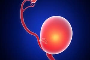
- Fertilization usually occurs in the uterine tubes.
- Sperm are unable to fertilize the oocyte until about 7 hours after ejaculation.
- During this time, sperm undergo capacitation in the female reproductive tract, causing their tails to beat more vigorously and preparing their plasma membranes to fuse with the oocyte.
- Only capacitated sperm can respond to the chemical factors produced by the surrounding cells of the ovulated oocyte.
- Penetration requires a capacitated sperm to penetrate the corona radiata and the zona pellucida.
- Acrosomal enzymes and strong movements help with penetration
1st week - Fertilization events
- Fusion of sperm cell with a 2º oocyte sets in motion events that block polyspermy, fertilization by more than one sperm
- "Fast block to polyspermy" is when the oocyte cell membrane depolarizes preventing further sperm fusion.
- "Slow block to polyspermy" occurs as molecules released in the fast block harden the zona pellucida.
- Once a sperm cell enters a secondary oocyte, the oocyte must complete meiosis II.
- The oocyte divides into a larger ovum and a smaller second polar body.
1st week - Zygote and Cleavage
- The nucleus in the sperm develops into a male pronucleus, while the nucleus in the ovum develops into a female pronucleus.
- These pronuclei then fuse (syngamy) to produce a single diploid nucleus.
- The resulting cell is called a zygote, which is the first embryonic stage.
- Cleavage is the rapid mitotic division of the zygote, starting the first week of development.
- Blastomeres are progressively smaller cells produced by cleavage.
1st week - Morula, Blastocyst
- 3-4 days after fertilization, a solid ball of cells, the morula, forms.
- The morula is followed a day later by the blastocyst.
- The blastocyst is a hollow ball that enters the uterine cavity.
- The blastocyst consists of:
- trophoblast (outer cell layer)
- inner cell mass (or embryoblast)
1st week - Implantation
- The dividing ball of cells reaches the uterine cavity on day 4 or 5.
- Roughly 2 days later, the blastocyst attaches to the endometrium of the uterine wall.
- This attachment is called implantation.
- Developing inner cell mass is directed toward the endometrium.
- As the blastocyst burrows, the endometrium responds by vascularizing and endometrial glands enlarge.
- The decidua is the portion of the endometrium modified after implantation
2nd week - Blastocyst development
- At about 8 days, the trophoblast develops into syncytiotrophoblast and cytotrophoblast, both becoming part of the chorion as growth continues.
- The inner cell mass differentiates into two layers:
- epiblast (primitive ectoderm)
- hypoblast (primitive endoderm)
- These give rise to the amniotic cavity and yolk sac, respectively.
- The spot where the two layers touch is called the bilaminar embryonic disc, and it becomes the embryo.
2nd week - Chorion
- The chorion develops from the trophoblast and from extraembryonic mesoderm.
- The chorion becomes the principal embryonic part of the placenta.
- The chorion (and trophoblast) secretes human chorionic gonadotropin (hCG).
- Human chorionic gonadotropin rescues the corpus luteum from degeneration to sustain its function.
2nd week - Amniotic cavity and Yolk sac
- Amnion is a membrane that develops from the epiblast and protects
- Initially, the amnion overlies only the bilaminar embryonic disc; as the embryo grows it eventually surrounds the entire embryo creating the amniotic cavity.
- The amniotic cavity fills with amniotic fluid, protecting the developing fetus.
- Yolk sac develops from the hypoblast.
- Key functions of the yolk sac includes:
- supplying nutrients to the embryo
- serving as an early source of blood cell
- producing germ cells that will become spermatogonia and oogonia.
2nd week - Lacunae
- On the 9th day, syncytiotrophoblast expands and forms small spaces called lacunae.
- By the 12th day, the lacunae merge with each other and with dilated endometrial capillaries called maternal sinusoids.
- Maternal blood is now available to the embryo as a source of nutrition and waste removal.
2nd week - Connecting stalk
- By the end of the 2nd week, the bilaminar embryonic disc connects to the trophoblast via the connecting stalk.
- The connecting stalk is a band of extraembryonic mesoderm.
3rd week - Gastrulation and 3 Primary Germ Layers
- At about 15 days, gastrulation occurs, transforming the bilaminar embryonic disc into a trilaminar embryonic disc with 3 germ layers:
- Ectoderm: tissues of the brain and nerves, and epidermis of the skin
- Mesoderm: blood, muscles,bones, and other connective tissue derivatives
- Endoderm: epithelial lining of digestive tract, respiratory tract, blood vessels, and several other organs
3rd week - Gastrulation structures
- Key structures form during gastrulation:
- Notochord: plays a key role in induction
- Oropharyngeal membrane its later degeneration connects the mouth cavity to the pharynx and the remainder of the gastrointestinal tract.
- Cloacal membrane its later degeneration forms the openings of the anus and the urinary/reproductive tracts.
- Allantois - outpouching of the yolk sac that helps form the umbilical cord.
3rd week - Neurulation
- At about 17 days, neurulation begins, with the notochord inducing overlying ectodermal cells to form the neural tube.
- In the 4th and 5th weeks, the neural tube forms vesicles that become the brain and spinal cord.
- Neural tube defects (NTDs) are caused by abnormal development and closure of the neural tube including anencephaly and spina bifida
3rd week - Somites
- Somites are paired, cube-shaped mesoderm structures that develop next to the notochord and neural tube.
- A total of 42-44 pairs develop; each has 3 parts:
- myotome (becomes muscles)
- dermatome (forms CT, incl. dermis of skin)
- sclerotome (forms vertebrae and ribs).
- Other mesoderm tissue develops into pericardial, pleural, and peritoneal cavities, heart portions, respiratory/digestive systems, and limb bones/ligaments.
3rd week - Cardiovascular development
- At the start of the 3rd week, angiogenesis (blood vessel formation) begins in extraembryonic mesoderm of the yolk sac, connecting stalk, and chorion.
- On days 18 and 19, the heart forms from mesoderm cells in the head end of the embryo.
- Mesodermal cells form a pair of endocardial tubes, which fuse into a single primitive heart.
3rd week - Chorionic villi and Placenta
- Chorionic villi develop as projections of the cytotrophoblast, eventually containing blood-filled capillaries.
- These capillaries connect to the embryonic heart via 2 umbilical arteries and 1 umbilical vein.
- The placenta includes:
- a fetal part formed by chorionic villi
- a maternal part formed by decidua basalis of the endometrium.
- Placenta facts:
- nutrients and waste exchange between maternal and fetal blood, without mixing
- protective barrier
- stores nutrients
- secretes hormones to maintain pregnancy
- The umbilical cord connects the placenta and embryo.
4th week
- Embryonic folding converts the embryo from a flat, two-dimensional trilaminar disc to a three-dimensional cylinder.
- Somite and neural tube development continues.
- Pharyngeal (branchial) arches develop on each side of the future head and neck regions.
- The otic and lens placodes are the first signs of a developing ear and eye, respectively
- Upper limb buds appear mid-week, and lower limb buds appear at the end of the week.
5th through 8th weeks
- During the fifth week:
- rapid brain development
- considerable head growth
- During the sixth week:
- the head grows larger
- substantial limb growth
- neck and trunk begin to straighten
- the heart becomes four-chambered
- During the seventh week the various regions of the limbs become distinct and the beginnings of the digits appear.
- By the end of the eighth week all regions of the limbs are apparent, the digits are distinct, the eyelids come together, the tail disappears, and the external genitals begin to differentiate.
Fetal Period
- The fetal period occupies the remainder of pregnancy, from the 9th through the 40th week
- Tissues and organs that developed during embryonic period continue to grow and differentiate
Fetal Period
- Very few new structures appear; consequently, the fetus is less vulnerable to the damaging effects of drugs, radiation, and microbes.
- The rate of fetal growth is remarkable, with half of the full-term weight added in the last 2.5 months of intrauterine life.
- The head becomes more proportionate to the rest of the body.
- By week 34, the fetus usually assumes an upside-down position in preparation for labor and delivery.
Teratogens
- A teratogen is any agent or influence that causes developmental defects in the embryo.
- The placenta is a porous barrier between maternal and fetal circulations so any drug or chemical dangerous to an infant may be considered potentially dangerous to the fetus when given to the mother.
- Alcohol is the number one fetal teratogen; alcohol exposure can result in fetal alcohol syndrome.
- Cigarette smoking is implicated as a cause of low birth weight and a higher fetal and infant mortality rate. It can also cause cardiac abnormalities and anencephaly.
Prenatal Diagnostic Tests - Fetal Ultrasonography
- In fetal ultrasonography, an image of the fetus, the sonogram, is displayed on a screen.
- It is used to:
- determine true fetal age
- evaluate fetal viability and growth
- determine fetal position
- ascertain multiple pregnancies
- identify fetal-maternal abnormalities
- serve as an adjunct to special procedures such as amniocentesis and chorionic villus sampling
Prenatal Diagnostic Tests - Amniocentesis
- Amniocentesis is a medical procedure that samples amniotic fluid through a puncture of the amniotic sac
- It is performed between 14-16 weeks gestation
- Sloughed-off fetal cells within the fluid can be examined for genetic abnormalities.
- Up to 350 abnormalities can be identified
Prenatal Diagnostic Tests - Chorionic villi sampling
- Chorionic villi sampling (CVS) involves withdrawal of chorionic villi for chromosomal analysis. CVS can be done earlier than amniocentesis (at 8-10 weeks gestation), and the results are quickAvailable more
- Sampling is normally done transvaginally using an ultrasound-guided catheter
- Transabdominal sampling, similar to amniocentesis, is also possible.
Prenatal Diagnostic Tests - Maternal alphafetoprotein (AFP)
- The maternal alphafetoprotein (AFP) test is a noninvasive prenatal test.
- It analyzes maternal blood for the presence of AFP
- A high level of AFP after 16 weeks indicates that the fetus has a neural tube defect.
- Quad AFP plus test (AFP + 3 other markers) is used to screen for Down syndrome, trisomy 18, and neural tube defects
- It also helps predict delivery date and may reveal the presence of twins.
Maternal Changes during Pregnancy - Hormones of Pregnancy
- During the first 3-4 months, the corpus luteum secretes the estrogen and progesterone necessary to maintain the lining of the uterus and prepare the mammary glands to secrete milk.
- Early on, the developing chorion begins to secrete hCG to maintain the corpus luteum. But from the 3rd month on, the placenta takes over.
- The placenta produces its own levels of hormone, hCG levels fall, corpus luteum degenerates.
- Human chorionic somatomammotropin (hCS) is also from the Chorion.
- Corticotropin-releasing hormone (CRH) is also secreted by the placenta.
- Relaxin is produced by the corpus luteum and then later by the placenta.
Maternal Changes During Pregnancy
- In full-term pregnancy, the uterus fills nearly the entire abdominal cavity.
- Major physiological changes include:
- Weight gain
- Storage of proteins, triglycerides and minerals
- Breast enlargement
- Pronounced spinal lordosis
Organ System Changes During Pregnancy
- The cardiovascular system experiences a substantial increase in stroke volume.
- Approximate rise in cardiac output
- An increase in heart rate
- Augmentation of minute ventilation to supply extra oxygen
- Appetite increases in the digestive system, drop in GI motility can lead to constipation, delayed gastric emptying.
- increase in kidney filtration rate in the urinary system so extra pressure on the urinary bladder
Exercise and Pregnancy
- Moderate physical activity does not endanger the fetuses of healthy females who have a normal pregnancy and it may improve well-being and reduce physical complaints.
- Exercise may need to be modified during pregnancy to accommodate changes in the female's body.
- Fatigue, morning sickness, weight gain and posture changes, and loss of joint stability may all affect the ability to exercise.
Labor
- Labor is the bringing of the fetus to birth (delivery)
- A decrease in progesterone levels and elevated levels of estrogens, prostaglandins, oxytocin, and relaxin are all involved Labor.
- The effects of progesterone diminished for contractions to occur
- High levels of estrogens overcome the inhibiting effects of progesterone and increase the number of oxytocin.
- The process of labor is accentuated through positive feedback mechanisms.
- Labor is divided into 3 distinct stages:
Labor - Stage 1 (the Stage of dilation:)
- This stage lasts from the onset of labor until the complete dilation of the cervix
- Stage 1 can last 6-12 hours or longer.
- There are regular contractions of the uterus
- There is rupturing of the amniotic sac
- There is complete dilation of the cervix (~10cm) of the cervix.
Labor - Stage 2 (the Stage of expulsion)
- The Stage of expulsion lasts from complete cervical dilation to delivery of the baby (10 min to several hours).
- Contractions force the fetal head into the cervix, stretching it.
- Stretch receptors cause release of more oxytocin, which in turn leads to stronger, more frequent contractions and the cycle is broken only when delivery is accomplished.
Labor - Stage 3 (the Placental Stage)
- The Placental Stage lasts the 5-30 minutes after delivery until the placenta or afterbirth" expelled by powerful.
Puerperium
- After delivery of the baby and placenta, the puerperium is the period of time for about six weeks
- Reproductive organs and maternal physiology return to the prepregnancy state.
- Uterus reduction is called iinvolution and uterine discharge (lochia) of four weeks
Adjustments of the Infant at Birth
- At birth, important cardio-respiratory changes occur in the neonate.
- Respiratory adjustments occur in the infant at birth.
- first deep, vigorous inspiration inflates the collapsed lungs.
- The foramen ovale between the atria of the fetal heart closes at the moment of birth diverting deoxygenated blood to the lungs
Physiology of Lactation
- Lactation is ejectinging milk from the mammary glands.
- Promote milk production with Prolactin (PHL)
- no milk production occurs because progesterone inhibits the and the inhibition is removed delivery
- Release of oxytocin can cause Physiology of Lactation
Physiology of Lactation
- The milk ejection reflex is initiated through touch receptors to increase
- Oxytocin is carried by the blood-stream to the mammaryglands
- Oxytocin stimulates contraction of duct
Inheritance
- Inheritance is the passage of hereditary traits from one generation to another.
- The branch of it is called genetics
- Offers health advice on such is genetic counseling
Chromosome and Gene Review
- Nuclei of all human cells except gametes contain 23 chromosome pairs.
- from each parent
- two chromosomes contain same genes
- At same location
- Mutation produces different traits
Genotype and Phenotype
- genetic make is genotype, physical out is phenotype
- alleles control is recessive, inhibted by recessive dominate.
- with same is homozygous with different is heterozygous
Genomic Imprinting
- is gene related
- case of willi syndrom
Punnett Square
- charts called p-unnet squares used.
- construct alleles written top
Aneuploidy
- with more or less
- loss is monosomy addition is trisomy
- disorder sex chromsome aneuploidy
Normal traits not always dominant
- that code is dominant, six finger poldactly
- recessive are homozygous
Studying That Suits You
Use AI to generate personalized quizzes and flashcards to suit your learning preferences.