Podcast
Questions and Answers
What is the primary characteristic of a burrow in the skin?
What is the primary characteristic of a burrow in the skin?
- A small red or purple spot caused by minor bleeding
- A tunnel created by the movement of a specific mite (correct)
- A swelling caused by inflammation of blood vessels
- An area of discoloration resulting from extravasation
Which lesion is defined as a red or purple spot less than 4 mm, resulting from minor bleeding?
Which lesion is defined as a red or purple spot less than 4 mm, resulting from minor bleeding?
- Purpura
- Telangiectasia
- Petechia (correct)
- Ecchymosis
During a dermatological examination, which aspect is NOT typically assessed?
During a dermatological examination, which aspect is NOT typically assessed?
- The specific blood type of the patient (correct)
- The name and morphology of the lesion
- The site of the lesion on the body
- The pattern of the lesion's distribution
In what condition do red or purple discolored spots appear on the skin and measure between 4 to 10 mm?
In what condition do red or purple discolored spots appear on the skin and measure between 4 to 10 mm?
Which examination would NOT typically be included in a general health check?
Which examination would NOT typically be included in a general health check?
What role do dead keratinocytes play in the stratum corneum?
What role do dead keratinocytes play in the stratum corneum?
Which layer of the epidermis contains melanocytes?
Which layer of the epidermis contains melanocytes?
Which layer is specifically noted for containing keratohyalin granules?
Which layer is specifically noted for containing keratohyalin granules?
What is the primary component of the stratum corneum?
What is the primary component of the stratum corneum?
Which statement about the stratum lucidum is true?
Which statement about the stratum lucidum is true?
What type of cells are primarily found in the stratum spinosum?
What type of cells are primarily found in the stratum spinosum?
Which function of the skin is related to thermoregulation?
Which function of the skin is related to thermoregulation?
The primary cells responsible for producing keratin in the epidermis are known as?
The primary cells responsible for producing keratin in the epidermis are known as?
Which cell type is primarily responsible for the production of keratin in the epidermis?
Which cell type is primarily responsible for the production of keratin in the epidermis?
What is the primary function of melanocytes within the skin?
What is the primary function of melanocytes within the skin?
Which type of cells in the epidermis are responsible for the first-line immune defense?
Which type of cells in the epidermis are responsible for the first-line immune defense?
What type of organelle is responsible for melanin production in melanocytes?
What type of organelle is responsible for melanin production in melanocytes?
What is the function of the dermal-epidermal junction?
What is the function of the dermal-epidermal junction?
Which structure is NOT a component of the dermal-epidermal junction?
Which structure is NOT a component of the dermal-epidermal junction?
Where are Merkel cells primarily located?
Where are Merkel cells primarily located?
Which type of cell is involved in calcium regulation through UVB absorption?
Which type of cell is involved in calcium regulation through UVB absorption?
What type of granules do Langerhans cells contain?
What type of granules do Langerhans cells contain?
Which of the following is NOT a function of the dermal-epidermal junction?
Which of the following is NOT a function of the dermal-epidermal junction?
Which layer of the dermis is primarily composed of loose connective tissue?
Which layer of the dermis is primarily composed of loose connective tissue?
What is the primary function of the reticular layer of the dermis?
What is the primary function of the reticular layer of the dermis?
Which cell type is NOT typically found in the dermis?
Which cell type is NOT typically found in the dermis?
What structural feature differentiates a macule from a patch?
What structural feature differentiates a macule from a patch?
What is the primary characteristic of a papule?
What is the primary characteristic of a papule?
In the context of skin lesions, which is considered a primary skin lesion?
In the context of skin lesions, which is considered a primary skin lesion?
Which of the following is NOT a component found in the hypodermis?
Which of the following is NOT a component found in the hypodermis?
Which group of lesions includes vesicles and pustules?
Which group of lesions includes vesicles and pustules?
Which option describes a significant aspect of the Fitzpatrick seven questions?
Which option describes a significant aspect of the Fitzpatrick seven questions?
What is the primary purpose of conducting a systemic review during a dermatological history taking?
What is the primary purpose of conducting a systemic review during a dermatological history taking?
What is a characteristic feature of a pustule?
What is a characteristic feature of a pustule?
Which lesion is characterized by localized, transient erythema and edema?
Which lesion is characterized by localized, transient erythema and edema?
What defines a bulla in dermatological terms?
What defines a bulla in dermatological terms?
Which secondary skin lesion involves the complete loss of the epidermis and part of the dermis?
Which secondary skin lesion involves the complete loss of the epidermis and part of the dermis?
A cyst is defined as which of the following?
A cyst is defined as which of the following?
Which skin lesion is associated with healing without scar formation?
Which skin lesion is associated with healing without scar formation?
What describes lichenification?
What describes lichenification?
Which of the following best describes atrophy?
Which of the following best describes atrophy?
What is the visual characteristic of scale in skin lesions?
What is the visual characteristic of scale in skin lesions?
What condition does a comedone refer to?
What condition does a comedone refer to?
Flashcards
Stratum corneum
Stratum corneum
The outermost layer of skin composed of dead keratinocytes, providing a tough, protective barrier.
Melanocyte
Melanocyte
A specialized cell found in the stratum basale, responsible for producing melanin, the pigment that gives skin its color.
Stratum basale
Stratum basale
A layer of the epidermis containing actively dividing stem cells that create new keratinocytes, constantly renewing the skin.
Stratum spinosum
Stratum spinosum
Signup and view all the flashcards
Hypodermis
Hypodermis
Signup and view all the flashcards
Dermis
Dermis
Signup and view all the flashcards
Dermatology
Dermatology
Signup and view all the flashcards
Dermatological examination
Dermatological examination
Signup and view all the flashcards
Macule
Macule
Signup and view all the flashcards
Patch
Patch
Signup and view all the flashcards
Papule
Papule
Signup and view all the flashcards
Plaque
Plaque
Signup and view all the flashcards
Vesicle
Vesicle
Signup and view all the flashcards
Bulla
Bulla
Signup and view all the flashcards
Pustule
Pustule
Signup and view all the flashcards
What are keratinocytes?
What are keratinocytes?
Signup and view all the flashcards
What is the function of a melanocyte?
What is the function of a melanocyte?
Signup and view all the flashcards
What are Merkel cells?
What are Merkel cells?
Signup and view all the flashcards
What are Langerhans cells?
What are Langerhans cells?
Signup and view all the flashcards
What is the dermo-epidermal junction?
What is the dermo-epidermal junction?
Signup and view all the flashcards
What is the function of attachment devices or hemidesmosomes?
What is the function of attachment devices or hemidesmosomes?
Signup and view all the flashcards
What is the function of lamina densa?
What is the function of lamina densa?
Signup and view all the flashcards
What is the function of lamina lucida?
What is the function of lamina lucida?
Signup and view all the flashcards
What is the function of lamina fibroreticularis?
What is the function of lamina fibroreticularis?
Signup and view all the flashcards
Burrow (Scabies)
Burrow (Scabies)
Signup and view all the flashcards
Ecchymosis
Ecchymosis
Signup and view all the flashcards
Petechiae
Petechiae
Signup and view all the flashcards
Purpura
Purpura
Signup and view all the flashcards
Telangiectasia
Telangiectasia
Signup and view all the flashcards
Nodule
Nodule
Signup and view all the flashcards
Wheal
Wheal
Signup and view all the flashcards
Lichenification
Lichenification
Signup and view all the flashcards
Cyst
Cyst
Signup and view all the flashcards
Scar
Scar
Signup and view all the flashcards
Atrophy
Atrophy
Signup and view all the flashcards
Erosion
Erosion
Signup and view all the flashcards
Study Notes
Lecture 1: Basic Anatomy and Physiology of The Skin
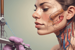
- Dermatology is the branch of medicine dealing with the skin and related diseases.
- The skin is the largest organ of the body, making up about 15% of the adult body weight (approximately 4 kg).
- It performs vital functions, including protection against physical, chemical, and biological agents.
- It also prevents excess water loss and regulates body temperature.
Skin Layers
- The skin has three layers: epidermis, dermis, and hypodermis.
Epidermis Layers
- Stratum basale (stratum germinativum): The deepest layer, composed of a single layer of columnar, mitotically active cells that produce keratinocytes. It also contains melanocytes.
- Stratum spinosum (prickle cell layer): Composed of 8-10 layers of cells with cytoplasmic processes (spines) connected by desmosomes. Contains Langerhans cells.
- Stratum granulosum: Contains 3-5 cell layers of diamond-shaped cells with keratohyalin and lamellar granules. Keratohyalin facilitates keratin aggregation; lamellar granules secrete glycolipids maintaining cellular cohesion.
- Stratum lucidum: A thin, clear layer present in thick skin (e.g., palms, soles). Consists of eleidin, a transformation product of keratohyalin.
- Stratum corneum: The outermost layer comprised of 20-30 layers of dead, keratinized cells (anucleate squamous cells) that form horny scales. They release defensins for immune defense.
Epidermal Cells
- Keratinocytes: The predominant cells (90%) originating from the basal layer. These cells produce keratin and lipids for the epidermal water barrier and contribute to calcium regulation by enabling UVB light absorption.
- Melanocytes: Dendritic pigment-synthesizing cells primarily in the basal layer responsible for melanin production and transfer to keratinocytes. Melanin is produced within melanosomes (membrane-bound organelles).
- Langerhans cells: Dendritic cells acting as the skin's first-line immune defenders. They contain Birbeck granules and tennis racket-shaped cytoplasmic organelles, expressing MHC I and MHC II molecules, taking up antigens, and transporting them to lymph nodes.
- Merkel cells: Oval-shaped modified epidermal cells in the stratum basale, serving as mechanoreceptors for light touch, found in the palms, soles, and oral/genital mucosa, especially in fingertips.
Dermo-Epidermal Junction
- The interface between epidermis and dermis is a porous basement membrane that allows cell and fluid exchange, holding the layers together.
- Basal keratinocytes are crucial components.
- The junction comprises the basal cell plasma membrane with hemidesmosomes, lamina lucida, lamina densa, and lamina fibroreticularis.
- Functions include epidermal-dermal adherence, mechanical support for the epidermis, and a barrier against large molecule exchange.
Dermis
- The dermis consists of two connective tissue layers: papillary (thinner) and reticular (thicker).
- The papillary layer contacts the epidermis, composed of loose connective tissue.
- The reticular layer has dense connective tissue with collagen fiber bundles, housing sweat glands, hair follicles, muscles, sensory neurons, and blood vessels.
- Cell types include fibroblasts, macrophages, mast cells, and white blood cells.
Hypodermis
- Also known as the subcutaneous fascia, this is the deepest layer beneath the dermis.
- It contains adipose lobules, sensory neurons, blood vessels, and sparse skin appendages (e.g., hair follicles).
History taking
- Attitude: Greet patient, introduce yourself, ask for permission.
- Personal Data: Name, age, sex, occupation, residence, marital status, and origin.
- Complaint: Symptoms (use patient's words), name of lesion, site of lesion, duration, and example: itchy pimples and bumps on both legs for one month.
- History of Present Illness (HPI): Fitzpatrick seven questions: onset (sudden or gradual), location of first appearance, symptom analysis, disease evolution, single lesion evolution, aggravating/relieving factors, and current medications.
- Systemic Review, Past History, Family History, Drugs History, and Social History
Dermatological Examination
- Skin Lesions:
- Primary skin lesions: Macules (circumscribed discoloration < 5mm), Patch (circumscribed discoloration > 5mm), Papules (<5mm solid elevation), Plaque (solid elevation >1cm), Vesicle/Bulla (< or > 5mm fluid-filled), Nodule (>5mm solid elevation), Wheal (transient edematous), and Pustule (pus-filled).
- Secondary skin lesions: Scar, Atrophy, Erosion, Ulcer, Crust, Scale, Fissure, Lichenification, and Excoriation.
- Special skin lesions (e.g., Comedone, Burrow, Dandruff, Ecchymosis, Petechiae, Purpura, Telangiectasia).
- Examination of the Lesions: Includes location, color, shape, surface, size, margin, and distribution (e.g., linear, annular, dermatomal).
- Examination of Special Sites: Examines scalp, palms, soles, mucous membranes (eyes, nose, genitalia).
- Eliciting Disease-Related Signs: Observe for any additional signs related to the condition being investigated.
Studying That Suits You
Use AI to generate personalized quizzes and flashcards to suit your learning preferences.