Podcast
Questions and Answers
What is the primary characteristic of the papillary dermis?
What is the primary characteristic of the papillary dermis?
- Is deeper than the reticular dermis.
- Is responsible for structural support.
- Constitutes 9/10th of the dermis.
- Contains oval shaped cells. (correct)
Which layer of the dermis is responsible for the majority of the dermal structure?
Which layer of the dermis is responsible for the majority of the dermal structure?
- Reticular dermis (correct)
- Papillary dermis
- Stratum corneum
- Dermo-Epidermal Junction
What is a feature of the dermo-epidermal junction?
What is a feature of the dermo-epidermal junction?
- It connects the epidermis and dermis. (correct)
- It is characterized by hyperkeratosis.
- It contains spongiosis.
- It is the thickest part of the epidermis.
Which of the following conditions describes the pathological thickening in hyperkeratosis?
Which of the following conditions describes the pathological thickening in hyperkeratosis?
What type of degeneration is characterized by intracellular edema within the stratum spinosum?
What type of degeneration is characterized by intracellular edema within the stratum spinosum?
What characterizes acantholytic cells seen in certain skin diseases?
What characterizes acantholytic cells seen in certain skin diseases?
What does the diascope examine in dermatological conditions?
What does the diascope examine in dermatological conditions?
Which skin condition is associated with a genetic basis linked to X-linked dominant inheritance?
Which skin condition is associated with a genetic basis linked to X-linked dominant inheritance?
What type of lines on the abdomen are characterized by their pathway of epidermal cell migration during embryonic development?
What type of lines on the abdomen are characterized by their pathway of epidermal cell migration during embryonic development?
Which term corresponds to the collagen fiber orientation in the dermis that does not relate to vascular structures?
Which term corresponds to the collagen fiber orientation in the dermis that does not relate to vascular structures?
In the context of epidermal migration, which line is described as not corresponding to arteries, veins, or nerves?
In the context of epidermal migration, which line is described as not corresponding to arteries, veins, or nerves?
When pressed with a glass slide, which condition demonstrates the staining of blood vessel walls due to RBC extravasation?
When pressed with a glass slide, which condition demonstrates the staining of blood vessel walls due to RBC extravasation?
Which of the following conditions is NOT typically linked with acantholysis?
Which of the following conditions is NOT typically linked with acantholysis?
What is the primary characteristic of the Stratum Corneum?
What is the primary characteristic of the Stratum Corneum?
Which layer of the skin is primarily involved in keratin synthesis?
Which layer of the skin is primarily involved in keratin synthesis?
What significant component is found in the Stratum Granulosum?
What significant component is found in the Stratum Granulosum?
What is the average epidermal turnover time (ETT) from the Stratum Basale until shedding?
What is the average epidermal turnover time (ETT) from the Stratum Basale until shedding?
Langerhans cells are primarily located in which layer of the skin?
Langerhans cells are primarily located in which layer of the skin?
Which structure provides intercellular attachment between keratinocytes?
Which structure provides intercellular attachment between keratinocytes?
Which layer of skin is characterized by the presence of refractile granules of eleidin?
Which layer of skin is characterized by the presence of refractile granules of eleidin?
What type of cell is primarily responsible for the production of melanin?
What type of cell is primarily responsible for the production of melanin?
What is the location of Merkel cells within the skin?
What is the location of Merkel cells within the skin?
Which of the following is NOT a known component of the Stratum Granulosum?
Which of the following is NOT a known component of the Stratum Granulosum?
Flashcards are hidden until you start studying
Study Notes
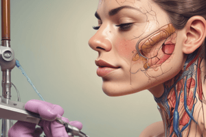
Dermatology Basics: Layers of Skin
- Stratum Corneum: Composed of dead, flat cells with no nuclei.
- Stratum Lucidum: Present only in palms and soles; contains refractile granules of Eleidin.
- Stratum Granulosum: Contains keratohyalin granules, which produce Filaggrin, and lamellar granules (Odland bodies) that store lipids.
- Stratum Spinosum: Contains desmosomes for keratinocyte attachment; site of keratin synthesis.
- Stratum Basale (Germinativum): Highly mitotically active layer responsible for skin proliferation.
- Epidermal Turnover Time (ETT): Time for keratinocytes to move from stratum Basale to shedding; averages 8 weeks (42–75 days). In psoriasis, turnover is accelerated to 4 days.
Cells of Epidermis
- Keratinocytes: Primary cells originated from ectoderm, produce keratin.
- Non-Keratinocytes: Includes Langerhans cells (immune function), melanocytes (pigment production), and Merkel cells (mechanotransduction).
- Langerhans Cells: Located in the spinosum; derived from bone marrow; characterized by Birbeck granules, serving as antigen-presenting cells.
- Melanocytes: Located in the basale; produce melanin and are derived from the neural crest.
- Merkel Cells: Located in the basale; neurosecretory cells acting as mechanoreceptors for touch.
Dermatopathology
- Basement Membrane Junction: Connects epidermis and dermis; key in maintaining skin integrity.
- Conditions:
- Hyperkeratosis: Thickening of the stratum corneum.
- Parakeratosis: Retention of nuclei in the stratum corneum.
- Ballooning Degeneration: Intracellular edema in the stratum spinosum.
- Spongiosis: Intercellular edema in the epidermis.
Layers of Dermis
- Papillary Dermis: Superficial layer making up 1/10th of the dermis; contains oval-shaped cells.
- Reticular Dermis: Deep layer constituting 9/10ths of the dermis and provides structural support.
Acantholytic Cells
- Seen in conditions such as pemphigus; prepared via Tzanck smear (Giemsa stain).
- Characterized by an oval shape, large nucleus, and perinuclear halo.
- Associated with autoimmune, bacterial (bullous impetigo, Staphylococcal Scalded Skin Syndrome), viral (HSV), and genetic disorders (Hailey-Hailey disease, Darier disease).
Lines in Dermatology
- Blaschko’s Lines: Pathways of cell migration during embryonic development.
- Langer Lines: Collagen fiber orientation in the dermis; important for incision placement to minimize scarring.
- Example: Incontinentia pigmenti, an X-linked dominant condition.
Diascopy / Vitro Pressure Test
- Technique involves pressing lesions with a glass slide.
- Used to differentiate types of skin lesions:
- Erythema: Redness blanches with pressure.
- Purpura: Extravasation of red blood cells; does not blanch.
- Apple Jelly Nodules: Granulomas become more visible and do not blanch.
Studying That Suits You
Use AI to generate personalized quizzes and flashcards to suit your learning preferences.