Podcast
Questions and Answers
A thorough clinical examination is not essential for a good case history.
A thorough clinical examination is not essential for a good case history.
False (B)
Pain perception is the phenomenon of deviation from the normal, and is indicative of an illness.
Pain perception is the phenomenon of deviation from the normal, and is indicative of an illness.
False (B)
Antibiotics are used to kill microorganisms on skin.
Antibiotics are used to kill microorganisms on skin.
False (B)
Differential diagnosis is the process of determining the nature of a disease.
Differential diagnosis is the process of determining the nature of a disease.
Chief complaint is not essential for a good case history.
Chief complaint is not essential for a good case history.
Duration is not an important aspect of pain history.
Duration is not an important aspect of pain history.
Temperature is an example of a subjective symptom.
Temperature is an example of a subjective symptom.
Pain assessment is not essential for correct clinical diagnosis.
Pain assessment is not essential for correct clinical diagnosis.
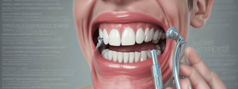
Laser Doppler flowmetry is used to determine the extent of caries in the pulp.
Laser Doppler flowmetry is used to determine the extent of caries in the pulp.
The heat test can be performed using a rubber cup to polish the crown.
The heat test can be performed using a rubber cup to polish the crown.
Anesthetic test is used to localize the affected tooth when the pain source cannot be differentiated.
Anesthetic test is used to localize the affected tooth when the pain source cannot be differentiated.
The cold test uses a hot burnisher to stimulate the pulp.
The cold test uses a hot burnisher to stimulate the pulp.
Electric pulp testing is a technique that is uncomfortable for patients.
Electric pulp testing is a technique that is uncomfortable for patients.
Transilluminaton test is used to detect vertical fractures in teeth.
Transilluminaton test is used to detect vertical fractures in teeth.
The electric pulp tester provides a qualitative reading of the pulp response.
The electric pulp tester provides a qualitative reading of the pulp response.
Biting test is used to diagnose reversible pulpitis.
Biting test is used to diagnose reversible pulpitis.
Thermal and electric pulp tests are used to diagnose periodontal diseases.
Thermal and electric pulp tests are used to diagnose periodontal diseases.
Guttapercha point tracing is used to diagnose periodontal lesions.
Guttapercha point tracing is used to diagnose periodontal lesions.
The correct placement of the electric pulp tester requires contact with the adjacent gingival tissue.
The correct placement of the electric pulp tester requires contact with the adjacent gingival tissue.
Pulse Oximetry is used to determine the vascularity of the pulp.
Pulse Oximetry is used to determine the vascularity of the pulp.
Lateral canals are not a portal of entry for toxins.
Lateral canals are not a portal of entry for toxins.
Radiographs are used to assess the PDL status of the tooth.
Radiographs are used to assess the PDL status of the tooth.
Staining is used to diagnose reversible pulpitis.
Staining is used to diagnose reversible pulpitis.
The intensity of the stimulus in electric pulp testing is not controlled.
The intensity of the stimulus in electric pulp testing is not controlled.
A radiograph can accurately determine the state of pulpal health.
A radiograph can accurately determine the state of pulpal health.
Early vertical root fracture can be diagnosed with radiographic examination.
Early vertical root fracture can be diagnosed with radiographic examination.
The extent of caries is always accurately represented on a radiograph.
The extent of caries is always accurately represented on a radiograph.
Anatomic structures can never be misinterpreted as periapical pathology on a radiograph.
Anatomic structures can never be misinterpreted as periapical pathology on a radiograph.
Cone beam computerized tomography (CBCT) is not used for evaluating periapical areas.
Cone beam computerized tomography (CBCT) is not used for evaluating periapical areas.
Periapical pathology is always evident on a radiograph.
Periapical pathology is always evident on a radiograph.
Radiovisiography (RVG) is a type of radiographic examination.
Radiovisiography (RVG) is a type of radiographic examination.
Magnetic Resonance Imaging (MRI) is commonly used for evaluating periapical areas.
Magnetic Resonance Imaging (MRI) is commonly used for evaluating periapical areas.
A patient with recent myocardial infarction within 6 months requires modification of dental treatment.
A patient with recent myocardial infarction within 6 months requires modification of dental treatment.
Facial asymmetry is not an important observation during extra-oral examination.
Facial asymmetry is not an important observation during extra-oral examination.
Pulpitis can be reversible or irreversible depending on the extent of inflammation.
Pulpitis can be reversible or irreversible depending on the extent of inflammation.
Peptic ulcer disease does not require modification of dental treatment.
Peptic ulcer disease does not require modification of dental treatment.
Visual and tactile inspection is not essential for clinical examination of soft tissue.
Visual and tactile inspection is not essential for clinical examination of soft tissue.
End-stage renal disease requires modification of dental treatment and consultation.
End-stage renal disease requires modification of dental treatment and consultation.
Hard tissue examination is not important for diagnosing pulpitis.
Hard tissue examination is not important for diagnosing pulpitis.
Lymphadenopathy is not an important observation during extra-oral examination.
Lymphadenopathy is not an important observation during extra-oral examination.
In external resorption, the resorptive area will appear more radiolucent than the root canal.
In external resorption, the resorptive area will appear more radiolucent than the root canal.
Super-EBA is a type of zinc oxide eugenol cement reinforced with ethoxy benzoic acid (EBA) used for root canal treatment.
Super-EBA is a type of zinc oxide eugenol cement reinforced with ethoxy benzoic acid (EBA) used for root canal treatment.
Anachoretic pulpitis is a type of pulpitis caused by bacterial invasion through a chemical or mechanical injury to the pulp.
Anachoretic pulpitis is a type of pulpitis caused by bacterial invasion through a chemical or mechanical injury to the pulp.
Internal resorption is a type of external resorption that occurs on the crown of the tooth.
Internal resorption is a type of external resorption that occurs on the crown of the tooth.
In apical root resorption, the mechanism is different from internal resorption.
In apical root resorption, the mechanism is different from internal resorption.
Pulpitis can be reversible or irreversible depending on the extent of inflammation.
Pulpitis can be reversible or irreversible depending on the extent of inflammation.
Aging is a contributing factor to idiopathic pulpitis.
Aging is a contributing factor to idiopathic pulpitis.
Reversible pulpitis is a severe inflammatory condition of the pulp.
Reversible pulpitis is a severe inflammatory condition of the pulp.
Extreme heat can cause internal bleaching of the tooth.
Extreme heat can cause internal bleaching of the tooth.
Endodontic treatment can arrest the progression of internal resorption.
Endodontic treatment can arrest the progression of internal resorption.
Pulp necrosis is a type of pulp degeneration.
Pulp necrosis is a type of pulp degeneration.
Calcium hydroxide paste is placed in the canal until the defect is repaired by a calcific barrier.
Calcium hydroxide paste is placed in the canal until the defect is repaired by a calcific barrier.
Local anesthesia is required for the treatment of generalized external resorption.
Local anesthesia is required for the treatment of generalized external resorption.
In irreversible pulpitis, the pain is sharp and of short duration.
In irreversible pulpitis, the pain is sharp and of short duration.
Trauma is a possible contributing factor to internal resorption.
Trauma is a possible contributing factor to internal resorption.
The exact cause of internal resorption is well understood.
The exact cause of internal resorption is well understood.
The pulp of a tooth can become necrotic in severe cases.
The pulp of a tooth can become necrotic in severe cases.
Pain during the releasing of biting pressure is a sign of a healthy pulp.
Pain during the releasing of biting pressure is a sign of a healthy pulp.
A J-shaped lesion is a characteristic radiological manifestation of pulpitis in the early stage.
A J-shaped lesion is a characteristic radiological manifestation of pulpitis in the early stage.
High-speed engines and poor coolant systems can contribute to thermal injuries during cavity preparation.
High-speed engines and poor coolant systems can contribute to thermal injuries during cavity preparation.
Polishing with dry powders can lead to a decrease in temperature.
Polishing with dry powders can lead to a decrease in temperature.
Galvanic current from dissimilar metal fillings is a common cause of pulpitis.
Galvanic current from dissimilar metal fillings is a common cause of pulpitis.
Direct invasion through dentin is one of the pathways of bacterial invasion of the pulp.
Direct invasion through dentin is one of the pathways of bacterial invasion of the pulp.
Bacterial causes are the least common cause of pulpitis.
Bacterial causes are the least common cause of pulpitis.
Trauma to the tooth is a common cause of pulp necrosis in children.
Trauma to the tooth is a common cause of pulp necrosis in children.
Habits such as opening hard objects with the teeth and compulsive bruxism are not risk factors for pulp injury.
Habits such as opening hard objects with the teeth and compulsive bruxism are not risk factors for pulp injury.
Dental procedures such as crown preparation can never injure the pulp.
Dental procedures such as crown preparation can never injure the pulp.
Pathologic wear and tear of the tooth surface is only caused by abrasion.
Pathologic wear and tear of the tooth surface is only caused by abrasion.
Erosion of the tooth surface is caused by physical wear and tear.
Erosion of the tooth surface is caused by physical wear and tear.
Cracked tooth syndrome is a type of pulpitis.
Cracked tooth syndrome is a type of pulpitis.
Abfraction is a type of tooth wear caused by faulty brushing.
Abfraction is a type of tooth wear caused by faulty brushing.
The incidence of pulp necrosis is 1% after certain dental procedures.
The incidence of pulp necrosis is 1% after certain dental procedures.
Pulp anatomy is simple with a single root canal.
Pulp anatomy is simple with a single root canal.
The interface between the dentine and pulp is protected from irritation by an intact layer of enamel in the root.
The interface between the dentine and pulp is protected from irritation by an intact layer of enamel in the root.
Accessory canal diverges at a right angle from the main canal.
Accessory canal diverges at a right angle from the main canal.
Mechanical injuries can cause pulp disease.
Mechanical injuries can cause pulp disease.
Pulp reacts to irritants by always becoming necrotic.
Pulp reacts to irritants by always becoming necrotic.
The pulp has a hard, bony consistency.
The pulp has a hard, bony consistency.
Lateral canals are a type of accessory canal.
Lateral canals are a type of accessory canal.
Periapical pathosis is a type of pulp disease.
Periapical pathosis is a type of pulp disease.
Flashcards are hidden until you start studying
Study Notes
Pulpal Disease Diagnosis
- Lateral canals are portals of entry for toxins, and pulpal degeneration can occur if not diagnosed correctly.
- Thermal and electric pulp tests must be performed along with periodontal examination to distinguish between disease of pulpal and periodontal origin.
Thermal Test Techniques
- Heat test can be performed using different techniques such as:
- Hot water
- Hot burnisher
- Hot gutta-percha
- Hot compound
- Polishing of crown with a rubber cup
- Cold test involves isolating the quadrant with the tooth to be tested and using:
- Cold water
- Cold air from a 3-way syringe directed against the crown of previously dried tooth
- Ethyl chloride spray
- Ice stick
- CO2 snow
Electric Pulp Testing (EPT)
- The test should be first described to the patient, and teeth to be tested should be isolated with cotton rolls, saliva ejector, and air dried.
- The EPT tester should be checked for proper functioning, and an electrolyte (toothpaste) should be applied to the tooth surface.
- Avoid contact of the electrolyte or electrode with any restorations or the adjacent gingival tissue as this could lead to a false response.
Advantages of EPT
- Intensity of stimulus is comfortable to patients.
- Digital display of many EPT testers provides instant, easy, and reliable information.
- In some EPT testers, a red indicator light flashes on and off when maximum stimulus is reached.
- Gives a quantitative reading and can be compared with the normal reading of control tooth.
Anesthetic Test
- Restricted to patients who are in pain at the time of the test and when the usual tests have failed to help identify or localize the offending tooth.
- The objective is to anesthetize a single tooth at a time until the pain disappears and is localized to a specific tooth.
- If the source of pain cannot be differentiated (i.e., maxillary/mandibular), then a mandibular block is implemented.
- Further localization of the affected tooth is done by an intraligament injection.
Transillumination Test
- Light from a fiberoptic is applied from the buccal surface to illuminate the tooth to detect fractured lines when present.
- From the contact area to detect contact caries.
Biting Test
- Placing the pointed end on each cusp and then having the patient bite and release.
- A normal, healthy tooth will have no pain.
- An orangewood stick is placed on the occlusal/incisal aspect (on each cusp in case of posteriors) of the tooth, and the patient is asked to bite.
Staining
- Remove the filling from the suspected tooth and place 2% Iodine (or other stain) in the cavity preparation.
- The iodine stains the:
- Fracture line dark.
- Canal orifice.
Guttapercha Point Tracing
- Can localize the endodontic lesion to the specific tooth.
- Aids in the differential diagnosis between a periodontal and an endodontic lesion.
- Placing a guttapercha point through the sinus/fistula tract and taking a radiograph.
Radiographs Importance
- Provides information on the extent of caries into the pulp.
- Number of root canals and accessories.
- Course and shape of the canals.
- Length of the root.
- Calcifications.
- Resorptions.
- PDL status.
Diagnosis
- Is a process of determining the nature of a disease.
- It is very important for proper treatment.
- Differential diagnosis: is the process of differentiating between similar diseases.
- Sequential agreement is very important for correct differential diagnosis:
- Proper knowledge of the disease.
- Skill and art on how to apply proper diagnostic methods.
Clinical Examination
- Extra oral examination:
- Facial asymmetry.
- Localized swellings.
- Changes in color, bruises/ scars.
- Lymphadenopathy (groups of lymph nodes).
- Visual and tactile inspection:
- Soft tissue:
- Color.
- Contour.
- Consistency.
- Hard tissue:
- Color.
- Contour.
- Soft tissue:
Medical Conditions Requiring Modification of Treatment and Consultation
- Cardiovascular:
- High & moderate risk of Endocarditis.
- Pathologic heart Murmurs.
- Hypertension.
- Unstable angina.
- Recent myocardial infarction (6 months).
- Arrythmias.
- Poorly managed congestive heart failure.
- Pulmonary: COPD, Asthma, Tuberculosis.
- GIT & Renal: End stage renal disease, Hemodialysis, Viral hepatitis (A,B,C,E), Alcoholic liver disease, Peptic ulcer disease, Endocrine & Hematologic: STDs: HIV & AIDS, Diabetes mellitus, Adrenal insufficiency, Pregnancy, Bleeding disorders, Cancer & leukemia, Osteoarthritis, rheumatoid arthritis, SLE.
Limitations of Radiographs
- 2D image of a 3D object.
- State of pulpal health cannot be ascertained.
- P/A pathology is evident only after much destruction (33%).
- Early vertical root fracture cannot be diagnosed.
- Bony trabeculae misinterpreted for horizontal root #.
- Extend of caries is usually less than the actual extent as is true for P/A pathology.
Anatomic Structure Can Mimic P.A. Pathology
- Canal seems to disappear due to narrowing of canal space or overlapping of the roots on the radiograph.
Other Radiographic Methods
- Xeroradiography.
- Radiovisiography (RVG).
- Cone beam computerized tomography (CBCT).
- Magnetic Resonance Imaging (MRI).
- Ultrasound imaging.
CBCT
- Basic 3 sections.
- Check the cervical cross-sectional shape.
Trauma and Habits
- Trauma is a common cause of pulp disease in children, resulting from violent blows to the tooth during fights, sports, or household accidents.
- Habits such as opening hard objects with teeth, compulsive bruxism, nail biting, and thread biting can also lead to pulp disease.
Dental Procedures
- Certain dental procedures can injure the pulp, including crown preparation, over-cutting with dull burs, and reduced cooling system of turbines.
- Removing dentin for a crown preparation, curing materials needed for provisionalization, and microleakage under a temporary crown can contribute to possible pre-existing pulpitis.
Pathologic Wear
- Attrition is natural tooth-to-tooth friction that occurs when you clench or grind your teeth, often involuntarily during sleep.
- Abrasion is physical wear and tear of the tooth surface that happens with faulty brushing or hard toothbrushes, fingernails, and chewing tobacco.
- Abfraction is the pathological loss of tooth substance caused by biomechanical loading forces, resulting in flexure and failure of enamel and dentin at a location away from the loading.
- Erosion occurs chemically when acidic content hits the tooth surface, such as with certain medications, highly acidic foods, and frequent vomiting.
Cracked Tooth Syndrome
- Incomplete fracture through the body of the tooth can be due to excessive masticatory forces, causing symptoms ranging from mild pain to severe pain.
- The pulp of the tooth may become necrotic in severe cases.
- Patients may feel pain when biting on a cotton or rubber wheel, and radiological manifestations may include a J-shaped lesion in the late stage.
Thermal Injuries
- Thermal injuries can occur due to cavity preparation, including:
- Deeper cavity preparation
- High-speed engines
- Poor coolant system
- Extensive cavity
- Excessive cutting of dentin
- Dull burs
- High pressure with cutting
Polishing Injuries
- Polishing with dry powders can lead to temperature rise and heat conduction by restoration.
- Heat conduction can occur through very deep metallic restorations close to the pulp without any intermediate cement base.
Electrical Injuries
- Galvanic current from dissimilar metal fillings can cause pulp disease.
Chemical Causes
- Pulpal inflammation can occur due to:
- Citrus ingestion
- Acid etchants used to expose dentin
- Dehydrating chemicals for sterilizing/drying
- Gastrointestinal problems that produce repeated exposure of teeth to gastric acids
Bacterial Causes
- Bacterial invasion can occur through:
- Direct invasion through dentin
- Invasion through open blood vessels or lymphatics associated with periodontal disease
- Invasion through blood (anachoretic effect) during infectious diseases or transient bacteremia
Idiopathic Causes
- Aging can cause pulp disease, including:
- Atrophy of the pulp
- Decrease in the number and size of cells
- Increase in collagen fiber content
- Internal resorption can occur due to:
- Trauma
- Caries
- Ortho treatment
- Infection/pulpitis
- Extreme heat
Internal Resorption
- Internal resorption is a localized internal dentin destruction due to odontoclastic activity.
- Clinical appearance includes a pink spot on the crown.
- Treatment involves endodontic treatment to arrest the progression of the resorption.
External Resorption
- External resorption occurs when the resorptive tissue is supplied by blood vessels coming through the apex.
- Treatment involves removal of the cause, and in cases where it is accessible, RCT and composite restoration.
- In cases where it is non-accessible, surgical access and correction by MTA or BIODENTINE are necessary.
Pulpitis
- Pulpitis is inflammation of the pulp, and can be classified as:
- Reversible pulpitis (acute or chronic)
- Irreversible pulpitis (acute or chronic)
- Pulp degeneration (calcific or atrophic)
- Pulp necrosis
Reversible Pulpitis
- Reversible pulpitis is a mild to moderate inflammatory condition that can return to its normal position after removal of stimuli.
- Characteristics include:
- Sharp pain of short duration
- Pain subsides after stimulus is removed
- The affected tooth responds to cold
- Tooth responds to electric pulp testing at very low levels of current than control tooth
Studying That Suits You
Use AI to generate personalized quizzes and flashcards to suit your learning preferences.


