Podcast
Questions and Answers
Why is the presence of a positive proximal contact area (PCA) between adjacent teeth important for maintaining dental health?
Why is the presence of a positive proximal contact area (PCA) between adjacent teeth important for maintaining dental health?
- It prevents food impaction between teeth and helps stabilize the dental arch through combined anchorage. (correct)
- It allows for minor tooth movement to prevent excessive force on individual teeth during chewing.
- It facilitates the flow of nutrients to the periodontal ligament, enhancing the teeth's shock-absorbing capabilities.
- It directly stimulates saliva production, which aids in the digestion of food particles trapped between teeth.
How do the anatomic and clinical crowns of a tooth differ from each other?
How do the anatomic and clinical crowns of a tooth differ from each other?
- The anatomic crown is only present in anterior teeth, while the clinical crown is exclusive to posterior teeth.
- The anatomic crown refers to the portion covered by enamel, while the clinical crown includes both enamel and cementum.
- The anatomic crown remains constant in size extending to below the gumline, whereas the clinical crown can vary in visible size. (correct)
- The clinical crown is the part of the tooth embedded in the jawbone, while the anatomic crown is the visible part above the gumline.
What is the primary role of teeth in relation to the tissues that support them?
What is the primary role of teeth in relation to the tissues that support them?
- To stimulate the production of saliva, which contains enzymes that break down food particles and protect supporting tissues from bacterial infections.
- To filter out harmful bacteria from the bloodstream, preventing inflammation and infection in the surrounding gingival tissues.
- To help sustain themselves in the dental arches by assisting in the development and protection of these supporting tissues. (correct)
- To generate stem cells that repair damaged periodontal ligaments, ensuring continuous tissue regeneration.
Which characteristic is associated with the roots of teeth?
Which characteristic is associated with the roots of teeth?
How do well-developed teeth and jaws contribute to overall well-being beyond physical appearance?
How do well-developed teeth and jaws contribute to overall well-being beyond physical appearance?
Which periodontal fiber group is embedded in the cementum of adjacent teeth and extends interproximally over the alveolar crest?
Which periodontal fiber group is embedded in the cementum of adjacent teeth and extends interproximally over the alveolar crest?
What is the primary function of cementum?
What is the primary function of cementum?
Which part of the alveolar bone immediately surrounds the root of a tooth?
Which part of the alveolar bone immediately surrounds the root of a tooth?
What is the origin/derivation of dentin?
What is the origin/derivation of dentin?
How far away (mm) is considered 2nd degree furcation involvement?
How far away (mm) is considered 2nd degree furcation involvement?
Which of the following structures is NOT found within the pulp?
Which of the following structures is NOT found within the pulp?
Which periodontal fiber group extends at right angles to the long axis of the tooth?
Which periodontal fiber group extends at right angles to the long axis of the tooth?
In multirooted teeth, where are interradicular fibers located?
In multirooted teeth, where are interradicular fibers located?
When viewing a tooth from the incisal aspect, what information can be gathered regarding contact areas?
When viewing a tooth from the incisal aspect, what information can be gathered regarding contact areas?
What is the typical separation distance between the enamel and the alveolar bone within the interproximal space?
What is the typical separation distance between the enamel and the alveolar bone within the interproximal space?
What is the primary function of embrasures formed by the curvatures of adjacent teeth?
What is the primary function of embrasures formed by the curvatures of adjacent teeth?
What is the approximate normal curvature from the cementoenamel junction (CEJ) to the crest of contour on the facial and lingual surfaces of a tooth at the cervical third?
What is the approximate normal curvature from the cementoenamel junction (CEJ) to the crest of contour on the facial and lingual surfaces of a tooth at the cervical third?
How does excessive curvature in the cervical ridges potentially affect gingival health?
How does excessive curvature in the cervical ridges potentially affect gingival health?
How does the curvature of the cervical line (CEJ) typically differ between the mesial and distal aspects of a tooth?
How does the curvature of the cervical line (CEJ) typically differ between the mesial and distal aspects of a tooth?
Which of the following structures is NOT considered a supporting structure of the tooth?
Which of the following structures is NOT considered a supporting structure of the tooth?
What is the approximate thickness range of the periodontal ligament (PDL)?
What is the approximate thickness range of the periodontal ligament (PDL)?
Considering the functions of the periodontal ligament (PDL), what role does it play in response to increased occlusal forces?
Considering the functions of the periodontal ligament (PDL), what role does it play in response to increased occlusal forces?
Which function of the Periodontal Ligament (PDL) enables the ability to discern the amount of pressure during chewing and identify which tooth is being tapped?
Which function of the Periodontal Ligament (PDL) enables the ability to discern the amount of pressure during chewing and identify which tooth is being tapped?
Which of the following is the primary function of odontoblasts within the dental pulp?
Which of the following is the primary function of odontoblasts within the dental pulp?
What is the main characteristic of the sulcular epithelium (SE) that lines the gingival sulcus?
What is the main characteristic of the sulcular epithelium (SE) that lines the gingival sulcus?
In which region of the gingiva would you typically observe a 'stippled' appearance?
In which region of the gingiva would you typically observe a 'stippled' appearance?
Which component of the dental pulp decreases in both quantity and quality as a person ages?
Which component of the dental pulp decreases in both quantity and quality as a person ages?
Which of the following groups of gingival fibers runs from the cementum of one tooth to the cementum of an adjacent tooth?
Which of the following groups of gingival fibers runs from the cementum of one tooth to the cementum of an adjacent tooth?
Where do the nerve fibers and blood vessels enter the pulp?
Where do the nerve fibers and blood vessels enter the pulp?
Which layer of gingival epithelium is non-keratinized and forms a collar-like band around the tooth?
Which layer of gingival epithelium is non-keratinized and forms a collar-like band around the tooth?
What is the primary function of the interdental gingiva (or papilla)?
What is the primary function of the interdental gingiva (or papilla)?
Which of the following best describes the function of the dentoperiosteal group of gingival fibers?
Which of the following best describes the function of the dentoperiosteal group of gingival fibers?
What is the clinical significance of 'lost stippling' on the gingiva?
What is the clinical significance of 'lost stippling' on the gingiva?
Where are the collagen fibers of the dentogingival group embedded?
Where are the collagen fibers of the dentogingival group embedded?
Which feature characterizes the contour of healthy gingiva?
Which feature characterizes the contour of healthy gingiva?
Which of the following best describes the composition of the lamina propria of the gingiva?
Which of the following best describes the composition of the lamina propria of the gingiva?
What is the role of melanocytes in the gingiva?
What is the role of melanocytes in the gingiva?
Which pulpal cell type is MOST responsible for initiating an immune response?
Which pulpal cell type is MOST responsible for initiating an immune response?
Flashcards
Functions of Teeth
Functions of Teeth
Teeth incise food and aid in mastication, sustaining themselves in dental arches.
Crown of the Tooth
Crown of the Tooth
The visible part of the tooth; differs as anatomic (below gum) and clinical crown (visible part).
Proximal Contact Area (PCA)
Proximal Contact Area (PCA)
Contact area between adjacent teeth, preventing food from packing.
Importance of Good Teeth
Importance of Good Teeth
Signup and view all the flashcards
Root of the Teeth
Root of the Teeth
Signup and view all the flashcards
Interradicular Artery
Interradicular Artery
Signup and view all the flashcards
Principal Periodontal Fiber Groups
Principal Periodontal Fiber Groups
Signup and view all the flashcards
Transseptal Fibers
Transseptal Fibers
Signup and view all the flashcards
Cementum
Cementum
Signup and view all the flashcards
Cementoblasts
Cementoblasts
Signup and view all the flashcards
Alveolar Bone Proper
Alveolar Bone Proper
Signup and view all the flashcards
Functions of the Pulp
Functions of the Pulp
Signup and view all the flashcards
Hypercementosis
Hypercementosis
Signup and view all the flashcards
PCA Aspects
PCA Aspects
Signup and view all the flashcards
Interproximal Space
Interproximal Space
Signup and view all the flashcards
Embrasures
Embrasures
Signup and view all the flashcards
Types of Embrasure
Types of Embrasure
Signup and view all the flashcards
Cervical Ridges
Cervical Ridges
Signup and view all the flashcards
Physiologic Importance of Cervical Ridges
Physiologic Importance of Cervical Ridges
Signup and view all the flashcards
Curvature of the CEJ
Curvature of the CEJ
Signup and view all the flashcards
Periodontal Ligament (PDL)
Periodontal Ligament (PDL)
Signup and view all the flashcards
Functions of PDL
Functions of PDL
Signup and view all the flashcards
Blood Supply in PDL
Blood Supply in PDL
Signup and view all the flashcards
Composition of Dental Pulp
Composition of Dental Pulp
Signup and view all the flashcards
Cells of Pulp
Cells of Pulp
Signup and view all the flashcards
Ground Substance of Pulp
Ground Substance of Pulp
Signup and view all the flashcards
Collagen Fibers in Pulp
Collagen Fibers in Pulp
Signup and view all the flashcards
Blood Vessels in Pulp
Blood Vessels in Pulp
Signup and view all the flashcards
Nerves in Pulp
Nerves in Pulp
Signup and view all the flashcards
Aging Pulp Response
Aging Pulp Response
Signup and view all the flashcards
Gingiva Definition
Gingiva Definition
Signup and view all the flashcards
Marginal Gingiva
Marginal Gingiva
Signup and view all the flashcards
Attached Gingiva
Attached Gingiva
Signup and view all the flashcards
Layers of Gingival Epithelium
Layers of Gingival Epithelium
Signup and view all the flashcards
Dentogingival Group Fibers
Dentogingival Group Fibers
Signup and view all the flashcards
Gingival Morphology - Color
Gingival Morphology - Color
Signup and view all the flashcards
Gingival Consistency
Gingival Consistency
Signup and view all the flashcards
Location of Gingiva
Location of Gingiva
Signup and view all the flashcards
Study Notes
Physiology of Teeth and Supporting Structures
- Teeth are crucial for mastication, aiding in food reduction and incision.
- Teeth support themselves within dental arches by participating in the development and protection of supporting tissues. An example is physiological mesial drifting.
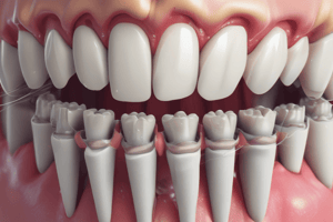
- Tooth anatomy distinguishes a crown, neck, and root.
- The enamel is the hardest outer layer of the tooth.
- Dentin is beneath the enamel, making up most of the tooth structure.
- The pulp is the soft tissue within the tooth, containing nerves and blood vessels.
- Tooth cementum covers the root.
Tooth Anatomy
- The crown is the visible part of a tooth.
- The anatomic crown extends below the gum line.
- The clinical crown is what's visible.
- Crown size varies.
- Anterior teeth have incisal edges, while posterior teeth have cusps.
- Incisors are for cutting and punching food during mastication.
- Canines, or cuspids, are for shearing and tearing food, supporting incisors and premolars.
- Premolars (bicuspids) are used to grind food.
- Molars have multiple cusps for grinding and trituration of food.
- Roots are covered in cementum, with varying lengths and numbers depending on tooth size and function.
- Teeth are classified as single-rooted (incisors, canines, and certain premolars) or multi-rooted (molars).
Protective Functional Forms of the Tooth Crown
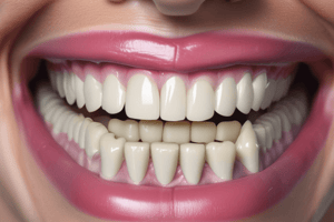
- Proximal contact area (PCA) assures one tooth contacts another mesially and distally.
- PCA prevents food from packing between teeth.
- It stabilizes dental arches via the combined anchorage of all teeth.
- PCA can be viewed via labial/buccal and incisal/occlusal aspects.
Interproximal Space
- Interproximal spaces are triangular shapes between teeth, usually filled with gingival tissue.
- A typical separation of 1-1.5 mm exists between enamel and alveolar bone.
Embrasures
- Spaces created by the curvature of adjacent teeth are termed embrasures.
- Embrasures provide pathways for food during mastication to escape.
- They prevent food from being trapped between teeth.
Cervical Ridges
- Facial and lingual contours of the cervical third of teeth create cervical ridges.
- Typically, the curvature is roughly 0.5 millimeters from the cementoenamel junction (CEJ) to the contour.
- Cervical ridges hold the gingiva under tension and deflect food away from gingival margins.
- Insufficient or excessive curvature may cause gum recession or food packing, respectively.
Supporting Structures
- The supporting structures of a tooth include the periodontal ligament, cementum, alveolar process, pulp, and gingiva.
Periodontal Ligament
- A dense connective tissue that anchors the tooth to the alveolar bone.
- It ranges between 0.1-0.25mm thick.
- It provides shock absorption to manage occlusal forces and prevent tooth breakage.
- It's involved in formative reactions, activating osteoblasts, cementoblasts, and fibroblasts for bone/tooth maintenance.
- Contains sensory nerve endings for pressure assessment and tooth identification during chewing.
Blood Supply of Periodontal Ligaments
- Blood supply arises from alveolar artery branches.
- Additional supply from interradicular and gingival vessels.
- Vessels nourish the crestal area.
Periodontal Fiber Groups
- Transseptal fibers connect adjacent tooth cementum.
- Alveolar crest fibers extend obliquely from the cementum and reach the alveolar crest.
- Horizontal fibers run at right angles to the tooth's long axis through cementum and bone tissues.
- Oblique fibers compose a major group, running obliquely from cementum to bone.
- Apical fibers stem from the apical cementum to the base of the socket.
- Interradicular fibers span furcations in multi-rooted teeth.
Cementum
- A mineralized connective tissue covering tooth roots.
- It contains cementoblasts, cementoclasts, and cementocytes.
- It's essential for anchoring periodontal ligament fibers to the tooth.
Classification of Cementum
- Cementum is classified based on location (radicular, coronal), cellularity (cellular, acellular), and collagen fibril presence (fibrillar, afibrillar).
Types of Cementum
- Acellular fibrillar cementum covers the crown portion, lacking cells.
- Cellular fibrillar cementum covers the root portion, possessing cells.
Developmental and Acquired Anomalies Associated with Cementogenesis
- Anomalies involve enamel projections, enamel pearls, cementicles, and hypercementosis.
Alveolar Bone
- This jawbone part supports and houses teeth.
- Consists of osteoblasts (bone-forming cells), osteoclasts (bone-resorbing cells), and osteocytes.
Parts of Alveolar Bone
- Alveolar bone proper directly surrounds tooth roots, forming a thin lamella.
- Supporting bone envelops the alveolar bone proper, enhancing its functional role.
Bone Cells
- Cells involved in bone formation and structure include osteoblasts, osteoclasts, and osteocytes.
Pulp
- Pulp is a vascular connective tissue contained within dentin walls.
- It encompasses fibroblasts for collagen formation, odontoblasts, and defensive cells.
Functions of the Pulp
- Formative (forming new dentin).
- Nutritive (providing nourishment).
- Sensory (detecting stimuli).
- Defensive (protecting against pathogens).
Composition of Pulp
- Cells comprise fibroblasts, odontoblasts, and defensive cells (e.g., histiocytes, leukocytes).
- Pulp's ground substance consists of proteins and glycosaminoglycans.
Pulp Response to Aging
- Age-related pulp changes include reduced cellular components, dentinal sclerosis, decreased blood vessel quality, decreased pulp size, increased collagen fiber thickness, and pulp stone/dystrophic mineralization increases.
Gingiva
- Gingiva is a part of the oral mucosa covering alveolar processes and surrounding tooth cervical regions.
Regions of Gingiva
- Marginal gingiva (free gingiva): borders teeth like a collar.
- Attached gingiva: tight to the alveolar periosteum.
- Interdental papilla: fills spaces between adjacent teeth.
Layers of Gingival Epithelium
- Oral epithelium: outer layer encompassing crest and superficial attached gingiva; keratinized.
- Sulcular epithelium: non-keratinized, lining gingival sulcus.
- Junctional epithelium: non-keratinized band attaching gingiva to tooth; reinforced by gingival fibers.
- Gingival epithelium varies throughout.
Groups of Gingival Fibers
- Denotgingival, Dentoperiosteal, Alveologingival, Circular, Semicircular and Transseptal fibers.
Lamina Propria
- A connective tissue component of gingiva, rich in collagen.
- It contains papillary and reticular layers to interact with underlying bone or epithelial.
Morphology of Gingiva
- Color: primarily coral pink, may have pigmentations from melanocytes (brown or purple).
- Contour: shaped by various factors, resembling a scalloped line.
- Consistency: firm and resilient in attached gingiva.
- Texture: often stippled, a bumpy surface characteristic of health, may vanish with gingivitis.
- Location: typically at the cementoenamel junction (CEJ).
Studying That Suits You
Use AI to generate personalized quizzes and flashcards to suit your learning preferences.