Podcast
Questions and Answers
What is the preferred test to visualize clots in the pulmonary artery?
What is the preferred test to visualize clots in the pulmonary artery?
- CT pulmonary angiogram (correct)
- VQ scan
- ECG
- D-dimer blood test
Which test is used for high probability of a pulmonary embolism?
Which test is used for high probability of a pulmonary embolism?
- CT pulmonary angiogram (correct)
- Chest x-ray
- PERC score with D-dimer
- D-dimer with VQ scan
In diagnosing a pulmonary embolism, what indicates a low-risk PE?
In diagnosing a pulmonary embolism, what indicates a low-risk PE?
- Perfusion defects on VQ scan
- Right heart strain on ECG
- Normal blood pressure (correct)
- Presence of RV dilation
What treatment is recommended for stable patients with pulmonary embolism?
What treatment is recommended for stable patients with pulmonary embolism?
Which test is used for diagnosing a pulmonary embolism in patients with moderate probability?
Which test is used for diagnosing a pulmonary embolism in patients with moderate probability?
What indicates a massive PE burden?
What indicates a massive PE burden?
In DVT treatment, what is the approach for massive proximal DVT?
In DVT treatment, what is the approach for massive proximal DVT?
How is PE further classified based on echocardiogram findings?
How is PE further classified based on echocardiogram findings?
What is recommended for hemodynamically unstable patients with massive PE?
What is recommended for hemodynamically unstable patients with massive PE?
What are the components of Virchow's Triad that contribute to clot formation in DVT?
What are the components of Virchow's Triad that contribute to clot formation in DVT?
Which condition is NOT a potential cause of hypercoagulability in DVT?
Which condition is NOT a potential cause of hypercoagulability in DVT?
Where does distal DVT commonly occur?
Where does distal DVT commonly occur?
What complication of DVT presents with a congested bluish limb?
What complication of DVT presents with a congested bluish limb?
Which scoring systems are commonly used to assess the likelihood of DVT or PE?
Which scoring systems are commonly used to assess the likelihood of DVT or PE?
What symptom distinguishes phlegmasia cerulea dolens from phlegmasia alba dolens?
What symptom distinguishes phlegmasia cerulea dolens from phlegmasia alba dolens?
Which condition is a direct cause of stasis of blood flow in deep vein thrombosis (DVT)?
Which condition is a direct cause of stasis of blood flow in deep vein thrombosis (DVT)?
What is the main consequence of pulmonary embolism (PE) leading to obstructive shock?
What is the main consequence of pulmonary embolism (PE) leading to obstructive shock?
What is an example of endothelial injury that can contribute to clot formation in DVT?
What is an example of endothelial injury that can contribute to clot formation in DVT?
Flashcards are hidden until you start studying
Study Notes
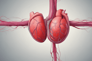
- Deep vein thrombosis (DVT) is characterized by the formation of blood clots (thrombus) in the veins, commonly affecting the legs.
- Proximal DVT occurs above the bifurcation, while distal DVT occurs below, with different veins like iliac, femoral, popliteal, tibial, and peroneal being involved.
- Virchow's Triad explains that stasis of blood flow, hypercoagulability, and endothelial injury contribute to clot formation in DVT.
- Factors leading to stasis include post-op states, paralysis, and prolonged travel, while endothelial injury can be caused by smoking and venous catheters.
- Hypercoagulability can be due to conditions like malignancy (pancreatic or lung), pregnancy, oral contraceptives, genetic mutations (Factor V Leiden), nephrotic syndrome, or antiphospholipid syndrome.
- Complications of DVT include phlegmasia cerulea dolens (congested bluish limb), phlegmasia alba dolens (pale limb due to arterial compression), post-thrombotic syndrome, and pulmonary embolism (PE).
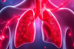
- PE occurs when a clot breaks off from DVT and blocks a pulmonary artery, leading to symptoms like pleuritic chest pain, dyspnea, and hemoptysis.
- PE can cause respiratory failure due to VQ mismatch resulting in hypoxemia, and obstructive shock from reduced right ventricular output leading to hypotension and shock.
- Wells Criteria and PERC score are used to assess the likelihood of DVT or PE based on risk factors, symptoms, and history.
- Diagnostic tests for PE include chest x-ray (often normal), ECG (may show signs of right heart strain), D-dimer blood test, and imaging like CT pulmonary angiogram or VQ scan.
- Treatment for DVT depends on the location and severity, ranging from anticoagulation for distal DVT to thrombectomy or catheter-directed thrombolysis for massive proximal DVT.- To diagnose a pulmonary embolism (PE), different tests are used based on the patient's probability: CTPA or VQ scan for high probability, D-dimer with CTPA or VQ scan for moderate probability, and PERC score with D-dimer for low probability.
- CTPA is the preferred test in most cases to visualize clots in the pulmonary artery, while a VQ scan may show perfusion defects.
- If a PE is confirmed, further classification is needed: low-risk PE if echocardiogram shows no right ventricle (RV) dilation, and submassive/massive PE if RV dilation or dysfunction is present.
- The presence of low blood pressure indicates a massive PE burden, while normal blood pressure suggests a submassive PE.
- Treatment for PE varies based on hemodynamic stability: anticoagulation (Warfarin, Heparin, Xarelto, Apixaban) for stable patients, IVC filter if anticoagulation is contraindicated, and thrombolytics or embolectomy for hemodynamically unstable patients with massive PE.
- Prevention of venous thromboembolism involves pharmacological prophylaxis with LMWH or subcutaneous Heparin for immobile hospitalized patients, or mechanical compression devices if anticoagulants are unsafe.
Studying That Suits You
Use AI to generate personalized quizzes and flashcards to suit your learning preferences.