Podcast
Questions and Answers
What happens to bile secretion when intrabiliary pressure rises?
What happens to bile secretion when intrabiliary pressure rises?
- It remains unchanged
- It increases
- It slows down
- It stops (correct)
What is the effect of vagal stimulation on the gallbladder?
What is the effect of vagal stimulation on the gallbladder?
- Stronger contraction and sphincter of Oddi relaxation
- Weaker contraction and sphincter of Oddi relaxation (correct)
- No effect on the gallbladder
- Stronger contraction and sphincter of Oddi constriction
What is the purpose of acidification of bile?
What is the purpose of acidification of bile?
- To decrease the absorption of bile salts
- To increase the consistency of bile
- To keep lecithin, cholesterol, and bile salts in solution (correct)
- To increase the pH of bile
What is the main function of the autonomic nervous system?
What is the main function of the autonomic nervous system?
What is the functional division of the nervous system responsible for transmitting sensory information?
What is the functional division of the nervous system responsible for transmitting sensory information?
What is the building block of the nervous system?
What is the building block of the nervous system?
What is the role of the nervous system in maintaining homeostasis?
What is the role of the nervous system in maintaining homeostasis?
What are the two control systems that integrate functions of the body?
What are the two control systems that integrate functions of the body?
What is the structure that envelops each muscle fiber?
What is the structure that envelops each muscle fiber?
What type of contraction occurs when a muscle contracts against a heavy load?
What type of contraction occurs when a muscle contracts against a heavy load?
What is the function of the motor end plate?
What is the function of the motor end plate?
What is the term for the transmission of nerve impulses from a motor neuron to skeletal muscle fibers?
What is the term for the transmission of nerve impulses from a motor neuron to skeletal muscle fibers?
What is the term for the branch of a motor neuron that approaches the muscle?
What is the term for the branch of a motor neuron that approaches the muscle?
What is the vesicle that contains acetylcholine in the nerve terminal?
What is the vesicle that contains acetylcholine in the nerve terminal?
What is the term for the union of the sarcolemma with a tendon fiber?
What is the term for the union of the sarcolemma with a tendon fiber?
What is the sequence of events during neuromuscular transmission?
What is the sequence of events during neuromuscular transmission?
What is the primary cause of cyanosis?
What is the primary cause of cyanosis?
What is the minimum amount of reduced Hb required to produce cyanosis?
What is the minimum amount of reduced Hb required to produce cyanosis?
What is the function of dendrites in a neuron?
What is the function of dendrites in a neuron?
What is the main cell body of a neuron?
What is the main cell body of a neuron?
What is the outermost layer of a peripheral nerve?
What is the outermost layer of a peripheral nerve?
Why may severely anemic patients not show cyanosis despite having hypoxia?
Why may severely anemic patients not show cyanosis despite having hypoxia?
What is the function of the connective tissue in a peripheral nerve?
What is the function of the connective tissue in a peripheral nerve?
Where is the bluish coloration of cyanosis most visible?
Where is the bluish coloration of cyanosis most visible?
Study Notes
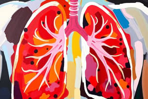
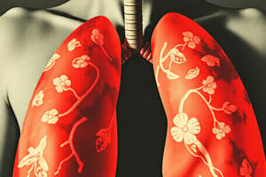
Cyanosis
- Cyanosis is the bluish coloration of skin and mucous membranes due to increased amounts of deoxygenated hemoglobin (Hb) in capillaries.
- The threshold of cyanosis is 5 gm reduced (deoxygenated) Hb / 100 ml capillary blood.
- Reduced Hb has an intense blue color, which is visible wherever the skin is thin, such as lips, mucous membrane, nail beds, and ear lobes.
Nerve and Muscle
Structure of Nerve
- Peripheral nerves are made up of axons of neurons that form groups of fibers in the center of the nerve.
- The nerve is covered by layers of connective tissue surrounding axons and also covering the necessary capillaries required to supply these axons.
The Neuron
- The neuron (or nerve cell) is the basic structural unit of the nervous system.
- Neurons are generally composed of three major parts: the soma (main cell body), dendrites (processes that project out from the soma for reception of signals from other neurons), and axons.
The Nervous System
- The nervous system is made up of the central nervous system (formed of brain and spinal cord) and peripheral nervous system (formed of nerves).
- The nervous system is responsible for intelligence, learning, memory, movement, different sensations, and basic body functions controlled by the autonomic nervous system.
- The nervous system coordinates the rapid activities of the body through fast transmission of impulses.
- The nervous system can be functionally divided into three main divisions: sensory, central (brain and spinal cord), and motor (voluntary and involuntary).
Skeletal Muscles
- The skeletal muscle is made up of individual muscle fibers bundled together by connective tissues and arranged in parallel.
- Each muscle fiber is a single cell enveloped by the cell membrane (sarcolemma).
- At each end of the muscle fiber, the sarcolemma fuses with a tendon fiber, and the tendon fibers collect into bundles to form the muscle tendons.
Types of Contraction of Skeletal Muscle
- There are two types of skeletal muscle contraction: isometric contraction and isotonic contraction.
- Isometric contraction: the muscle is stimulated to contract against a heavy load, but it will not be able to shorten because the load is too heavy for the muscle.
- Isotonic contraction: the muscle contracts against a smaller load, contraction will start isometrically, and muscle tension rises until the tension reaches a level that can lift the load.
Neuromuscular Transmission
- Neuromuscular transmission is the transmission of nerve impulses from a motor neuron to skeletal muscle fibers.
- Physiologic anatomy of neuromuscular junction: the motor neuron branches as it approaches the muscle, sending axon terminals (end feet) to several skeletal muscle fibers.
- Each skeletal muscle fiber receives only one axon terminal containing acetylcholine (Ach) vesicles.
- The nerve ending fits into a depression in the muscle membrane called the motor end plate.
- Sequence of events during neuromuscular transmission: 1. arrival of the nerve impulse at the motor end plate, 2. release of acetylcholine (Ach) from the nerve terminal, 3. binding of Ach to receptors on the muscle fiber membrane, and 4. generation of muscle contraction.
Studying That Suits You
Use AI to generate personalized quizzes and flashcards to suit your learning preferences.
Description
Learn about cyanosis, a medical condition characterized by a bluish coloration of skin and mucous membranes due to deoxygenated hemoglobin in capillaries.