Podcast
Questions and Answers
What characterizes significant urinary crystals?
What characterizes significant urinary crystals?
- They are commonly found in freshly voided urine.
- They are solely linked to dietary factors.
- They can indicate metabolic disorders. (correct)
- They always indicate infection.
In what conditions are crystals likely to form in urine specimens?
In what conditions are crystals likely to form in urine specimens?
- Refrigerated specimens with high solute concentration. (correct)
- High solute concentration and high temperature.
- Freshly voided urine with low specific gravity.
- Low pH and low temperature.
Which of the following crystals would be most concerning if found in a urine sample?
Which of the following crystals would be most concerning if found in a urine sample?
- Calcium oxalate.
- Struvite crystals.
- Cystine crystals. (correct)
- Amorphous urates.
How do some laboratories approach the reporting of urinary crystals?
How do some laboratories approach the reporting of urinary crystals?
Which of these factors does NOT affect crystal formation in urine?
Which of these factors does NOT affect crystal formation in urine?
What is the correct shape description of Calcium Phosphate crystals?
What is the correct shape description of Calcium Phosphate crystals?
Which type of crystals can be confused with bacteria due to similar shapes?
Which type of crystals can be confused with bacteria due to similar shapes?
What is the primary clinical significance of Ammonium Biurate crystals?
What is the primary clinical significance of Ammonium Biurate crystals?
Which condition is associated with the presence of Leucine crystals?
Which condition is associated with the presence of Leucine crystals?
What do Calcium Phosphate crystals do when glacial acetic acid is added?
What do Calcium Phosphate crystals do when glacial acetic acid is added?
In what urine condition are abnormal crystals typically found?
In what urine condition are abnormal crystals typically found?
Which characteristics distinguish Cystine crystals from Uric Acid crystals?
Which characteristics distinguish Cystine crystals from Uric Acid crystals?
What color are Ammonium Biurate crystals typically?
What color are Ammonium Biurate crystals typically?
What is the primary condition that can lead to the formation of urinary casts?
What is the primary condition that can lead to the formation of urinary casts?
What is the main component of urinary casts?
What is the main component of urinary casts?
What does an increase in urinary casts indicate?
What does an increase in urinary casts indicate?
Which factors would increase the rate of uromodulin excretion?
Which factors would increase the rate of uromodulin excretion?
What characteristic is true about urinary casts when viewed microscopically?
What characteristic is true about urinary casts when viewed microscopically?
What is the primary characteristic used for identifying different types of urine crystals?
What is the primary characteristic used for identifying different types of urine crystals?
Which of the following types of crystals can be found in acidic urine?
Which of the following types of crystals can be found in acidic urine?
What color are uric acid crystals typically?
What color are uric acid crystals typically?
What shape is primarily associated with Dihydrate Calcium Oxalate crystals?
What shape is primarily associated with Dihydrate Calcium Oxalate crystals?
Which factors can lead to increased levels of uric acid crystals?
Which factors can lead to increased levels of uric acid crystals?
How can you best differentiate between amorphous urates and amorphous phosphates in urine?
How can you best differentiate between amorphous urates and amorphous phosphates in urine?
What is a common factor associated with the presence of calcium oxalate crystals?
What is a common factor associated with the presence of calcium oxalate crystals?
What describes the appearance of amorphous phosphates after refrigeration?
What describes the appearance of amorphous phosphates after refrigeration?
What is a common characteristic of fatty casts in urine analysis?
What is a common characteristic of fatty casts in urine analysis?
Which method is used to confirm the presence of cholesterol in urine sediment?
Which method is used to confirm the presence of cholesterol in urine sediment?
In which condition would one primarily find fatty casts in urine?
In which condition would one primarily find fatty casts in urine?
What staining method is used to identify triglycerides in urine sediment?
What staining method is used to identify triglycerides in urine sediment?
When should a microscopic examination of urine sediment be performed?
When should a microscopic examination of urine sediment be performed?
Flashcards are hidden until you start studying
Study Notes
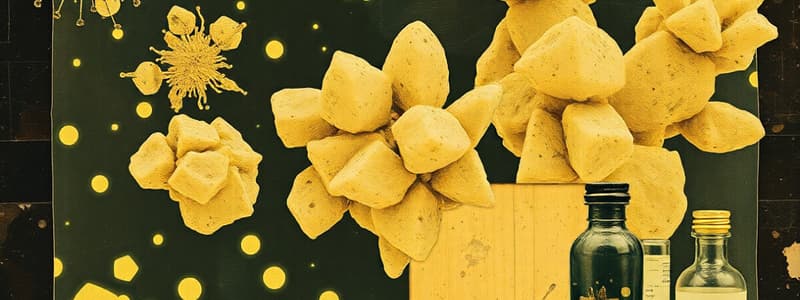
Crystals in Urine
- Crystals are not usually found in freshly voided urine.
- Many crystals have little clinical significance.
- Clinically significant crystals can indicate metabolic disorders, stone formation, or medication regulation.
- Examples of clinically significant crystals include cystine and tyrosine.
- To identify crystals, consider their appearance, the pH of the urine, and solubility characteristics.
- Most crystals are not clinically significant; some laboratories report all crystals, while others only report clinically significant ones.
- Crystals can appear as true geometrically formed structures or as amorphous material.
- Differentiate between the few abnormal crystals that indicate liver disease, inborn errors of metabolism, and damage to tubules.
- Report the number of crystals seen using a scale of 1+ to 4+ based on laboratory policy.
Crystal Formation
- Crystals form through precipitation of urine solutes like salts, organic compounds, and medications.
- Formation is affected by temperature, solute concentration, and pH.
- Many crystals form in refrigerated specimens.
- Crystals found in freshly voided urine are usually associated with concentrated (high specific gravity) specimens.
- Most crystals have characteristic shapes and colors.
- Urine pH is the most valuable aid in identifying crystals.
- Crystals are classified as normal acid, normal alkaline, or abnormal acid.
- All abnormal crystals are found in acid urine.
- Polarized microscopy and solubility characteristics are also valuable in identifying crystals.
Normal Crystals in Acid Urine
Uric Acid Crystals
- Shapes include rhombic, four-sided flat plates (whetstones), wedges, and rosettes.
- Color is typically yellow-brown.
- Can be colorless and six-sided, similar to cystine crystals.
- Highly birefringent under polarized light, unlike cystine crystals.
- Increased levels of uric acid crystals are associated with increased purine and nucleic acid levels, leukemia patients receiving chemotherapy, and patients with gout.
Amorphous Urates
- Shape is granular sediment.
- Color is yellow-brown or pink due to the pigment uroerythrin.
- Found in acidic urine with a pH greater than 5.5.
- Can obscure the microscopic field and make it difficult to see other elements.
- Common in refrigerated specimens and may dissolve if warmed.
Normal Crystals in Acid/Neutral Urine
Calcium Oxalate Crystals
- Two possible forms:
- Dihydrate Calcium Oxalate: Most common, envelope-shaped.
- Monohydrate Calcium Oxalate: Oval or dumbbell-shaped.
- Colorless.
- Birefringent under polarized light.
- Increased levels associated with:
- Renal calculi.
- Oxalate-rich foods: Tomatoes, asparagus, spinach, oranges.
- Ethylene glycol poisoning (Noticeable presence of the monohydrate form).
Normal Crystals in Alkaline Urine
- Amorphous Phosphates
- Ammonium Biurate
- Calcium Carbonate
- Calcium Phosphate
- Triple Phosphate
Amorphous Phosphates
- Shape: Granular sediment.
- Color: White or colorless.
- Present in large quantities after refrigeration.
- Do not dissolve when warmed.
How to differentiate between amorphous urates and amorphous phosphates:
| pH | Color | |
|---|---|---|
| Urates | Acid | Pink |
| Phosphates | Alkaline | White |
Calcium Phosphate
- Shape: Flat rectangular plates or rosettes.
- Color: Colorless.
- Can be confused with sulfonamide crystals.
- Use acetic acid to differentiate: Calcium phosphate crystals will dissolve, but sulfonamide crystals will not.
- No clinical significance.
Triple Phosphate (Ammonium Magnesium Phosphate)
- Shape: "Coffin-lid" prism or fern leaf shaped.
- Color: Colorless.
- Birefringent under polarized light.
- No clinical significance.
- Found in highly alkaline urine and in patients with UTIs due to the presence of urea-splitting bacteria.
Ammonium Biurate
- Shape: Spicule-covered spheres ("thorny apples").
- Color: Yellow-brown.
- Dissolve at 60°C.
- Convert to uric acid crystals when glacial acetic acid is added.
- Occur in old specimens.
- Associated with the presence of ammonia produced by urea-splitting bacteria.
Calcium Carbonate
- Shape: Small dumbbell or spherical.
- Color: Colorless.
- Can be confused with:
- Bacteria: Calcium carbonate birefringent, bacteria is not.
- Amorphous material: Gas is formed after the addition of acetic acid to calcium carbonate but not after addition to amorphous material.
- No clinical significance.
Abnormal Crystals
- Always found in acidic urine or rarely in neutral urine.
- If abnormal crystals are suspected, confirm with chemical testing before reporting (if applicable).
- Check patient history, including disorders and medications, to correlate findings.
- Sulfa drugs and ampicillin can crystallize out in concentrated specimens.
Abnormal Crystals
- Cystine: Metabolic disorder.
- Cholesterol: Nephrotic syndrome.
- Leucine, Tyrosine, Bilirubin: Liver diseases.
- Sulfonamides, Radiographic Dye, Ampicillin: Medications
Cystine
- Shape: Hexagonal plates (thick or thin).
- Color: Colorless.
- Differentiation from uric acid crystals: Uric acid crystals are highly birefringent, but only thick cystine crystals polarize.
Ampicillin
- Shape: Needles that form bundles after refrigeration.
- Color: Colorless.
- Found in patients following large doses of ampicillin who are not adequately hydrated.
Urinary Casts
-
The major constituent of casts is uromodulin (Tamm-Horsfall protein).
-
Uromodulin is excreted at a constant rate under normal conditions.
-
The rate of excretion increases during stress and exercise.
-
Uromodulin gels more readily under conditions of urine-flow stasis, acidity, and the presence of sodium and calcium.
-
The size and shape of the cast depends on the tubule where it was formed.
-
The cast matrix may become embedded with cells, bacteria, granules, pigments, and crystals from the tubular filtrate.
-
Shape: Cylindrical with parallel sides and rounded edges
-
May contain inclusions from the tubular filtrate (cells, granules, pigments, etc.)
-
Casts DO NOT have dark edges.
-
Find casts under low power and identify under high power.
-
Low light is essential - casts have a low refractive index.
-
Report the number of casts seen per low power field (lpf).
Urinary Casts: Composition and Formation
- Uromodulin is secreted by renal tubular epithelial cells of the distal convoluted tubule and collecting ducts.
- Secretion is increased during stress and exercise.
- The formation of a matrix occurs from protein fibrils during urine stasis, acid pH, and the presence of sodium and calcium.
- Uromodulin is not detected by reagent strips.
- An increase in protein is due to albumin and underlying renal conditions.
- An increase in urinary casts is called cylindruria, also called Renal Failure Casts.
Fatty Casts
- Will have fat droplets and oval fat bodies attached to the protein matrix.
- Highly refractile under bright-field microscopy.
- Confirmation is done by:
- Polarized microscopy: Cholesterol will have a maltese cross.
- Sudan III or Oil Red O stains: Triglycerides will stain orange.
- Free fat droplets and oval fat bodies should be seen in the urine sediment as well.
Fatty Casts: Clinical Significance
- Primarily Nephrotic syndrome: A collection of symptoms that indicate kidney damage and include excess levels of protein in the urine.
- Also seen in:
- Tubular necrosis.
- Diabetes mellitus.
- Crush injuries.
Oval Fat Bodies
- Lipid-containing renal tubular epithelial cells (RTE cells).
- RTE cells absorb lipids in the glomerular filtrate (cholesterol, triglycerides or neutral fat).
- Cholesterol will have a maltese cross appearance under polarized light.
- Triglycerides and neutral fats can be stained orange with Sudan III or Oil Red O.
Criteria for Microscopics
- Laboratories will have specific criteria on when to perform a microscopic examination of urine sediment.
- Generally, a microscopic is performed if any of the following reagent strip results are positive:
- Blood
- Leukocyte esterase
- Protein
- Nitrite
- If all reagent strip results are negative or normal, no microscopic examination is performed.
Studying That Suits You
Use AI to generate personalized quizzes and flashcards to suit your learning preferences.


