Podcast
Questions and Answers
Which cranial nerve is primarily responsible for the perception of smell?
Which cranial nerve is primarily responsible for the perception of smell?
- Vagus nerve
- Optic nerve
- Facial nerve
- Olfactory nerve (correct)
What type of axons does the facial nerve include?
What type of axons does the facial nerve include?
- Neither sensory nor motor axons
- Only sensory axons
- Only motor axons
- Both sensory and motor axons (correct)
Where are the nerve cell bodies for somatic motor fibers located in cranial nerves?
Where are the nerve cell bodies for somatic motor fibers located in cranial nerves?
- In the spinal cord
- In the peripheral ganglia
- Within the nuclei in the brain (correct)
- In the dorsal root of the spinal nerve
Which cranial nerve is involved in visual perception?
Which cranial nerve is involved in visual perception?
What is the functional classification of the olfactory nerve?
What is the functional classification of the olfactory nerve?
What kind of fibers does the vagus nerve carry?
What kind of fibers does the vagus nerve carry?
What is the role of the optic chiasm?
What is the role of the optic chiasm?
What type of function does the special visceral afferent serve?
What type of function does the special visceral afferent serve?
What is the result of a lesion at the optic chiasm regarding visual perception?
What is the result of a lesion at the optic chiasm regarding visual perception?
Which cranial nerve is responsible for the blink response mechanism?
Which cranial nerve is responsible for the blink response mechanism?
Which structure is essential for the blink response to occur?
Which structure is essential for the blink response to occur?
What is the primary function of the oculomotor nerve (CN III)?
What is the primary function of the oculomotor nerve (CN III)?
What would likely occur if there is damage to the oculomotor nerve?
What would likely occur if there is damage to the oculomotor nerve?
In the visual pathway, which structure processes signals after the lateral geniculate nucleus (LGN)?
In the visual pathway, which structure processes signals after the lateral geniculate nucleus (LGN)?
When evaluating the pupillary light reflex (PLR), what is indicative of damage to CN III?
When evaluating the pupillary light reflex (PLR), what is indicative of damage to CN III?
What role does the cerebellum play in the blink response?
What role does the cerebellum play in the blink response?
What is the primary structure where the majority of optic tract fibers terminate?
What is the primary structure where the majority of optic tract fibers terminate?
What is the expected visual outcome from damage to the right optic nerve before the optic chiasm?
What is the expected visual outcome from damage to the right optic nerve before the optic chiasm?
Which part of the brainstem is involved in the Pupillary Light Reflex (PLR)?
Which part of the brainstem is involved in the Pupillary Light Reflex (PLR)?
What is the primary role of the vestibular nerve?
What is the primary role of the vestibular nerve?
What type of vision pathway is primarily responsible for the Menace response?
What type of vision pathway is primarily responsible for the Menace response?
Which cranial nerves are involved in the motor component of the vestibular pathway?
Which cranial nerves are involved in the motor component of the vestibular pathway?
In the oculocephalic reflex test, what is observed when the head is moved laterally?
In the oculocephalic reflex test, what is observed when the head is moved laterally?
What would occur with a lesion at the optic chiasm?
What would occur with a lesion at the optic chiasm?
In the indirect PLR test, what is an abnormal result?
In the indirect PLR test, what is an abnormal result?
What does a head tilt indicate in the context of vestibular lesions?
What does a head tilt indicate in the context of vestibular lesions?
Which structure serves as the relay component for vestibular signals?
Which structure serves as the relay component for vestibular signals?
Which cranial nerve is primarily tested when examining the Pupillary Light Reflex?
Which cranial nerve is primarily tested when examining the Pupillary Light Reflex?
Which cranial nerve primarily carries vestibular sensory information?
Which cranial nerve primarily carries vestibular sensory information?
What indicates that the pupillary constrictor muscles are being activated?
What indicates that the pupillary constrictor muscles are being activated?
What does abnormal nystagmus indicate in a clinical examination?
What does abnormal nystagmus indicate in a clinical examination?
What are the signs of vestibular ataxia observed during an examination?
What are the signs of vestibular ataxia observed during an examination?
What muscle does the trochlear nerve (CN IV) innervate to aid in stabilizing vision during head tilts?
What muscle does the trochlear nerve (CN IV) innervate to aid in stabilizing vision during head tilts?
Which cranial nerve is responsible for the sensory innervation of the face, and includes the ophthalmic, maxillary, and mandibular branches?
Which cranial nerve is responsible for the sensory innervation of the face, and includes the ophthalmic, maxillary, and mandibular branches?
What type of strabismus can be observed in species with slit-like pupils due to trochlear nerve dysfunction?
What type of strabismus can be observed in species with slit-like pupils due to trochlear nerve dysfunction?
Which reflex is tested to assess the sensory function of the ophthalmic branch of the trigeminal nerve?
Which reflex is tested to assess the sensory function of the ophthalmic branch of the trigeminal nerve?
Which part of the trigeminal nerve primarily innervates the nasal region?
Which part of the trigeminal nerve primarily innervates the nasal region?
Which cranial nerve is primarily involved in motor function related to mastication?
Which cranial nerve is primarily involved in motor function related to mastication?
During a clinical exam, which branch of the trigeminal nerve is assessed by touching the skin over the mandible?
During a clinical exam, which branch of the trigeminal nerve is assessed by touching the skin over the mandible?
Which nerve contains a large ganglion that holds primary nerve cell bodies for its sensory component?
Which nerve contains a large ganglion that holds primary nerve cell bodies for its sensory component?
Flashcards are hidden until you start studying
Study Notes
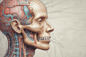
Cranial Nerves
- Some nerves are exclusively sensory, some exclusively motor and some are mixed
- 12 pairs of cranial nerves originate from a specific area of the brain and innervate the head or sometimes body
- Each cranial nerve has a unique function including special senses like taste, smell, hearing, balance, and vision
- Nerve cell bodies for somatic motor fibers in CN are located within nuclei in the brain, considered lower motor neurons
- Nerve cell bodies for the somatic sensory neurons (not special senses) are in ganglia outside of the CNS
- Cranial nerves are considered part of the PNS and CNS
Terminology
- General somatic afferent = sensory, pain, and temp to the face
- General somatic efferent = motor (muscles for chewing or facial movement)
- Special Somatic afferent = sensory fibers, vision, hearing, balance, has special afferent function
- Special Visceral Afferent = taste and smell
- General Visceral efferent = motor fibers from the ANS (PARA) that innervate smooth muscle
- Special Visceral efferent = innervates larynx, pharynx, and part of the esophagus to control swallowing and respiration
CN I: Olfactory Nerve
- Sensory only, responsible for the conscious perception of smell
- Chemoreceptors in the nasal mucosa detect odors and transmit sensory information to axons in CN I
- The nucleus for this nerve is found in the olfactory bulb and anterior olfactory nucleus in the forebrain
- Anosmia - absent sense of smell, hyposmia- decreased sense of smell
CN II: Optic Nerve
- Sensory for visual perception (VISION) and the sensory component of the pupillary light reflex
- Light activates photoreceptors in the retina, generating AP, which are relayed via retinal ganglion cells which form the axons in the optic nerves
- The optic nerves from each eye converge at the optic chiasm
- Optic nerve and retina are part of the CNS
- Cell bodies of neurons in the retina are the ganglion cells
- Conscious (response and vision pathway): lateral geniculate nuclei (80%)
- Subconscious (reflex pathway): pretectal nuclei (mesencephalon/midbrain, 20%)
- Retinal Ganglion cells → optic nerve → optic chiasm → optic tract → LGN of thalamus → visual cortex
Clinical Exam: CN II
- Pupillary Light Reflex (PLR)
- Direct PLR: Shine light into one eye and observe for pupillary constriction in the same eye. Abnormal result is lack of constriction
- Indirect (Consensual) PLR: Shine light into one eye and observe the other eye for pupillary constriction. Abnormal result is lack of constriction.
- Menace response:
- While covering one eye, make a threatening gesture at the uncovered eye, looking for a blink in response.
- Structures needed for blink: retina, CN II, LGN, visual cortex, cerebellum, CN VII
- Menace response is not a reflex, as the cortex is involved
Visual Pathway
- CN II: Retina → optic nerve → optic chiasm → contralateral optic tract → LGN → optic radiation → visual cortex
Motor Pathway
- Visual cortex impulses → motor cortex → facial motor nucleus → facial muscles around the eye (e.g., orbicularis oculi and levator palpebrae muscles)
Lesions: Optic Nerve
- Before optic chiasm: Blindness in the eye supplied by the damaged optic nerve
- At optic chiasm: Partial blindness in both eyes
- In optic tract: Partial blindness
CN III: Oculomotor Nerve
- Motor control of iris constrictor muscles (PARA, ANS - pupillary constriction)
- Somatic motor innervation (aka somatic nucleus) of some medial rectus and retractor bulbi muscle
- Originates from nuclei in the rostral (anterior) ventral area of the midbrain
- Fibers that control pupillary sphincter and ciliary muscles originate in the Edinger-Westphal nucleus
Clinical Exam: CN III
- Observe the eye and PLR
- If nerve or somatic nucleus is damaged: Ventrolateral strabismus results (down and out, side and downwards), ipsilateral damage
- If CN III nerve or EW nucleus is damaged: Abnormal PLR - an inappropriate lack of constriction of the pupil. Possible anisocoria
CN IV: Trochlear Nerve
- Motor innervation of the dorsal oblique muscle of the eye
- Helps keep the visual world "steady" by counter-rotating the eye when the head tilts
- Motor nucleus located caudally in the mesencephalon (midbrain)
Clinical Exam: CN IV
- Difficult to diagnose in dogs
- Mild dorsolateral strabismus
- Abnormal eye movements when the head is moved or gaze shifts laterally
- Rotation of fundus via ophthalmoscope
CN V: Trigeminal Nerve
- Both motor and sensory components
- Sensory portion innervates the face (pinnae, eyelids, cornea, oral cavity, mucosa of the nasal septum)
- Motor innervates the muscles involved with mastication
- Nuclei (sensory and motor) are located/originate in the pons
Clinical Exam: CN V Sensory
- Observe for blink response (palpebral reflex)
- Symmetrical response to stimulation of the nasal mucosa (use cotton bud to touch nasal mucosa on each side)
- Stimulate the medial canthus (ophthalmic branch), lateral canthus (maxillary branch), and skin over mandible (mandibular branch) and look for a blink
CN VIII: Vestibulocochlear Nerve
- Two parts: Vestibular and cochlear
- Vestibular portion: responsible for normal posture and coordination of the eye, head, neck, trunk, and limbs
- Cochlear nerve: travels with the vestibular portion to the medulla, responsible for hearing
Clinical Exam: CN VIII
- Oculocephalic reflex test (Doll’s Eye Reflex): Move head laterally from side to side and observe symmetrical, coordinated eye movement
- Normal nystagmus
- Abnormal nystagmus: eyes move repeatedly back and forth, up and down, vertical, or circles
- Observe for vestibular ataxia: stumbling, circling, head tilt
- Hearing is not normally assessed, noted in history
Pathway: CN VIII, CN III, CN IV, CN VI
- CN VIII (vestibulocochlear) (+ CN II optic): vestibular input from semicircular canals in the ear → vestibular nuclei in the medulla
- Medial Longitudinal Fasciculus (MLF): tract that connects the vestibular nuclei with the nuclei of cranial nerves III, IV, and VI
- Motor nuclei of individual nerves that innervate extrinsic eye muscles: III/IV/VI
CN IX: Glossopharyngeal Nerve
- Motor and Sensory
- Motor innervation of muscles of the pharynx
- PARA (ANS, motor) innervation to the parotid salivary gland
Studying That Suits You
Use AI to generate personalized quizzes and flashcards to suit your learning preferences.