Podcast
Questions and Answers
Which cranial nerve is responsible for controlling movements of the face and eye muscles?
Which cranial nerve is responsible for controlling movements of the face and eye muscles?
- Vestibulocochlear nerve
- Olfactory nerve
- Trigeminal nerve (correct)
- Optic nerve
Which cranial nerve is associated with smell (olfaction)?
Which cranial nerve is associated with smell (olfaction)?
- Facial nerve
- Trochlear nerve
- Olfactory nerve (correct)
- Optic nerve
Which cranial nerve is involved in hearing and balance?
Which cranial nerve is involved in hearing and balance?
- Glossopharyngeal nerve
- Vagus nerve
- Vestibulocochlear nerve (correct)
- Abducens nerve
Which cranial nerve aids in controlling eye movements?
Which cranial nerve aids in controlling eye movements?
What is the function of the glossopharyngeal cranial nerve?
What is the function of the glossopharyngeal cranial nerve?
Which cranial nerve is related to vision?
Which cranial nerve is related to vision?
Which cranial nerve is responsible for sensing smells?
Which cranial nerve is responsible for sensing smells?
Which imaging technique is considered the gold standard for evaluating cranial nerves?
Which imaging technique is considered the gold standard for evaluating cranial nerves?
Which cranial nerve carries motor fibers that control facial expressions and secretomotor fibers for salivation and lacrimation?
Which cranial nerve carries motor fibers that control facial expressions and secretomotor fibers for salivation and lacrimation?
Which cranial nerve is responsible for controlling eye movements and pupillary reflexes?
Which cranial nerve is responsible for controlling eye movements and pupillary reflexes?
Which cranial nerve is the largest mixed nerve, with both sensory and motor functions, supplying sensation to the face and jaw regions and innervating muscles involved in chewing and facial expressions?
Which cranial nerve is the largest mixed nerve, with both sensory and motor functions, supplying sensation to the face and jaw regions and innervating muscles involved in chewing and facial expressions?
Which cranial nerve splits into the vestibular and cochlear branches, responsible for hearing and balance functions, respectively?
Which cranial nerve splits into the vestibular and cochlear branches, responsible for hearing and balance functions, respectively?
Study Notes
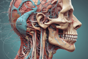
Head and Neck Anatomy: Focusing on Cranial Nerves
The human body features twelve pairs of cranial nerves, each responsible for controlling motor and sensory functions of the head and neck region. Understanding the structure, location, and functions of these nerves is essential for diagnosing and treating neurological conditions affecting this area. In this article, we will explore the cranial nerves in detail.
Cranial Nerve Functions
Cranial nerves play a significant role in sensory perception and motor control. Some cranial nerves are purely sensory, while others are motor or mixed (both sensory and motor). The cranial nerves are organized into four groups based on their functions:
- Olfactory: Responsible for smelling (olfaction)
- Optic: Related to sight (vision)
- Motor: Controls movements of the face and eye muscles
- Special senses: Involved in hearing, balance, speech, and taste
Each cranial nerve has a specific name and is assigned a number ranging from I to XII. These numbers follow the mnemonic "On Old Olympians Go Dancing Joyfully Exultant," which corresponds to the following sequence: olfactory, optic, oculomotor, trochlear, trigeminal, abducens, facial, vestibulocochlear, glossopharyngeal, vagus, accessory, and hypoglossal.
Cranial Nerve Anatomy
Cranial nerves originate from the brainstem and exit through various foramina located on the skull base. The anatomy of each nerve varies slightly, but they share certain general characteristics:
-
Olfactory Nerve: The olfactory nerve starts at the superior turbinals in the nasal cavity and runs along the floor of the anterior cranial fossa to terminate in the olfactory bulb. It is responsible for sensing smells.
-
Optic Nerve: The optic nerve originates from the lateral geniculate nucleus and carries visual signals from the retina to the visual cortex in the occipital lobe.
-
Ocular Motor Nerves: These include the oculomotor, trochlear, and abducens nerves, which control eye movements and pupillary reflexes.
-
Trigeminal Nerve: The trigeminal nerve is the largest mixed cranial nerve, with both sensory and motor functions. Its branches supply sensation to the face and jaw regions and innervate muscles involved in chewing and facial expressions.
-
Facial Nerve: The facial nerve carries motor fibers that control facial expression and secretomotor fibers for salivation and lacrimation. Additionally, it has taste fibers for the anterior two-thirds of the tongue.
-
Vestibulocochlear Nerve: This nerve splits into the vestibular and cochlear branches, each responsible for hearing and balance functions, respectively.
-
Glossopharyngeal and Vagus Nerves: These two nerves are closely related, with the glossopharyngeal nerve providing motor function to the larynx and taste sensation from the posterior third of the tongue. The vagus nerve controls several muscles in the head and neck region, such as those involved in speech production and facial expressions.
Imaging Techniques for Studying Cranial Nerves
Magnetic resonance imaging (MRI) is considered the gold standard technique for evaluating cranial nerves due to its excellent soft tissue contrast and ability to provide detailed information about their anatomical relationships with surrounding structures. Computed tomography (CT) scans can also be useful for visualizing intraosseous segments of cranial nerves, skull base foramina, and bony traumatic lesions.
In summary, the cranial nerves play a crucial role in controlling various aspects of head and neck function. Understanding their structure and function is essential for diagnosing and treating conditions affecting these regions.
Studying That Suits You
Use AI to generate personalized quizzes and flashcards to suit your learning preferences.
Description
Test your knowledge on the structure, function, and anatomy of the twelve pairs of cranial nerves that control motor and sensory functions in the head and neck region. Learn about the roles of different cranial nerves, imaging techniques for studying them, and their significance in diagnosing neurological conditions.