Podcast
Questions and Answers
Which measurement technique allows for the indirect assessment of tear volume without utilizing any invasive methods?
Which measurement technique allows for the indirect assessment of tear volume without utilizing any invasive methods?
- Schirmer test
- Tear prism height (correct)
- Tear Break-Up-Time (TBUT)
- Phenol-red thread test
What is the normal range for tear prism height as specified by Port and Asaria?
What is the normal range for tear prism height as specified by Port and Asaria?
- 0.2 to 1.0 mm (correct)
- 1.0 to 2.0 mm
- 0.1 to 0.5 mm
- 0.3 to 0.8 mm
Which of the following is NOT an invasive method for assessing tear production or quality?
Which of the following is NOT an invasive method for assessing tear production or quality?
- Tear prism height measurement (correct)
- Schirmer test
- Tear Break-Up-Time (TBUT)
- Rose Bengal staining
When using the slit lamp to measure tear prism height, what adjustment is essential for accurate measurement?
When using the slit lamp to measure tear prism height, what adjustment is essential for accurate measurement?
Why should fluorescein not be utilized during the measurement of tear prism height?
Why should fluorescein not be utilized during the measurement of tear prism height?
What is the primary method for assessing corneal staining during a slit-lamp examination?
What is the primary method for assessing corneal staining during a slit-lamp examination?
Which of the following conditions is NOT associated with corneal staining aetiology?
Which of the following conditions is NOT associated with corneal staining aetiology?
What symptom is typically NOT associated with corneal staining?
What symptom is typically NOT associated with corneal staining?
What is the recommended magnification for observing corneal staining after initial assessment with a parallelepiped?
What is the recommended magnification for observing corneal staining after initial assessment with a parallelepiped?
Which staining pattern indicates a mechanical cause of corneal staining?
Which staining pattern indicates a mechanical cause of corneal staining?
Which type of corneal staining is characterized by delayed hypersensitivity reactions?
Which type of corneal staining is characterized by delayed hypersensitivity reactions?
What aspect of the tear film is evaluated during a slit-lamp examination?
What aspect of the tear film is evaluated during a slit-lamp examination?
Which of the following is characteristic of solution induced corneal staining (SICS)?
Which of the following is characteristic of solution induced corneal staining (SICS)?
What is the primary reason for using a Wratten 12 filter during the invasive tear break-up time assessment?
What is the primary reason for using a Wratten 12 filter during the invasive tear break-up time assessment?
Which step is NOT typically included in the slit-lamp examination flowchart using blue light?
Which step is NOT typically included in the slit-lamp examination flowchart using blue light?
What indicates the first break up of tears during the invasive tear break-up time assessment?
What indicates the first break up of tears during the invasive tear break-up time assessment?
Which component is essential to wet fluorescein strips before use?
Which component is essential to wet fluorescein strips before use?
During a slit-lamp examination, how should the pupil be treated to effectively assess the crystalline lens?
During a slit-lamp examination, how should the pupil be treated to effectively assess the crystalline lens?
What type of dry eye condition is characterized by aqueous tear deficiency?
What type of dry eye condition is characterized by aqueous tear deficiency?
When assessing the conjunctiva with blue light, which part is NOT usually examined?
When assessing the conjunctiva with blue light, which part is NOT usually examined?
Which illumination technique is commonly used to assess tear film status?
Which illumination technique is commonly used to assess tear film status?
What is the patient instructed to do after the last blink during the tear break-up time assessment?
What is the patient instructed to do after the last blink during the tear break-up time assessment?
What is NOT typically recorded during a routine anterior eye assessment?
What is NOT typically recorded during a routine anterior eye assessment?
What is the primary purpose of Tear Break-Up-Time testing?
What is the primary purpose of Tear Break-Up-Time testing?
Which method is considered non-invasive for assessing Tear Break-Up-Time?
Which method is considered non-invasive for assessing Tear Break-Up-Time?
What does the Schirmer test specifically measure?
What does the Schirmer test specifically measure?
For Invasive Tear Break-Up-Time assessment, which equipment setup is incorrect?
For Invasive Tear Break-Up-Time assessment, which equipment setup is incorrect?
Which interpretation of the Schirmer test indicates extremely dry eyes?
Which interpretation of the Schirmer test indicates extremely dry eyes?
What is the main advantage of the Phenol-Red Thread Test over the Schirmer test?
What is the main advantage of the Phenol-Red Thread Test over the Schirmer test?
What is a significant limitation of using Fluorescein in Invasive Tear Break-Up-Time assessments?
What is a significant limitation of using Fluorescein in Invasive Tear Break-Up-Time assessments?
Decreased lacrimation leading to cell degeneration is primarily assessed using which method?
Decreased lacrimation leading to cell degeneration is primarily assessed using which method?
What is a key reason for using grading scales in healthcare disciplines?
What is a key reason for using grading scales in healthcare disciplines?
What is one drawback of photographic grading scales?
What is one drawback of photographic grading scales?
What is the primary focus of lid eversion during an eye examination?
What is the primary focus of lid eversion during an eye examination?
Which grading scale designation indicates a condition that requires urgent clinical action?
Which grading scale designation indicates a condition that requires urgent clinical action?
What is the correct sequence for performing lid eversion?
What is the correct sequence for performing lid eversion?
Which of the following is NOT a method of assessing the tear film as mentioned?
Which of the following is NOT a method of assessing the tear film as mentioned?
What document helps in recording the staining pattern seen on the cornea?
What document helps in recording the staining pattern seen on the cornea?
Why might grading scales lead to reduced subjectivity?
Why might grading scales lead to reduced subjectivity?
What is the main issue with subjective descriptions of conditions?
What is the main issue with subjective descriptions of conditions?
What is a key factor in maintaining patient comfort during a slit-lamp examination?
What is a key factor in maintaining patient comfort during a slit-lamp examination?
Flashcards
Tear Prism Height
Tear Prism Height
A non-invasive method to assess tear volume by measuring the height of the tear reservoir along the lower eyelid margin.
Non-Invasive Tear Break-Up-Time (NIBUT)
Non-Invasive Tear Break-Up-Time (NIBUT)
The time it takes for the tear film to break up after blinking, measured with a special light source.
Tear Thinning Time
Tear Thinning Time
The thickness of the tear film, measured using a slit lamp and a special eyepiece.
Tear Break-Up-Time (TBUT)
Tear Break-Up-Time (TBUT)
Signup and view all the flashcards
Schirmer Test
Schirmer Test
Signup and view all the flashcards
Corneal staining
Corneal staining
Signup and view all the flashcards
Corneal staining - Mechanical cause
Corneal staining - Mechanical cause
Signup and view all the flashcards
Corneal staining - Exposure cause
Corneal staining - Exposure cause
Signup and view all the flashcards
Corneal staining - Metabolic cause
Corneal staining - Metabolic cause
Signup and view all the flashcards
Corneal staining - Solution induced corneal staining (SICS)
Corneal staining - Solution induced corneal staining (SICS)
Signup and view all the flashcards
Corneal staining - Toxic & Allergic cause
Corneal staining - Toxic & Allergic cause
Signup and view all the flashcards
Corneal staining - Pattern
Corneal staining - Pattern
Signup and view all the flashcards
Bulbar Conjunctival Staining
Bulbar Conjunctival Staining
Signup and view all the flashcards
Lid Eversion
Lid Eversion
Signup and view all the flashcards
Cobblestone Nodules
Cobblestone Nodules
Signup and view all the flashcards
Grading Scale
Grading Scale
Signup and view all the flashcards
Photographic Grading Scale
Photographic Grading Scale
Signup and view all the flashcards
Drawn Grading Scale
Drawn Grading Scale
Signup and view all the flashcards
Slit Lamp
Slit Lamp
Signup and view all the flashcards
Tear Film
Tear Film
Signup and view all the flashcards
Non-invasive Tear Film Assessment
Non-invasive Tear Film Assessment
Signup and view all the flashcards
Tear Break-Up-Time
Tear Break-Up-Time
Signup and view all the flashcards
Non-invasive Tear Break-Up-Time
Non-invasive Tear Break-Up-Time
Signup and view all the flashcards
Invasive Tear Break-Up-Time (ITBUT)
Invasive Tear Break-Up-Time (ITBUT)
Signup and view all the flashcards
Interpreting Schirmer Test Results
Interpreting Schirmer Test Results
Signup and view all the flashcards
Phenol-Red Thread Test
Phenol-Red Thread Test
Signup and view all the flashcards
Rose Bengal Staining
Rose Bengal Staining
Signup and view all the flashcards
Dry Eye
Dry Eye
Signup and view all the flashcards
Slit Lamp Examination Flowchart
Slit Lamp Examination Flowchart
Signup and view all the flashcards
White Light Assessment
White Light Assessment
Signup and view all the flashcards
Blue Light Assessment
Blue Light Assessment
Signup and view all the flashcards
Sodium Fluorescein (NaFl)
Sodium Fluorescein (NaFl)
Signup and view all the flashcards
Wratten Filter
Wratten Filter
Signup and view all the flashcards
Aqueous Tear-Deficient Dry Eye
Aqueous Tear-Deficient Dry Eye
Signup and view all the flashcards
Dry Eye with Decreased Wettability
Dry Eye with Decreased Wettability
Signup and view all the flashcards
Evaporative Dry Eye
Evaporative Dry Eye
Signup and view all the flashcards
Invasive Tear Break-Up Time (BUT)
Invasive Tear Break-Up Time (BUT)
Signup and view all the flashcards
Study Notes
Lecture Recording
- The lecture is being recorded as part of Plymouth University's Content Capture Project
- The recording will be available via the Panopto block on the module DLE pages shortly
- Students can ask questions during the lecture
- If a student asks a question, it may appear on the recording
- The lecturer can pause the recording if a student does not want their question recorded
Optometry Skills Lecture
- The lecture is about Specialist Optometry Skills (OPT506)
- The specific topic is Slit Lamp Biomicroscopy 3
- The lecturer is Dr. Asma Zahidi
- A one-time code (XV-RL-YT) is provided
Lecture Outline
- The lecture will cover a revision of the anterior eye assessment using white light
- Tear film assessment will also be covered using white light
- Routine anterior eye assessment with blue light techniques will be discussed
- Recording findings will be the final portion of the lecture
Slit-Lamp Examination Flowchart (White Light)
- The examination starts with observing the lids, margins & eyelashes
- Next, the tears/tear film, conjunctiva and bulbar are examined.
- Palpebral and limbus areas are also considered
- Further areas include cornea, anterior chamber, iris, and crystalline lens
Slit Lamp Examination Flowchart (Blue Light)
- The blue light examination includes Tears/tear film, Cornea & Conjunctiva (bulbar & palpebral)
- The light is combined with NaFl
Blue Light Assessment
- Fluorescein strips are used
- The strips require saline solution for wetting
- Fluorescein is often used with topical anesthetic.
Fluorescein Installation
- The correct installation technique is illustrated
- Incorrect installation techniques are indicated with a red "X"
Invasive Tear Break-Up Time
- The slit lamp is set up with broad beam illumination
- A cobalt blue (Wratten 12) filter improves fluorescein visibility
- Sufficient magnification for the cornea is mandatory
- Students should ask the patient to blink a few times before the test
- The patient's eyes are kept open for as long as possible
- Tear break-up time is determined by observing the first break in the tear film which appears as a black streak
Tear Break-Up Time
- Various images of different types of tear break-up time are shown
- Severity of dry eye is classified as Severe, Mild/Moderate (using Area Break-up, Spot Break-up, Random Break-up and others)
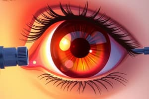
Corneal Staining
- Dot-shaped defects on the corneal epithelium is a particular characteristic when staining is observed
- Visual inspection is possible by using Fluorescein and adding a yellow filter, which stains the tear film
- Symptoms that occur alongside corneal staining could include pain, redness of the eye and increased tear flow
- Paraellelepiped (16x) and Optic section (25x-40x) are used for assessment magnification
Corneal Staining - Aetiology
- Mechanical factors including trauma, foreign bodies, damaged lenses can be an aetiology
- Exposure, metabolic (hypoxia, hypercapnia) and solution-induced (SICS) are additional aetiologies
- Toxic factors like care regimen hypersensitivity and allergic factors (immediate/delayed) can also be a factor
- Infectious disease and general health issues can also result in corneal staining.
Corneal Staining - Pattern
- Dryness (Decompensated cells and Corneal staining)
- Mechanical cause (Removal of cells e.g. 3 and 9 o'clock staining)
- Toxic cause (Decompensated Cells, SICS)
Corneal Abrasions: Staining Patterns
- Various patterns of corneal abrasions are illustrated
- The patterns are categorized and described in detail.
Bulbar Conjunctival Staining
- Bulbar conjunctival staining is used as an indicator of dry eye
- Certain dry eye drops might result in higher mucin secretion and improved staining scores.
- Comparison of before and after staining is often conducted
Lid Eversion
- The patient is asked to look down to grasp the upper eyelid at the base of the lashes using one hand (H1)
- This is done at a 45-degree angle.
- The other hand (H2) is used to press the upper lid downwards (and a cotton bud can also be used).
- The lashes are pulled gently upwards, following the motion of the eye downwards.
Why Lid Eversion?
- Cobblestone-like nodules in the palpebral conjunctiva are a potential cause for lid eversion procedures
- Small deposits of calcium can often result as a consequence of the procedure
Record Keeping
- The recording methods used include grading scales
- The grading scales allow accurate recording of data and are used to monitor disease
Record Cards (Contact Lens)
- Grading scales are used for recording information regarding various eye conditions such as: conjunctival redness, limbal redness in both the right (R) and left (L) eyes.
- Various conditions are graded and included such as corneal neovascularisation, corneal staining, conjunctiva staining, papillary conjunctivitis, blepharitis and others.
Areas of the Superior Palpebral Conjunctiva
- Different zones (areas) in the superior palpebral conjunctiva for detailed analysis are covered
- Image references are provided as guide as well
Reasons for Using Grading Scales
- In health care, accurate recording is needed for physiological and pathological traits
- Monitoring disease
- Allows proper care follow-up by other clinicians
- Tracks investigative processes.
- Documents decisions made during the process
Difficulties in Descriptions
- Severity description can be subjective using terms like mild/moderate and severe
- Grading scales provide objectivity and standardization for assessing these factors
Grading Scales
- Types of grading scales can include photographic and drawn grading scales
- The photographic grading scale provides various visual references of grading factors.
Drawbacks of Grading Scales
- Photographic grading scales require extensive libraries for comparison.
- Different patients, perspectives and conditions might produce variable results.
- Photographic results are not always realistic
Grading
- Grading between units should be determined.
- A system for grading is needed
Grading (General Summary)
- Grading 0-normal, no tissue change
- Grading 1-trace, no clinical action required
- Grading 2-mild, clinical action may be required
- Grading 3-moderate, clinical reaction usually required
- Grading 4-severe, action urgently required
- Clinicians need further guidance to implement this grading system properly.
The Slit-Lamp Examination: Tips for Patient Comfort
- Communicate clearly to the patient before the examination
- Instrument-patient-examiner should be well aligned
Assessment of the Tear Film
- Methods of assessing tear film includes Non-invasive and invasive methods
Assessment of the Tear Film (Non-invasive)
- Non-invasive methods include Non-Invasive Tear Break-Up-Time (NIBUT), Tear Thinning Time, Tear Prism Height and Lipid Layer Evaluation
Assessment of the Tear Film (Invasive)
- Invasive methods include Tear Break-Up-Time (TBUT), Schirmer test, Phenol-red thread test, Rose Bengal staining and Lissamine Green
Tear Prism Height (Non-invasive)
- The tear prism height is a tear reservoir in the lower eyelid margin.
- The height is dependent on the tear volume.
- The tear prism height allows for indirect measurement of tear volume.
Tear Prism Height (Non-invasive)
- Measurements are in mm
- Graticule (if available) may be used
- Adjusting the slit lamp beam height
- Provides norms for tear prism height (0.2 to 1.0 mm)
Tear Break-Up Time
- Measures tear film stability, including time taken for break up after blinking
- Methods can be non-invasive or invasive
- Techniques involve observing specular reflection for assessing tear film commencement and end
- Different grid systems are used to evaluate the process
Sehrimer Test (Invasive)
- This test assesses tear volume by using a strip of filter paper, which is bent into an L shape and inserted into the lower fornix
- The length of the wet filter paper after 5 minutes is measured and is used to establish the presence of dry eyes
Phenol-Red Thread Test (Invasive)
- This innovative technique assesses tear volume utilizing the same principle as the Schirmer test.
- A thread is dipped/wet into the tear and if it turns red, it could indicate the possible presence of dry eye(s)
- Measurement of the red thread's wetness after 15 seconds will gauge the tear condition
Rose Bengal Staining (Invasive)
- Lacrimal production decreases, which, in turn, lowers cell degeneration.
- Rose Bengal will stain the necrotic cells.
- Staining amount is assessed to gauge the extent of cell degeneration and evaluate the eye condition
Lissamine Green (Invasive)
- This method is used to stain dead or deteriorated cells and mucus.
- A strip is wetted with saline, and after being placed on the inferior cul-de-sac, the view and inspection occur after 1-4 minutes using specific filter types, such as Hoya 25A or Kodak Wratten 92
Questions?
- A standard closing question for lectures.
Studying That Suits You
Use AI to generate personalized quizzes and flashcards to suit your learning preferences.