Podcast
Questions and Answers
Which type of connective tissue fibers are known for their strength and ability to resist stretch?
Which type of connective tissue fibers are known for their strength and ability to resist stretch?
- Reticular fibers
- Yellow elastic fibers
- Blood fibers
- White collagenous fibers (correct)
Which connective tissue fiber is characterized by being yellow in fresh state and having elastic properties?
Which connective tissue fiber is characterized by being yellow in fresh state and having elastic properties?
- Reticular fibers
- White collagenous fibers
- Yellow elastic fibers (correct)
- Adipose fibers
What is one of the primary functions of fat cells (adipocytes)?
What is one of the primary functions of fat cells (adipocytes)?
- Synthesis of collagen
- Involved in defense mechanisms
- Storage of energy (correct)
- Production of antibodies
What distinguishes reticular fibers from other types of connective tissue fibers?
What distinguishes reticular fibers from other types of connective tissue fibers?
Which staining method is used to visualize yellow elastic fibers?
Which staining method is used to visualize yellow elastic fibers?
Which cells originate from the activation of B lymphocytes?
Which cells originate from the activation of B lymphocytes?
Where are reticular fibers commonly found in the body?
Where are reticular fibers commonly found in the body?
What is the primary characteristic of yellow elastic fibers that distinguishes them from white collagenous fibers?
What is the primary characteristic of yellow elastic fibers that distinguishes them from white collagenous fibers?
What is a primary function of mast cells in connective tissue?
What is a primary function of mast cells in connective tissue?
Which statement correctly describes the cytoplasm of fibroblasts?
Which statement correctly describes the cytoplasm of fibroblasts?
Macrophages are primarily derived from which type of blood cell?
Macrophages are primarily derived from which type of blood cell?
What type of leukocytes primarily respond during acute infections?
What type of leukocytes primarily respond during acute infections?
Which of the following components is found in the matrix of connective tissue?
Which of the following components is found in the matrix of connective tissue?
What occurs if there is an increase in the fluid component of connective tissue?
What occurs if there is an increase in the fluid component of connective tissue?
Which cells are considered the mother cells of all types of connective tissue?
Which cells are considered the mother cells of all types of connective tissue?
The primary function of heparin secreted by mast cells is to:
The primary function of heparin secreted by mast cells is to:
Flashcards
Collagenous Fibers
Collagenous Fibers
Strong, white fibers made of collagen that resist stretching, often in bundles.
Elastic Fibers
Elastic Fibers
Yellow fibers that allow stretching and returning to original shape, found in places needing flexibility.
Reticular Fibers
Reticular Fibers
Thin, branching fibers forming a network, important for supporting soft organs.
Adipocytes
Adipocytes
Signup and view all the flashcards
Plasma Cells
Plasma Cells
Signup and view all the flashcards
Connective Tissue Proper
Connective Tissue Proper
Signup and view all the flashcards
Connective Tissue Types
Connective Tissue Types
Signup and view all the flashcards
Connective Tissue Function
Connective Tissue Function
Signup and view all the flashcards
Mast Cells
Mast Cells
Signup and view all the flashcards
Histamine
Histamine
Signup and view all the flashcards
Heparin
Heparin
Signup and view all the flashcards
Fibroblasts
Fibroblasts
Signup and view all the flashcards
Macrophages
Macrophages
Signup and view all the flashcards
Phagocytosis
Phagocytosis
Signup and view all the flashcards
Connective Tissue Matrix
Connective Tissue Matrix
Signup and view all the flashcards
Loose Connective Tissue
Loose Connective Tissue
Signup and view all the flashcards
Study Notes
Connective Tissue
- Supports and connects tissues/organs
- Composed of cells, fibers, and intercellular matrix (ground substance)
- Rich in blood vessels and nerves
Types of Connective Tissue
- Connective tissue proper (soft)
- Cartilage (rigid)
- Bone (hard)
- Blood (fluid)
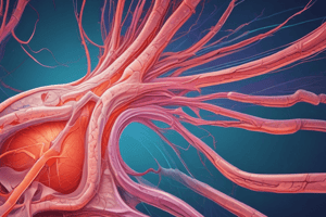
Connective Tissue Proper: Fibers
-
Collagenous Fibers (White):
- Made of collagen
- White in fresh state
- Strong, resist stretching
- Form wavy bundles
- Individual fibers don't branch, bundles do
- Digested by pepsin and collagenase
- Become gelatinous when boiled
- Types & Sites: Type I (white fibrocartilage, bone), Type II (hyaline and elastic fibrocartilage), Type III (reticular fibers)
- Staining: Acidophilic (H&E), red (Van Gieson's), blue (Mallory trichrome)
-
Elastic Fibers (Yellow):
- Elastic in nature
- Yellow in fresh state
- Thin, long, highly refractile
- Branch and form membranes (e.g., aorta)
- Digested by elastase
- Not affected by boiling
- Staining: Acidophilic (H&E), yellow (Van Gieson's), black (Ver-Hoff's)
- Sites: Arterial walls, trachea, ligaments of the back
-
Reticular Fibers:
- Very thin
- Branch and anastomose to form networks
- Staining: Not stained (H&E), black (silver stain)
- Sites: Stroma of organs (liver, spleen, lymph nodes)
Connective Tissue Proper: Cells
- Rounded/Oval Cells:
- Fat cells (adipocytes)
- Plasma cells
- Mast cells
- Blood leukocytes
- Branched Cells:
- Fibroblasts
- Macrophages
- Pigment cells
- Undifferentiated mesenchymal cells (UMCs)
Connective Tissue Proper: Adipocytes
- Origin: UMCs
- Site: Adipose and loose connective tissue
- Shape: Oval
- Nucleus: Flattened, peripheral
- Cytoplasm: Large fat globule, pushes nucleus
- Staining: Signet ring (H&E), orange (Sudan III), black (Sudan black, osmic acid)
- Functions: Energy storage, heat insulation
Connective Tissue Proper: Plasma Cells
- Origin: Activation of B lymphocytes by antigens
- Site: Lymphatic organs
- Shape: Oval
- Nucleus: Rounded, eccentric, "cart-wheel" chromatin
- Cytoplasm: Deep basophilic, negative Golgi image
- Function: Antibody secretion (humoral immunity)
Connective Tissue Proper: Mast Cells
- Site: Mucosa of the GI tract and respiratory system
- Shape: Oval
- Nucleus: Rounded, central
- Cytoplasm: Large basophilic granules
- Histamine
- Heparin (sulfated glycosaminoglycans)
- Staining: Toluidine blue (metachromatic purple)
- Functions: Histamine secretion (allergy), heparin secretion (anticoagulant)
Connective Tissue Proper: Blood Leukocytes
- Can leave blood and appear in connective tissue under certain conditions
- Examples: Eosinophils (allergy), neutrophils (acute infections), monocytes/lymphocytes (chronic infections)
Connective Tissue Proper: Fibroblasts
- Origin: Pericytes (from UMCs)
- Most common CT cell
- Abundant in loose CT
- Shape: Branched, spindle-shaped
- Nucleus: Oval, eccentric
- Cytoplasm: Basophilic, negative Golgi image
- Functions: Synthesize CT fibers, matrix, growth/wound healing
Connective Tissue Proper: Macrophages
- Origin: Blood monocytes
- Shape: Large, branched (pseudopodia)
- Nucleus: Eccentric, oval/kidney-shaped
- Cytoplasm: Not clear
- Staining: Trypan blue (vital stain)
- Functions: Phagocytosis, foreign body giant cell formation, collagenase/elastase secretion
Connective Tissue Proper: Pigment Cells
- Melanin-containing cells in skin and eyes
Connective Tissue Proper: Undifferentiated Mesenchymal Cells (UMCs)
- Mother cells of all CT cells
- Shape: Branched
- Nucleus: Large, oval
- Cytoplasm: Basophilic
Connective Tissue Proper: Matrix
- Soft material in which cells and fibers lie
- Components:
-
Organic (Amorphous): Proteins, glycoproteins, proteoglycans (GAGs) - secreted by fibroblasts
-
Fluid (Inorganic): Primarily water (60-70%)
-
Fluid component:
- Increased fluid leads to edema
- Decreased fluid leads to dehydration
-
Connective Tissue Proper: Types (Loose CT)
- Widely distributed (around blood vessels, submucosa of GI tract). - Structure details omitted.
Studying That Suits You
Use AI to generate personalized quizzes and flashcards to suit your learning preferences.