Podcast
Questions and Answers
What is the primary tissue type that is conspicuously absent within the central nervous system (CNS)?
What is the primary tissue type that is conspicuously absent within the central nervous system (CNS)?
- Connective tissue (correct)
- Muscle tissue
- Epithelial tissue
- Glandular tissue
- Nervous tissue
Which of the following is a key characteristic feature of neurons, distinguishing them from glial cells?
Which of the following is a key characteristic feature of neurons, distinguishing them from glial cells?
- Isolating individual neurons
- Repairing damaged tissue
- Conducting nerve impulses (correct)
- Supporting other cells
- Providing nutrition
Which neuronal component is primarily responsible for receiving stimuli from the environment or other cells?
Which neuronal component is primarily responsible for receiving stimuli from the environment or other cells?
- Cell body (perikaryon)
- Dendrites (correct)
- Axon
- Nucleus
- Nissl bodies
What is the Nissl substance observed within the cytoplasm of neurons primarily composed of?
What is the Nissl substance observed within the cytoplasm of neurons primarily composed of?
Multipolar neurons, characterized by multiple dendrites extending from the cell body, are most commonly exemplified by which type of neuron?
Multipolar neurons, characterized by multiple dendrites extending from the cell body, are most commonly exemplified by which type of neuron?
Which type of neuron is predominantly involved in sensory functions such as smell, sight, and balance?
Which type of neuron is predominantly involved in sensory functions such as smell, sight, and balance?
Pseudo-unipolar neurons are uniquely characterized by:
Pseudo-unipolar neurons are uniquely characterized by:
Neuropil, the 'feltwork' of nerve tissue, is composed of:
Neuropil, the 'feltwork' of nerve tissue, is composed of:
Which of the following is NOT a primary function of glial cells?
Which of the following is NOT a primary function of glial cells?
Astrocytes, a type of glial cell, are characterized by which of the following?
Astrocytes, a type of glial cell, are characterized by which of the following?
Astrocytes contribute significantly to the blood-brain barrier (BBB) by:
Astrocytes contribute significantly to the blood-brain barrier (BBB) by:
Which glial cells are primarily responsible for scar formation in the central nervous system following injury?
Which glial cells are primarily responsible for scar formation in the central nervous system following injury?
Oligodendrocytes are analogous to which cells in the peripheral nervous system (PNS) in terms of function?
Oligodendrocytes are analogous to which cells in the peripheral nervous system (PNS) in terms of function?
The myelin sheath produced by oligodendrocytes is primarily composed of:
The myelin sheath produced by oligodendrocytes is primarily composed of:
Which glial cell type is characterized as 'phagocytic' and derived from mononuclear cells of the blood?
Which glial cell type is characterized as 'phagocytic' and derived from mononuclear cells of the blood?
Ependymal cells are best described as:
Ependymal cells are best described as:
In certain brain locations, ependymal cells are modified to produce:
In certain brain locations, ependymal cells are modified to produce:
Which of the following is the most immediate cellular response in the CNS to tissue injury?
Which of the following is the most immediate cellular response in the CNS to tissue injury?
Gliosis, a common response to CNS damage, primarily involves the proliferation and hypertrophy of:
Gliosis, a common response to CNS damage, primarily involves the proliferation and hypertrophy of:
Chromatolysis, a reversible cellular change in neurons, is characterized by:
Chromatolysis, a reversible cellular change in neurons, is characterized by:
Vasogenic edema is primarily caused by:
Vasogenic edema is primarily caused by:
Cytotoxic edema is characterized by:
Cytotoxic edema is characterized by:
Which of the following cellular components is NOT a part of the blood-brain barrier (BBB)?
Which of the following cellular components is NOT a part of the blood-brain barrier (BBB)?
The primary function of tight junctions in the cerebral endothelium, a component of the BBB, is to:
The primary function of tight junctions in the cerebral endothelium, a component of the BBB, is to:
Considering the clinical case of a 14-year-old female with meningitis, which cellular component of the brain is MOST likely to be directly affected by the inflammatory process and pathogen?
Considering the clinical case of a 14-year-old female with meningitis, which cellular component of the brain is MOST likely to be directly affected by the inflammatory process and pathogen?
In the context of brain tumors, astrocytoma and glioblastoma multiforme originate from which cell type?
In the context of brain tumors, astrocytoma and glioblastoma multiforme originate from which cell type?
The 'fried-egg' appearance, often described in histology, is characteristic of which glial cell type?
The 'fried-egg' appearance, often described in histology, is characteristic of which glial cell type?
Which of the following diseases is NOT directly associated with clinical significance of neuropil dysfunction?
Which of the following diseases is NOT directly associated with clinical significance of neuropil dysfunction?
In a histological section stained with H&E, white matter appears lighter than grey matter primarily because of:
In a histological section stained with H&E, white matter appears lighter than grey matter primarily because of:
Which of the following is a characteristic feature of pyramidal neurons found in the cerebral cortex?
Which of the following is a characteristic feature of pyramidal neurons found in the cerebral cortex?
The cerebellum is characterized by a unique layered structure with 'white matter inside, grey outside'. Which layer of the cerebellar cortex is known for containing Purkinje cells?
The cerebellum is characterized by a unique layered structure with 'white matter inside, grey outside'. Which layer of the cerebellar cortex is known for containing Purkinje cells?
Cerebellar granule cells are notable for being:
Cerebellar granule cells are notable for being:
In the cerebellum, the molecular layer is characterized by:
In the cerebellum, the molecular layer is characterized by:
What percentage of potential drug treatments for brain disorders are estimated to be unable to effectively cross the blood-brain barrier?
What percentage of potential drug treatments for brain disorders are estimated to be unable to effectively cross the blood-brain barrier?
Which cell type is considered to play a significant role in the formation of 'white matter hyperintensities' observed on MRI scans in patients with mild traumatic brain injury (MTBI), indicative of brain scars?
Which cell type is considered to play a significant role in the formation of 'white matter hyperintensities' observed on MRI scans in patients with mild traumatic brain injury (MTBI), indicative of brain scars?
A tumor with a 'fried egg' appearance histologically is most likely to originate from:
A tumor with a 'fried egg' appearance histologically is most likely to originate from:
In a patient presenting with sudden onset right-sided weakness and confusion, and a CT scan showing loss of grey/white matter differentiation, which type of cerebral edema is MOST likely?
In a patient presenting with sudden onset right-sided weakness and confusion, and a CT scan showing loss of grey/white matter differentiation, which type of cerebral edema is MOST likely?
Histological features in an image showing cells with swollen cell bodies and pale cytoplasm, indicative of Nissl substance loss, best represent:
Histological features in an image showing cells with swollen cell bodies and pale cytoplasm, indicative of Nissl substance loss, best represent:
A histological image showing neurons, glial cells and capillaries is MOST likely representing:
A histological image showing neurons, glial cells and capillaries is MOST likely representing:
In the context of CNS histology, what is the main function of dendrites in neurons?
In the context of CNS histology, what is the main function of dendrites in neurons?
Which of the following best describes the primary function of the perikaryon (cell body) of a neuron?
Which of the following best describes the primary function of the perikaryon (cell body) of a neuron?
What is the primary role of the axon in a neuron?
What is the primary role of the axon in a neuron?
Which type of neuron is characterized by having multiple dendrites projecting from the cell body, making it the most common type of neuron in the CNS?
Which type of neuron is characterized by having multiple dendrites projecting from the cell body, making it the most common type of neuron in the CNS?
Which type of neuron is best suited to rapidly relay sensory information related to smell, sight and balance?
Which type of neuron is best suited to rapidly relay sensory information related to smell, sight and balance?
Which feature is most characteristic of pseudo-unipolar neurons?
Which feature is most characteristic of pseudo-unipolar neurons?
What is the neuropil primarily composed of?
What is the neuropil primarily composed of?
Which of the following is a key function of astrocytes that contributes to the maintenance of CNS homeostasis?
Which of the following is a key function of astrocytes that contributes to the maintenance of CNS homeostasis?
What is a critical function of astrocytes in the central nervous system regarding structural support and signaling?
What is a critical function of astrocytes in the central nervous system regarding structural support and signaling?
Which glial cells are characterized by their 'fried-egg' appearance under a microscope?
Which glial cells are characterized by their 'fried-egg' appearance under a microscope?
In CNS pathology, which glial cells, upon activation after tissue damage, can proliferate and differentiate into phagocytic cells to remove debris?
In CNS pathology, which glial cells, upon activation after tissue damage, can proliferate and differentiate into phagocytic cells to remove debris?
What is the primary function of ependymal cells in the central nervous system (CNS)?
What is the primary function of ependymal cells in the central nervous system (CNS)?
Considering the high proportion of drugs unable to cross the blood-brain barrier (BBB), which cellular component of the BBB presents the most significant obstacle to drug delivery into the CNS?
Considering the high proportion of drugs unable to cross the blood-brain barrier (BBB), which cellular component of the BBB presents the most significant obstacle to drug delivery into the CNS?
If a patient presents with loss of grey/white matter differentiation on a CT scan, indicative of swelling of both neurons, glial cells and endothelial cells, which type of cerebral edema is MOST likely?
If a patient presents with loss of grey/white matter differentiation on a CT scan, indicative of swelling of both neurons, glial cells and endothelial cells, which type of cerebral edema is MOST likely?
A researcher is investigating new therapeutic targets for neurodegenerative diseases and aims to modulate astrocyte function to promote neuronal survival. Which specific astrocyte function would be the MOST promising target for enhancing neuronal resilience?
A researcher is investigating new therapeutic targets for neurodegenerative diseases and aims to modulate astrocyte function to promote neuronal survival. Which specific astrocyte function would be the MOST promising target for enhancing neuronal resilience?
Flashcards
What is meningitis?
What is meningitis?
Inflammation of the meninges, often diagnosed by noting inflammatory cells in the cerebral spinal fluid after lumbar puncture.
What are the histological components of the Central Nervous System (CNS)?
What are the histological components of the Central Nervous System (CNS)?
The key components include neurons and glial cells.
What are neurodegenerative diseases?
What are neurodegenerative diseases?
Progressive degeneration and death of nerve cells, examples include Alzheimer's and Parkinson's.
What are demyelinating diseases?
What are demyelinating diseases?
Signup and view all the flashcards
What is a stroke?
What is a stroke?
Signup and view all the flashcards
What are dendrites?
What are dendrites?
Signup and view all the flashcards
What is a cell body or perikaryon?
What is a cell body or perikaryon?
Signup and view all the flashcards
What is the axon?
What is the axon?
Signup and view all the flashcards
What are Multipolar neurons?
What are Multipolar neurons?
Signup and view all the flashcards
What are bipolar neurons?
What are bipolar neurons?
Signup and view all the flashcards
What are Pseudo-unipolar neurons?
What are Pseudo-unipolar neurons?
Signup and view all the flashcards
What is neuropil?
What is neuropil?
Signup and view all the flashcards
What are Glial cells?
What are Glial cells?
Signup and view all the flashcards
What are astrocytes?
What are astrocytes?
Signup and view all the flashcards
What roles do astrocytes play in metabolic exchange?
What roles do astrocytes play in metabolic exchange?
Signup and view all the flashcards
What role do astrocytes play in scar formation?
What role do astrocytes play in scar formation?
Signup and view all the flashcards
What role do astrocytes play in signal propagation?
What role do astrocytes play in signal propagation?
Signup and view all the flashcards
What role do astrocytes play in ionic and transmitter balance?
What role do astrocytes play in ionic and transmitter balance?
Signup and view all the flashcards
What role do astrocytes play in the blood-brain barrier (BBB)
What role do astrocytes play in the blood-brain barrier (BBB)
Signup and view all the flashcards
What are oligodendrocytes?
What are oligodendrocytes?
Signup and view all the flashcards
What are Microglial cells?
What are Microglial cells?
Signup and view all the flashcards
What are Ependymal cells?
What are Ependymal cells?
Signup and view all the flashcards
What is Chromatolysis?
What is Chromatolysis?
Signup and view all the flashcards
What usually happens due to a lack of fibroblasts in CNS?
What usually happens due to a lack of fibroblasts in CNS?
Signup and view all the flashcards
What is gliosis?
What is gliosis?
Signup and view all the flashcards
What is the blood-brain barrier (BBB)?
What is the blood-brain barrier (BBB)?
Signup and view all the flashcards
What are the cellular elements of BBB?
What are the cellular elements of BBB?
Signup and view all the flashcards
How do endothelial cells help form the BBB?
How do endothelial cells help form the BBB?
Signup and view all the flashcards
How do astrocytes help form the BBB?
How do astrocytes help form the BBB?
Signup and view all the flashcards
How do pericytes help form the BBB?
How do pericytes help form the BBB?
Signup and view all the flashcards
Why is it hard to treat brain dieases?
Why is it hard to treat brain dieases?
Signup and view all the flashcards
Study Notes
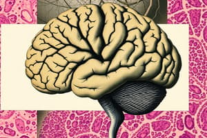
Overview: CNS Histology and Pathology - Part 1
- This lecture provides an overview of the histology and pathology of the central nervous system (CNS).
- Diagrams and photos are sourced from "Atlas of Functional Histology" by Jeffrey B Kerr, "Wheater's Functional Histology", and "Blue Histology (online)" from the University of Western Australia.
Clinical Importance: Meningitis Case
- A 14-year-old female was admitted to the emergency department with severe headache, fever, and vomiting.
- The diagnosis was meningitis, based on the presence of inflammatory cells in the cerebral spinal fluid obtained via lumbar puncture.
- The cellular components most likely to be damaged by the pathogen are:
- Endothelial cells
- Astrocytes
- Pericytes
Key Histological Components of the Nervous System
- The central nervous system (CNS) mainly comprises neurons and glial cells (non-neuronal cells).
- The peripheral nervous system (PNS) consists of ganglion cells and nerve plexuses.
The Central Nervous System (CNS)
Options include:
- The CNS anatomically includes the brain and spinal cord.
- The nervous tissue of the CNS lacks connective tissues, giving it a soft, jelly-like consistency.
- This is why there is no scar tissue from collagen
- There are scars from something called gliosis
- Histologically, the CNS consists of two main cell types: nerve cells (neurons) and glial cells.
Histology and Clinical Significance: CNS Diseases
- Examples of neurodegenerative disorders:
- Alzheimer's disease
- Parkinson's disease
- Motor neuron disease
- All involve progressive degeneration and death of nerve cells.
- Demyelinating diseases involve damage to insulating myelin forming cells.
- This leads to impaired nerve signal transmission
- An example is multiple sclerosis.
- Glial cells secrete myelin
- Damage to brain nerve cells can be caused by blockage of blood vessels or bleeding – a stroke.
- Damage to the blood-brain barrier (BBB) disrupts substance diffusion into the brain, preventing certain drugs from entering.
Histology of Neurons within the CNS
- Neurons consist of three parts:
- Dendrites which are specialized for receiving external stimuli
- Cell body (perikaryon), containing the trophic center for the whole nerve cell
- Axon, a single process specialized for generating/conducting nerve impulses to other cells.
- Neurons vary greatly in morphology
Types of Neurons
- Neurons come in many sizes and shapes.
- Multipolar neurons are the most common form, characterized by numerous dendrites projecting from the cell body; motor neurons are an example.
- Bipolar neurons have a single dendrite arising from the cell body opposite the axon's origin, functioning as receptor neurons for senses like smell and sight; examples include olfactory neurons.
- Pseudo-unipolar neurons feature a single dendrite and axon arising from a common stem of the cell body, typically acting as sensory neurons.
Microscopic Features of Neurons
- Most neurons have a light, large nucleus with a distinct nucleolus.
- The cytoplasm contains significant amounts of rough endoplasmic reticulum, forming Nissl bodies.
- The feltwork of neuronal and glial cell processes is the neuropil.
- Change of expression of proteins happen in the Neurpil.
- The clinical significance of neuropil relates to Schizophrenia and Alzheimer's disease.
- Astocyte scar is related to gliosis
- Inflammatory-encephalitis
Glial Cells in the CNS
- Glial cells provide support in the CNS.
- Glial cells are non-neuronal cells
- They offer support, nutrition, repair, defense, and isolation for neurons.
- Glial cells include:
- Astrocytes
- Oligodendrocytes
- Microglia
- Ependymal cells
Astrocytes
- These are stellate cells, 8-12µm in diameter, that contact neuron surfaces not contacted by synapses.
- Astrocytes cover about 99% of the brain capillary surface area.
- They bind neurons to capillaries for extracellular fluid maintenance.
- These hold up blood vessels - capillaries
Astrocyte Functions
- Specific functions include;
- scar*-forming cells of the CNS.
- Control of signal propagation between neurons;
- Maintenance of ionic and transmitter metabolism; and
- Induction of vascular endothelial cells to form a seal or selective filter, called the blood – brain barrier.
Clinical Significance of Astrocytes
- Astrocytoma is benign
- Glioblastoma Multiforme- Malignant
- Astrocyte modulation with therapeutic potential works by targeting astrocytic functions for neurodegenerative diseases, stroke and epilepsy.
- There is ongoing research on astrocyte transplantation for CNS repair in spinal cord injury.
Oligodendrocytes
- These produce the myelin sheath for axon insulation in the CNS, similar to Schwann cells in the PNS.
- Myelin consists mainly of myelin basic protein and phospholipids.
- Oligodendrocytes are predominant in white matter.
- One oligodendrocyte sends out glial processes forming forming compact myelin around an axon.
Identifying Oligodendrocytes Under a Microscope
- Look for a White halo, or peri-nuclear halo which will give a fried-egg appearance
- Should be areas with little to no inflammation
Microglial Cells
- These are small, elongated cells with short processes, found in small numbers with
- They are phagocytic cells derived from mononuclear blood cells.
- They can proliferate and differentiate after tissue damage.
- They act as macrophage like cells in the CNS to remove cellular debris and damaged cells.
Glial Cells: Identification
- Glial cell nuclei are observed in relatively small numbers.
- Immunochemical staining identifies microglial cell activity.
Ependymal Cells
- These are Low columnar cells that line the fluid filled cavities of the CNS.
- Ependymal cells are ciliated
- They transport fluid
- In the choroid plexus they produce cerebrospinal fluid.
- A lack of tight junctions allows free exchange between cerebrospinal fluid and nervous tissue.
Summary of Glial Cells
- Glial cells maintain CNS homeostasis and interact with neurons.
- Glial cells respond to lesions for repair
- Glial cells proliferate and can form tumors
Histological Responses in CNS Injury
- Neurons are less robust/adaptable than astrocytes or microglial cells in response to injury.
- Chromatolysis refers to neurons undergoing reversible cell damage which results in them becoming recognizable histologically because of swelling of the cell body associated with loss of Nissl substance
- Necrosis in brain tissue usually results in liquefaction of damaged tissues.
- An initial response is exudative. There will be there activation of local microglia and recruitment of phagocytic monocytes.
- Astrocytic scar is a common end product to damage to the specialised structures of the CNS.
The Impact of Brain Injuries
- Lack of Fibroblasts stops healing through granulation tissue so, results in fibrous scarring
- An exudative response activates local microglia
- An astrocytic scar may form.
- Reactive changes with inflammation, Cerebral oedema can occur.
Cerebral Edema
- Swelling of neurons, glial cells, and endothelial cells occurs due to failure of cellular ion pumps.
- The swelling of neurons or glial cells due to injuries or toxins is "Cytotoxic "
- Protein and fluid leakage from vessel permeability is "Vasogenic"
- Lesions will be localized in Cerebral edema.
- The surfaces of the gyri look flattened due to the expansion of the brain
Blood-Brain Barrier Explained
- It is composed of: endothelial cells, astrocyte end-feet, and pericytes.
- Endothelial cells are connected via tight junctions for excluding blood borne substances
- Astrocytic endfeet tightly enshorth the vessel wall and ensheath the vessel wall.
- Pericytes surround endothelial cells.
The Blood-Brain Barrier: Structure and Function
- Normal capillaries have fenestrations with an intact basal membrane, enabling selective transfer of metabolites.
- They connect Via Tight junctions
- The BBB contains intercellular space
- Blood-brain barrier (BBB): Composed of endothelial cells, astrocyte foot processes, and pericytes held together by the tight junctions .
- Astrocytes can also between the capillaries
BBB Clinical Implications
- An estimated 98% of drugs can't penetrate the BBB to treat diseases.
- Researchers temporarily open the BBB for drug delivery.
- BBB impairment complicates neurological diseases like stroke and neuroinflammatory disorders.
Brain-Histology
- Spinal cord has a white matter and gray matter.
- Glial cells are shown in both the white matter and gray matter
- Neuron Somata are shown in the gray matter
Brain Histology: Cerebral Cortex
- Pyramidal neurons are identified the character of basophilic(blue) and located in cytoplasm.
- Large nuclei and prominent nucleoli shown the charateristics of neuron
Brain Histology: Cerebellum
- The white matter inside is the medulla with plenty of neurons in the grey matter around.
- Three layers compose cerebellum which include outer molecular layer, purkinje cell layer and Inner Granular Layer.
Cerebellum Cell types
- The molecular layer: this dendrites occupy most of the layer
- Cerebellar granule cells account for half of the neurons in the CNS.
Self Assessment 1
- Microglial cells plays a signicant role in MRI hyperintensities, which is indicative of brain scars
Self Assessment 2
- Oligodendrocytes are indicated in fired egg from a 40 year old woman.
Self Assessment 3
- Astrocyte contributes in maintenance of the blood-brain barrier
Self Assessment 4
- In Cytotoxic oedea There is no white/grey matter differentiation
Self Assessment 5
- Some breakdown of chromatin is shown in the Astrocytic scar
Self Assessment 6
- Neurons, Glial cells and capillaries show features in Histology
Studying That Suits You
Use AI to generate personalized quizzes and flashcards to suit your learning preferences.