Podcast
Questions and Answers
What are the two main histological components of the central nervous system (CNS)?
What are the two main histological components of the central nervous system (CNS)?
- Ganglion cells and Nerve plexuses
- Astrocytes and Oligodendrocytes
- Ependymal cells and Microglia
- Neurons and Glial cells (correct)
In a patient diagnosed with meningitis, which brain structure is most susceptible to damage due to inflammatory cells?
In a patient diagnosed with meningitis, which brain structure is most susceptible to damage due to inflammatory cells?
- Ganglion
- Nerve plexuses
- Blood-brain barrier (correct)
- Ependymal cells
Which of the following is a characteristic of the nervous tissue within the CNS?
Which of the following is a characteristic of the nervous tissue within the CNS?
- High concentration of fibroblasts
- Absence of connective tissues (correct)
- Extensive collagen network
- Presence of dense connective tissues
What is the primary function of dendrites in a neuron?
What is the primary function of dendrites in a neuron?
Which of the following best describes the function of the cell body, also known as the perikaryon, of a neuron?
Which of the following best describes the function of the cell body, also known as the perikaryon, of a neuron?
What is the main function of an axon?
What is the main function of an axon?
Motor neurons are an example of which type of neuron based on its structure?
Motor neurons are an example of which type of neuron based on its structure?
Which type of neuron primarily functions as a receptor for the senses of smell, sight, and balance?
Which type of neuron primarily functions as a receptor for the senses of smell, sight, and balance?
Sensory neurons are most commonly classified as which type of neuron?
Sensory neurons are most commonly classified as which type of neuron?
What are Nissl bodies, found in the cytoplasm of neurons, primarily composed of?
What are Nissl bodies, found in the cytoplasm of neurons, primarily composed of?
What is the term used to describe the network of nerve fibers, neuronal and glial cell processes in the CNS?
What is the term used to describe the network of nerve fibers, neuronal and glial cell processes in the CNS?
Damage or changes to the neuropil is clinically significant in which of the following diseases?
Damage or changes to the neuropil is clinically significant in which of the following diseases?
Which of the following is NOT a type of glial cell found in the central nervous system?
Which of the following is NOT a type of glial cell found in the central nervous system?
What is the primary function of glial cells within the central nervous system?
What is the primary function of glial cells within the central nervous system?
Which glial cell type is responsible for surrounding and contacting approximately 99% of brain capillaries?
Which glial cell type is responsible for surrounding and contacting approximately 99% of brain capillaries?
Which of the following is a key function of astrocytes?
Which of the following is a key function of astrocytes?
What is the primary function of oligodendrocytes in the central nervous system?
What is the primary function of oligodendrocytes in the central nervous system?
Which area of the nervous system contains the most oligodendrocytes?
Which area of the nervous system contains the most oligodendrocytes?
Which glial cell type is known for being phagocytic and is derived from mononuclear cells of the blood?
Which glial cell type is known for being phagocytic and is derived from mononuclear cells of the blood?
What is the primary function of ependymal cells in the CNS?
What is the primary function of ependymal cells in the CNS?
Which of the following best describes a key feature of ependymal cells?
Which of the following best describes a key feature of ependymal cells?
Neurons, when subjected to sub-lethal injury, may undergo what process recognizable by swelling and loss of Nissl substance?
Neurons, when subjected to sub-lethal injury, may undergo what process recognizable by swelling and loss of Nissl substance?
What is gliosis?
What is gliosis?
Which of the following is true regarding the brains ability to heal?
Which of the following is true regarding the brains ability to heal?
What primary type of cell is found in astrocytic scars?
What primary type of cell is found in astrocytic scars?
Which of the following describes the role of microglia in response to damage within the CNS?
Which of the following describes the role of microglia in response to damage within the CNS?
What is cerebral edema?
What is cerebral edema?
What distinguishes vasogenic edema from cytotoxic edema?
What distinguishes vasogenic edema from cytotoxic edema?
Which cellular elements are critical components of the blood-brain barrier (BBB)?
Which cellular elements are critical components of the blood-brain barrier (BBB)?
What is the primary role of tight junctions in endothelial cells of the blood-brain barrier?
What is the primary role of tight junctions in endothelial cells of the blood-brain barrier?
Why are brain tumors difficult to treat with drugs delivered via the bloodstream?
Why are brain tumors difficult to treat with drugs delivered via the bloodstream?
In the context of traumatic brain injury (MTBI), what type of cell plays a key role in the formation of radiological findings such as white matter hyperintensities?
In the context of traumatic brain injury (MTBI), what type of cell plays a key role in the formation of radiological findings such as white matter hyperintensities?
A 40-year-old woman is diagnosed with a tumor in the temporal lobe, and histological analysis reveals cells with a 'fried egg' appearance. What is the most likely cellular origin?
A 40-year-old woman is diagnosed with a tumor in the temporal lobe, and histological analysis reveals cells with a 'fried egg' appearance. What is the most likely cellular origin?
In which of the following scenarios would cytotoxic edema most likely be the primary type of cerebral edema observed?
In which of the following scenarios would cytotoxic edema most likely be the primary type of cerebral edema observed?
A researcher is investigating a new drug that must cross the blood-brain barrier to treat a neurological disorder. Which strategy is MOST likely to succeed in facilitating drug passage across the BBB?
A researcher is investigating a new drug that must cross the blood-brain barrier to treat a neurological disorder. Which strategy is MOST likely to succeed in facilitating drug passage across the BBB?
A non-contrast CT scan of a 65-year-old patient presenting with sudden right-sided weakness and confusion reveals diffuse cerebral edema, obliterating the distinction between grey and white matter. Which type of cerebral edema is MOST likely present?
A non-contrast CT scan of a 65-year-old patient presenting with sudden right-sided weakness and confusion reveals diffuse cerebral edema, obliterating the distinction between grey and white matter. Which type of cerebral edema is MOST likely present?
The presence of which feature within a tissue sample would suggest a diagnosis of chromatolysis?
The presence of which feature within a tissue sample would suggest a diagnosis of chromatolysis?
Which of the following histological characteristics is MOST indicative of reactive astrocytosis in a brain tissue sample?
Which of the following histological characteristics is MOST indicative of reactive astrocytosis in a brain tissue sample?
Which of the following cell types contributes to the maintenance of the blood-brain barrier?
Which of the following cell types contributes to the maintenance of the blood-brain barrier?
A researcher is examining a brain tissue sample and observes a region with a high density of neuronal cell bodies and relatively few myelinated axons. According to the organization of grey and white matter, which area is the researcher looking at?
A researcher is examining a brain tissue sample and observes a region with a high density of neuronal cell bodies and relatively few myelinated axons. According to the organization of grey and white matter, which area is the researcher looking at?
In the cerebellum, Purkinje cells exhibit a distinct arrangement where their dendrites primarily occupy the molecular layer. If a histological section of the cerebellum shows a significant sparseness of nuclei within the molecular layer, what is the primary reason for this?
In the cerebellum, Purkinje cells exhibit a distinct arrangement where their dendrites primarily occupy the molecular layer. If a histological section of the cerebellum shows a significant sparseness of nuclei within the molecular layer, what is the primary reason for this?
Imagine a novel neurotoxin that selectively impairs the function of pericytes within the CNS vasculature. Which downstream effect is MOST likely to be observed in the initial stages following exposure to this toxin?
Imagine a novel neurotoxin that selectively impairs the function of pericytes within the CNS vasculature. Which downstream effect is MOST likely to be observed in the initial stages following exposure to this toxin?
What is the primary effect of demyelinating diseases on nerve cells?
What is the primary effect of demyelinating diseases on nerve cells?
Which of the following best describes the potential consequence of blood-brain barrier (BBB) damage?
Which of the following best describes the potential consequence of blood-brain barrier (BBB) damage?
What specialized feature do dendrites possess to facilitate their function?
What specialized feature do dendrites possess to facilitate their function?
Motor neurons are classified under which structural category of neurons?
Motor neurons are classified under which structural category of neurons?
What is the defining characteristic of the neuropil?
What is the defining characteristic of the neuropil?
Which of the following best describes the function of glial cells in the central nervous system?
Which of the following best describes the function of glial cells in the central nervous system?
What is a primary function of astrocytes related to capillaries in the brain?
What is a primary function of astrocytes related to capillaries in the brain?
What is the main function of ependymal cells within the central nervous system?
What is the main function of ependymal cells within the central nervous system?
What contributes to the selective permeability of the blood-brain barrier?
What contributes to the selective permeability of the blood-brain barrier?
A researcher observes a brain tissue sample characterized by a high density of oligodendrocytes. Which area of the nervous system is the researcher most likely examining?
A researcher observes a brain tissue sample characterized by a high density of oligodendrocytes. Which area of the nervous system is the researcher most likely examining?
Following an injury to the central nervous system, what is the primary role of astrocytes?
Following an injury to the central nervous system, what is the primary role of astrocytes?
In cases of cerebral edema, which of the following processes is most characteristic of cytotoxic edema?
In cases of cerebral edema, which of the following processes is most characteristic of cytotoxic edema?
Which condition is characterized by reversible cell damage, swelling of the cell body and loss of Nissl substance in neurons?
Which condition is characterized by reversible cell damage, swelling of the cell body and loss of Nissl substance in neurons?
Granule cells are found primarily in the cerebellum, what proportion of the neurons in the central nervous system do they make up?
Granule cells are found primarily in the cerebellum, what proportion of the neurons in the central nervous system do they make up?
Which alteration in brain tissue is most suggestive of cytotoxic edema?
Which alteration in brain tissue is most suggestive of cytotoxic edema?
Flashcards
CNS Cell Types
CNS Cell Types
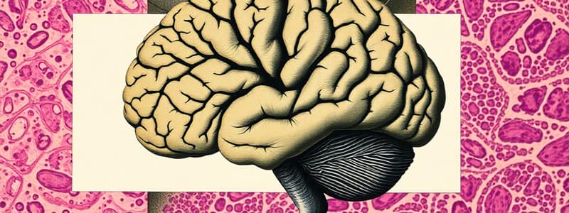
Histologically, the CNS consists of nerve cells (neurons) and glial cells.
Neurodegenerative Diseases
Neurodegenerative Diseases
Progressive degeneration and death of nerve cells, examples are Alzheimer's and Parkinson's.
Demyelinating Disease
Demyelinating Disease
Damage to myelin forming cells, impairing effective nerve signaling, example: Multiple sclerosis.
Dendrites
Dendrites
Signup and view all the flashcards
Cell Body or Perikaryon
Cell Body or Perikaryon
Signup and view all the flashcards
Axon
Axon
Signup and view all the flashcards
Multipolar neuron
Multipolar neuron
Signup and view all the flashcards
Bipolar neuron
Bipolar neuron
Signup and view all the flashcards
Pseudo-unipolar neuron
Pseudo-unipolar neuron
Signup and view all the flashcards
Neuropil
Neuropil
Signup and view all the flashcards
Glial Cells
Glial Cells
Signup and view all the flashcards
Astrocytes
Astrocytes
Signup and view all the flashcards
Scar Formation
Scar Formation
Signup and view all the flashcards
Blood-Brain Barrier (BBB)
Blood-Brain Barrier (BBB)
Signup and view all the flashcards
Oligodendrocytes
Oligodendrocytes
Signup and view all the flashcards
Microglial Cells
Microglial Cells
Signup and view all the flashcards
Ependymal Cells
Ependymal Cells
Signup and view all the flashcards
Chromatolysis
Chromatolysis
Signup and view all the flashcards
Gliosis
Gliosis
Signup and view all the flashcards
Vasogenic Edema
Vasogenic Edema
Signup and view all the flashcards
Cytotoxic Edema
Cytotoxic Edema
Signup and view all the flashcards
BBB elements
BBB elements
Signup and view all the flashcards
Endothelial cells
Endothelial cells
Signup and view all the flashcards
Cerebral edema
Cerebral edema
Signup and view all the flashcards
Study Notes
Clinical Importance
- A 14-year-old female was rushed to the emergency department with severe headache, fever, and vomiting
- Meningitis was diagnosed after noting inflammatory cells in the cerebral spinal fluid following lumbar puncture
- A likely point of damage through a pathogen is the blood-brain barrier
Key Histological Components
- The central nervous system (CNS) comprises neurons and glial cells
- The Peripheral nervous system (PNS) consists of Ganglion cells and Nerve plexuses
Central Nervous System (CNS)
- Anatomically, the CNS consists of the brain and spinal cord
- Nervous tissue in the CNS does not contain connective tissues
- This results in a soft jelly-like consistency of brain tissues
- Histologically, the CNS consists of two cell types which are nerve cells (neurons) and glial cells
Histology and Clinical Significance
- Neurodegenerative diseases involve progressive degeneration and death of nerve cells
- Examples include Alzheimer's and Parkinson's disease, and motor neuron disease
- Demyelinating diseases involve damage to myelin-forming cells, which can lead to a loss of effective signalling between nerve cells
- An example is multiple sclerosis
- Stroke is damage to nerve cells due to blockage of blood vessels or bleeding
- Blood-brain barrier (BBB) damage results in disruption to the diffusion of substances from the blood into the brain
CNS Histology: Neurons
- Most neurons consist of three parts, which are dendrites, cell body, and axon
- Dendrites are specialized for receiving stimuli from the environment, sensory epithelial cells, or other neurons
- The cell body or perikaryon represents the trophic center for the whole nerve cell and is also receptive to stimuli
- The axon is a single process specialized for generating or conducting nerve impulses to other cells which include nerve, muscle or gland cells
Neurons Size and Shape
- Neurons and their processes are extremely variable in size and shape
- Multipolar neurons are the most common form, with numerous dendrites projecting from the cell body
- An example of this is motor neurons
- Bipolar neurons have a single dendrite which arises from the pole of the cell body opposite to the origin of the axon
- These neurons act as receptor neurons for the senses of smell, sight, and balance
- Pseudo-unipolar neurons have a single dendrite and axon that arises from a common stem of the cell body
- An example of this consists of sensory neurons
Neuron Features
- Most neurons have a light, large nucleus with a distinct nucleolus
- The cytoplasm of many neurons contains fairly large amounts of rough endoplasmic reticulum
- This aggregates within the cytoplasm of the neuron to form Nissl-bodies
- The feltwork of nerve fibres, neuronal and glial cell processes consists of neuropil
Clinical Significance of Neuropil.
- Clinical significance of the neuropil involves Schizophrenia and Alzheimer's disease
CNS Histology: Glial Cells
- Glial cells are non-neuronal supporting cells in the CNS
- Glial cells offer support, nutrition, repair of damaged tissue, defense, isolation of individual neurons, and phagocytosis
- Glial cells include astrocytes, oligodendrocytes, microglia, and ependymal cells
Glial Cells: Astrocytes
- Astrocytes are stellate cells (8-12µm diameter) which send their processes to surround or contact surfaces of neurons not contacted by synapses
- Astrocytes surround or contact about 99% of the brain capillary surface area
- Astrocytes help in binding neurons to capillaries, thus play an important role in maintenance of the composition of the extracellular fluid
Astrocytes Functions
- Astrocytes provide metabolic exchange that facilitates the exchange of nutrients and metabolites between neurons and blood
- Astrocytes undergo scar formation which acts as the primary scar-forming cells of the CNS after injury
- Astrocytes regulate signal propagation and control signal transmission between neurons
- They also maintain ionic and transmitter balance, ionic homeostasis, and neurotransmitter metabolism to ensure optimal neuronal function
- Astrocytes induce vascular endothelial cells to form the selective barrier that protects the brain by controlling substance entry which also consists of the blood-brain barrier
Astrocyte Clinical Significance and Therapeutic Treatment
- Astrocytoma is benign
- Glioblastoma multiforme (Glioblastoma) is malignant
- Astrocyte modulation targets astrocytic functions, glutamate uptake, and inflammatory cytokine release
- An emerging therapeutic approach for neurodegenerative diseases, epilepsy, and stroke
- Astrocyte transplantation involves research into transplanting healthy astrocytes for CNS repair, specifically in spinal cord injury, which is ongoing
Glial Cells: Oligodendrocytes
- Oligodendrocytes produces the myelin sheath which insulates axons in the CNS, similar to Schwann cells in the PNS.
- Myelin basic protein and phospholipids consist of the major components of myelin
- Oligodendrocytes are predominant in white matter
- An oligodendrocyte sends a glial process forming compact myelin spiraling around an axon
- Each oligodendrocyte can make differing numbers of myelin segments with variable lengths along similar axon tracts
Glial Cells: Microglial cells
- Microglial cells are small, elongated, with short processes which are discovered in small amounts
- Microglial cells are phagocytic cells derived from mononuclear cells of the blood
- In the case of tissue damage, microglia can proliferate and differentiate into phagocytotic cells
- Microglial cells are macrophage-like cells that help remove cellular debris and damaged cells
Glial Cells: Ependymal cells
- Ependymal cells are low columnar ciliated cells that line the fluid-filled cavities of the CNS
- Ependymal cells transport fluid which contributes to circulation and homeostasis of cerebrospinal fluid (CSF)
- In several locations in the brain, specifically the choroid plexuses, they are specialized to produce cerebrospinal fluid (CSF)
- The lack of tight junctions between ependymal cells allows a free exchange between cerebrospinal fluid and nervous tissue
CNS Glial Cells Summary
- Glial cells are involved with CNS homeostasis and interactions with neurons
- They respond to lesions, leading to scar formation
- Glial cells may proliferate to form brain tumors which include astrocytoma and oligodendrocytoma
Clinical Significance: Histological Responses in CNS Injury/Damage
- Neurons have a limited ability to survive significant changes in their metabolic or physical environment compared to astrocytes or microglial cells
- Chromatolysis occurs when neurons undergo reversible cell damage, histologically recognizable by swelling of the cell body associated with loss of Nissl substance
- This is seen in the cell body of a neuron after damage to the axonal process
- Necrosis of brain tissue usually results in liquefaction, leaving a fluid-filled space
- Due to lack of fibroblasts, healing through granulation tissue and fibrous scarring following an injury or necrosis is an uncommon event in the CNS
- An initial response to injury includes an exudative response with activation of local microglia and recruitment of phagocytic monocytes to phagocytose dead tissue
- This is followed by proliferation of astrocytes to form an astrocytic scar which is also known as gliosis
- Gliosis and astrocytic scarring of the CNS is a common end product of damage to the specialized structures of CNS
Edema
- Cerebral edema involves accumulation of excess fluid within the brain parenchyma
- Leads to a higher intracranial pressure
- Potential neurological deficits
- Vasogenic edema involves this involves the disruption of the BBB and leads to high permeability of the vascular
-Common causes is inflammation, neoplasms, abscesses, and trauma.
- Primary Impacts white matter.
- Cytotoxic edema is swelling of neurons, glial,and endothelial cells due to the decline of the cell ion pumps due to stroke or toxins with intracellular accumulation of sodium
- this means involves white and grey matter
Blood-brain barrier elements
- Three cellular elements of the brain microvasculature compose the BBB: endothelial, astrocyte end-feet and pericytes
- Forms a diffusion barrier that prevents blood born subtances from going through
- Endothelial cells have junctions to cerebral endoethelial cells that creates the fusion barrier, keeping many subtances outside of the brain
- Astocytic end feet maintain the tight junctions
- Drug treatment has limited affect on brain tumors because of it ability to penetrate the blood brain barrier
- Opening of the barrier has been an evolution by drugs to reach their targets
- Neurological diseases like stroke, are an affect that can disrupt the BBB barrier
Self Assessment
- For a image consisting of a MRI in a male whom suffered a blast- the cell mostly associated is the formation of astrocytic/white matter hyper-intensities known as astrocytes
- If histology shows Fried egg cells known as oligodendrocyte, could be right side temperol
- Astrocytes contributes to which of the following cell types contributes to maintenance of the blood-brain barrier?
- For cytotoxic damage and white/grey matter damage- it's cytotoxic
- Histology showing neuron and glials and capillaries
Studying That Suits You
Use AI to generate personalized quizzes and flashcards to suit your learning preferences.


