Podcast
Questions and Answers
Which of the following descriptions accurately portrays the clavicle?
Which of the following descriptions accurately portrays the clavicle?
- A short bone characterized by multiple foramina.
- A long bone possessing a body and two extremities. (correct)
- A flat bone that features three ends.
- A cuboidal bone with a uniform shape.
How is the clavicle typically divided for ease of study?
How is the clavicle typically divided for ease of study?
- Into anterior and posterior segments.
- Into a lateral third that is flat and a medial two-thirds that are cylindrical. (correct)
- Into superior and inferior halves.
- Into proximal and distal regions by a transverse fissure.
What surface features are found on the lateral third of the clavicle?
What surface features are found on the lateral third of the clavicle?
- Medial and lateral borders
- Anterior and posterior borders
- Superior and inferior surfaces
- Superior and inferior surfaces separated by anterior and posterior borders. (correct)
What is the characteristic of the anterior border of the lateral clavicle?
What is the characteristic of the anterior border of the lateral clavicle?
What is the deltoid tubercle?
What is the deltoid tubercle?
Which feature is located on the inferior surface, near the posterior border of the clavicle?
Which feature is located on the inferior surface, near the posterior border of the clavicle?
Where is the trapezoid line located in relation to the conoid tubercle?
Where is the trapezoid line located in relation to the conoid tubercle?
What is the orientation of the trapezoid line?
What is the orientation of the trapezoid line?
What are the characteristics of the medial two-thirds of the clavicle?
What are the characteristics of the medial two-thirds of the clavicle?
Which anatomical features are located on the clavicle's inferior surface, progressing from medial to lateral?
Which anatomical features are located on the clavicle's inferior surface, progressing from medial to lateral?
What type of a joint surface does the acromial end of the clavicle possess?
What type of a joint surface does the acromial end of the clavicle possess?
With which structures does the sternal end of the clavicle articulate?
With which structures does the sternal end of the clavicle articulate?
Why are clavicular fractures more likely to occur at the curves?
Why are clavicular fractures more likely to occur at the curves?
In clavicular fractures, where the weight of the limb cause displacement?
In clavicular fractures, where the weight of the limb cause displacement?
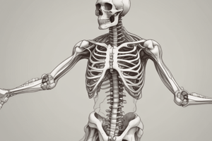
The scapula is best described as which shape?
The scapula is best described as which shape?
Where is the scapula located?
Where is the scapula located?
What are the main components of the scapula?
What are the main components of the scapula?
Which structure primarily constitutes the scapula?
Which structure primarily constitutes the scapula?
What are the names for the superior and inferior ends of the scapula?
What are the names for the superior and inferior ends of the scapula?
What are the names of the surfaces of the scapula?
What are the names of the surfaces of the scapula?
What type of angles forms the scapula?
What type of angles forms the scapula?
How are the borders of the scapula defined?
How are the borders of the scapula defined?
Which of the following describes glenoid cavity?
Which of the following describes glenoid cavity?
Where is the suprascapular notch located?
Where is the suprascapular notch located?
What is the description of the costal surface?
What is the description of the costal surface?
What does the spine of the scapula divide?
What does the spine of the scapula divide?
How are the supraspinous fossa and infraspinous fossa connected?
How are the supraspinous fossa and infraspinous fossa connected?
Which structure articulates with the glenoid cavity?
Which structure articulates with the glenoid cavity?
Where is the infraglenoid tubercle located?
Where is the infraglenoid tubercle located?
What is commonly referred to as the head of the scapula?
What is commonly referred to as the head of the scapula?
What structure is constricted distal to Region of gelnoid cavity?
What structure is constricted distal to Region of gelnoid cavity?
Where does the spine of the scapula blend with the dorsal surface?
Where does the spine of the scapula blend with the dorsal surface?
What term describes the free and thickened posterior border of the scapular spine?
What term describes the free and thickened posterior border of the scapular spine?
The root of the spine lies where?
The root of the spine lies where?
What does the acromion process represent?
What does the acromion process represent?
The surfaces of the acromion are separated by:
The surfaces of the acromion are separated by:
The upper angle is masked by?
The upper angle is masked by?
The intercostal space is determined when:
The intercostal space is determined when:
Flashcards
Clavicle
Clavicle
A long bone with a body and two ends.
Acromial End
Acromial End
The expanded end of the clavicle that articulates with the acromion of the scapula.
Sternal End
Sternal End
The medial end of the clavicle that articulates with the manubrium of the sternum.
Scapula
Scapula
Signup and view all the flashcards
Acromion
Acromion
Signup and view all the flashcards
Coracoid Process
Coracoid Process
Signup and view all the flashcards
Glenoid Cavity
Glenoid Cavity
Signup and view all the flashcards
Costal Surface
Costal Surface
Signup and view all the flashcards
Dorsal Surface
Dorsal Surface
Signup and view all the flashcards
Spine of Scapula
Spine of Scapula
Signup and view all the flashcards
Supraspinous Fossa
Supraspinous Fossa
Signup and view all the flashcards
Infraspinous Fossa
Infraspinous Fossa
Signup and view all the flashcards
Glenoid Notch
Glenoid Notch
Signup and view all the flashcards
Humerus
Humerus
Signup and view all the flashcards
Head of Humerus
Head of Humerus
Signup and view all the flashcards
Greater Tubercle
Greater Tubercle
Signup and view all the flashcards
Lesser Tubercle
Lesser Tubercle
Signup and view all the flashcards
Intertubercular Sulcus
Intertubercular Sulcus
Signup and view all the flashcards
Anatomical Neck
Anatomical Neck
Signup and view all the flashcards
Surgical Neck
Surgical Neck
Signup and view all the flashcards
Shaft of Humerus
Shaft of Humerus
Signup and view all the flashcards
Deltoid Tuberosity
Deltoid Tuberosity
Signup and view all the flashcards
Condyle
Condyle
Signup and view all the flashcards
Capitulum
Capitulum
Signup and view all the flashcards
Trochlea
Trochlea
Signup and view all the flashcards
Coronoid Fossa
Coronoid Fossa
Signup and view all the flashcards
Radial Fossa
Radial Fossa
Signup and view all the flashcards
Olecranon Fossa
Olecranon Fossa
Signup and view all the flashcards
Radius
Radius
Signup and view all the flashcards
Ulna
Ulna
Signup and view all the flashcards
Head of Radius
Head of Radius
Signup and view all the flashcards
Neck of Radius
Neck of Radius
Signup and view all the flashcards
Radial Tuberosity
Radial Tuberosity
Signup and view all the flashcards
Anterior Border
Anterior Border
Signup and view all the flashcards
Radial Notch
Radial Notch
Signup and view all the flashcards
Coronoid Process
Coronoid Process
Signup and view all the flashcards
Olecranon Process
Olecranon Process
Signup and view all the flashcards
Study Notes
-
Clavicle
-
Clavicle is a long bone with a body and two ends.
-
The clavicle is divided into two parts for ease of study: the outer 1/3, which is flat, and the inner 2/3, which is cylindrical.
-
The outer 1/3 has two surfaces, superior and inferior, which are separated by the anterior and posterior borders.
-
The anterior border is concave, and a small thickening called the deltoid tubercle is present.
-
The inferior surface has a prominent thickening near the posterior border called the conoid tubercle.
-
A rough line called the trapezoid line, which is oblique in its course, is also present.
-
The inner 2/3 has four surfaces: anterior, posterior, superior, and inferior, with no clear boundary between them.
-
A relatively large tuberosity is visible on the inferior surface.
-
A groove is visible on the inferior surface in the middle third.
-
The following structures are visible from the medial to the lateral side on the inferior surface of the clavicle: Impression for the costoclavicular ligament, subclavian groove (sulcus), nutrient foramen, conoid tubercle, and trapezoid line (ridge).
-
The acromial end of the bone has a small articular surface that articulates with the acromion of the scapula and forms a joint.
-
The sternal end articulates with the manubrium sterni and the first costal cartilage.
-
The clavicle has two curves: curves are more prone to fracture at this connection point.
-
Its lateral side shifts inferiorly relative to the weight on the side of the body.
-
Scapula
-
The scapula is a flat, triangular bone located in the back and upper chest.
-
It has two surfaces, three borders, three angles, three ridges, and three fossae.
-
Most of the bone is made of the body, which is flat and triangular.
-
The superior is called the base and the inferior is called the apex.
-
The body has two surfaces, anterior or costal surface and posterior or dorsal surface.
-
The body of the bone has three angles, superior, inferior, and lateral, which are separated by the anterior, posterior, and superior borders.
-
In addition to the body, three processes originate from the scapula: the spine of the scapula, the acromion process, and the coracoid process.
-
Between the superior and medial borders, there is a shallow notch called the glenoid cavity, which is the lateral angle of the bone.
-
A deep suprascapular notch is also located on the superior border.
-
The costal surface is concave and is in contact with the posterior wall of the chest.
-
A shock or spine is located on the dorsal surface, dividing it into two fossae: the supraspinous fossa, which is located above the spine, and the infraspinous fossa, which is located below the spine.
-
These two fossae communicate with each other via the spinoglenoid notch.
-
Humerus
-
The humerus is a long bone with a cylindrical middle part called the shaft or body, and two expanded ends.
-
The superior end can easily be distinguished from the inferior end by its relatively large, rounded head that faces medially.
-
A wide but shallow groove, the radial nerve groove, is visible in the upper part of the posterior surface of the body.
-
The radial nerve and deep brachial artery pass through it which meanders medially and inferiorly.
-
The nutrient foramen is visible where you find an anterior medial face.
-
The inferior end of the humerus has an irregular shape and is also known as the condyle.
-
The sides of the lower edges of the bone in this area form sharp edges known as the medial and lateral supracondylar ridges.
-
The distal ends of these sharp edges have two processes known as the medial and lateral epicondyles.
-
The area between the two epicondyles is an irregular articular surface divided into medial and lateral parts.
-
The lateral part is rounded and called the capitulum forming a joint with the radial head.
-
The medial part has a grooved structure called the trochlea.
-
Trochlear notch articulates with the superior ulna.
-
In addition to these anteriorly, two depressions can be seen in the distal humerus: The radial fossa is located above the capitulum, and the coronoid fossa is located above the trochlea.
-
From a posterior view of the humerus, a depression is visible in its distal end called the olecranon fossa,
-
The various parts of the forearm bones are able to move during flexion and extension in the elbow joint at the olecranon fossa.
-
Head has greater tubercle
-
At the head of the greater tubercle, there is a vast but shallow concavity known as the radial nerve groove.
-
Also known as the attachment for the lateral head of triceps brachii.
-
Ulna
-
The ulna is a long bone that constitutes the medial aspect of the forearm skeleton, having a body and two ends,
-
The distal end of the ulna is much smaller than the head, and the head has a disclike shape and a process called the styloid process.
-
Head has medial surface and articular disc
-
Head connected to ulnar styloid process
-
Head is a approximately round shaped surface on its distal aspect that is part of the wrist
-
The head forms a joint with the ulnar notch of the radius, which is named the inferior radioulnar joint.
-
In the anterior view, the bottom of head exhibits the articular
-
One styloid process is a small process that extends distally.
-
Shaft is a body where distal and distal intersect
-
Radial notch where the ulna notch articulates with radius
-
Ulnar process that extends laterally
-
Carpals
-
The skeleton of the hand includes the bones of the wrist, palm, and fingers.
-
The skeleton of the wrist, or carpus, includes eight small, irregular bones called carpal bones.
-
The skeleton of the palm, or metacarpus, contains five long bones called metacarpal bones.
-
Finally, the skeleton of the fingers, or digits, is made of small long bones called phalanges or bones of the fingers or toes
-
In each finger, there are three joints, distal middle distal distal bony bones
-
The Humerus: Clinical
-
Three nerves are in direct contact with the humerus and should be considered during humeral fractures.
-
Circumflex Axillary: N-1 near the surgical neck.
-
Radial nerves: N-2 along the radial groove.
-
Ulnar: N-3 in the Ulnar groove (posterior to Medial epicondly).
-
Humeral fractures usually occur in the surgical neck and supra-condylar in the surgical neck.
-
In the superior 1/3 portion 2/3 lower share, in which the bone is therefore more susceptible to fracture in this area.
-
Delayed union or non-union. Generally, with regard to humerus displacement, Capu or Head humeral bone, is usually lower. Generally, referred to as Inf.
-
Radius
-
The radius is a long bone which has a head, neck and radial tuberosity.
-
In the distal ends lies the styloid process, distal and anterior views.
-
It builds the lateral skeletal sides.
-
The radial head articulates with the capitulum of the humerus (creates the elbow), the radial notch of the ulna (creates the proximal radio-ulnar joint that allows pronation/supination) and the scaphoid, lunate (creates the wrist)
-
The radius also bears the brunt of force on the wrist joint.
-
Radial tuberosity attaches to the bicep.
Studying That Suits You
Use AI to generate personalized quizzes and flashcards to suit your learning preferences.