Podcast
Questions and Answers
What is the structural feature that separates the cervical enlargement from the rest of the spinal cord?
What is the structural feature that separates the cervical enlargement from the rest of the spinal cord?
- Posterior median septum
- Arachnoid layer
- Dorsal root ganglion
- Conus terminalis (correct)
Which layer of the spinal meninges is described as the innermost and highly vascular?
Which layer of the spinal meninges is described as the innermost and highly vascular?
- Pia mater (correct)
- Arachnoid
- Periosteum
- Dura mater
What fluid is found in the subarachnoid space?
What fluid is found in the subarachnoid space?
- Cerebrospinal fluid (correct)
- Lymphatic fluid
- Synovial fluid
- Interstitial fluid
What are the two types of paralysis mentioned in relation to spinal injuries?
What are the two types of paralysis mentioned in relation to spinal injuries?
Which of the following spaces is located between the dura mater and the arachnoid layer?
Which of the following spaces is located between the dura mater and the arachnoid layer?
What is the composition of the white matter in the spinal cord?
What is the composition of the white matter in the spinal cord?
Where is cerebrospinal fluid primarily produced?
Where is cerebrospinal fluid primarily produced?
What is true about the dorsal root ganglion?
What is true about the dorsal root ganglion?
What is the primary function of oligodendrocytes?
What is the primary function of oligodendrocytes?
What characteristic does not describe neuroglia?
What characteristic does not describe neuroglia?
What is the term for the conduction of nerve impulses in myelinated fibers?
What is the term for the conduction of nerve impulses in myelinated fibers?
Which cell type in the peripheral nervous system forms myelin sheaths around nerve fibers?
Which cell type in the peripheral nervous system forms myelin sheaths around nerve fibers?
What is the primary function of mixed nerves?
What is the primary function of mixed nerves?
Which structure carries out reflex actions without immediate involvement of the brain?
Which structure carries out reflex actions without immediate involvement of the brain?
Which part of the autonomic nervous system is responsible for mobilizing the body during extreme situations?
Which part of the autonomic nervous system is responsible for mobilizing the body during extreme situations?
Which factor is known to impair the conduction of nerve impulses?
Which factor is known to impair the conduction of nerve impulses?
What binds all fascicles together to form a cordlike nerve?
What binds all fascicles together to form a cordlike nerve?
How many pairs of cranial nerves are there in the human body?
How many pairs of cranial nerves are there in the human body?
What type of reflex involves skeletal muscles?
What type of reflex involves skeletal muscles?
How many pairs of spinal nerves are part of the spinal cord?
How many pairs of spinal nerves are part of the spinal cord?
What type of motor neuron is found in the ganglion of the autonomic nervous system?
What type of motor neuron is found in the ganglion of the autonomic nervous system?
Which spinal nerves form the intercostal nerves?
Which spinal nerves form the intercostal nerves?
What is the purpose of the perineurium in nerve structure?
What is the purpose of the perineurium in nerve structure?
Which of the following does not describe a function of the autonomic nervous system?
Which of the following does not describe a function of the autonomic nervous system?
What is the primary function of the choroid plexus?
What is the primary function of the choroid plexus?
Which part of the brain is responsible for the conscious movement of skeletal muscles?
Which part of the brain is responsible for the conscious movement of skeletal muscles?
What distinguishes a sulcus from a fissure in the cerebral cortex?
What distinguishes a sulcus from a fissure in the cerebral cortex?
Which structure in the midbrain is primarily involved in auditory and visual reflexes?
Which structure in the midbrain is primarily involved in auditory and visual reflexes?
Where is Broca's area located, and what is its function?
Where is Broca's area located, and what is its function?
What is one of the main functions of the pons in the brain stem?
What is one of the main functions of the pons in the brain stem?
Which of the following structures connects the third ventricle to the fourth ventricle?
Which of the following structures connects the third ventricle to the fourth ventricle?
Which of the following best describes the function of the medulla oblongata?
Which of the following best describes the function of the medulla oblongata?
What is another name for the sympathetic division?
What is another name for the sympathetic division?
Where are the preganglionic neurons of the parasympathetic division primarily located?
Where are the preganglionic neurons of the parasympathetic division primarily located?
What neurotransmitters are released by the postganglionic fibers of the sympathetic division?
What neurotransmitters are released by the postganglionic fibers of the sympathetic division?
Which division of the autonomic nervous system is primarily active during rest?
Which division of the autonomic nervous system is primarily active during rest?
What type of fibers do the sympathetic postganglionic fibers release?
What type of fibers do the sympathetic postganglionic fibers release?
What anatomical feature allows preganglionic axons to enter the sympathetic chain ganglia?
What anatomical feature allows preganglionic axons to enter the sympathetic chain ganglia?
Which term describes the sympathetic division's function during perceived threats?
Which term describes the sympathetic division's function during perceived threats?
In which region do pelvic splanchnic nerves primarily function?
In which region do pelvic splanchnic nerves primarily function?
Flashcards are hidden until you start studying
Study Notes
Neuroglia
- Glial cells do not transmit nerve impulses but aid in neuronal function.
- Neuroglia retain the ability to divide throughout life; most brain tumors stem from these cells (gliomas).
- Supporting cells in the Central Nervous System (CNS) include:
- Astrocytes: Anchor neurons to blood capillaries, providing structural support.
- Microglia: Act as scavengers that remove debris such as dead cells and bacteria.
- Ependymal Cells: Circulate cerebrospinal fluid (CSF) and form a protective cushion around the CNS.
- Oligodendrocytes: Produce the myelin sheath that insulates neuronal axons.
- Supporting cells in the Peripheral Nervous System (PNS):
- Schwann Cells: Generate myelin sheaths around nerve fibers.
- Satellite Cells: Provide protection and cushioning for neuronal cell bodies.
Physiology of Nerve Impulses
- Neurons exhibit two primary functional properties:
- Irritability: Ability to respond to stimuli and convert them into nerve impulses.
- Conductivity: Capability to transmit impulses to neurons, muscles, or glands.
- Saltatory Conduction: Fast transmission in myelinated fibers where impulses jump between nodes of Ranvier due to insulation from myelin.
- Impairments of Conduction: Influenced by alcohol, sedatives, anesthetics, cold temperatures, and sustained pressure.
Reflex Arc
- Reflex: An involuntary response to stimuli.
- Somatic Reflex: Involves skeletal muscles.
- Visceral Reflex: Involves smooth muscle, cardiac muscle, or glands.
- Spinal Reflex: Conducted by spinal cord neurons without immediate brain involvement.
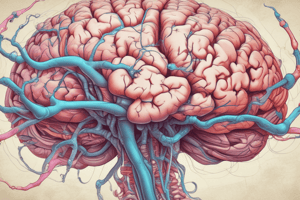
Anatomy of the Spinal Cord
- Comprises 31 pairs of spinal nerves, bridging the brain and most body parts.
- Functions:
- Connects the brain with the body.
- Facilitates spinal reflex actions (both somatic and visceral).
- It extends from the foramen magnum to the first lumbar vertebra, approximately 47 cm long.
- Contains significant anatomical features, including:
- Cervical and Lumbosacral enlargements: Indicate regions of increased neural processing.
- Meninges: Three protective layers surrounding the spinal cord:
- Dura Mater: Tough outer layer.
- Arachnoid: Middle layer that extends to S2 vertebra.
- Pia Mater: Innermost vascular layer.
- Spaces:
- Epidural Space: Contains blood vessels and fat.
- Subdural Space: Minimal space between dura and arachnoid, no cerebrospinal fluid (CSF).
- Subarachnoid Space: Contains CSF and blood vessels.
Cerebrospinal Fluid (CSF)
- A clear, watery fluid derived from blood, produced primarily in the choroid plexus of the brain.
- Provides cushioning and nutrient supply for the brain and spinal cord.
Internal Structure of the Spinal Cord
- Central Canal: Contains CSF.
- Gray Matter: Dark, H-shaped region including dorsal and ventral horns.
- White Matter: Composed of myelinated fiber tracts, categorized into:
- Posterior Column: Carries sensory input to the brain.
- Anterior and Lateral Columns: Responsible for motor output.
The Cerebral Hemispheres
- Gyri: Elevated ridges of tissue.
- Sulci: Shallow grooves between gyri; larger grooves are termed fissures.
- Critical areas:
- Somatic Sensory Area: Located in the parietal lobe, processes touch, pain, and temperature sensations.
- Primary Motor Area: Located in the frontal lobe, responsible for voluntary movement.
- Broca's Area: Associated with speech production.
Structures of the Brainstem
- Midbrain: Controls motor movement and processes auditory and visual information; consists of:
- Cerebral Aqueduct: Connects third and fourth ventricles.
- Cerebral Peduncles: Transmit impulses.
- Corpora Quadrigemina: Involved in reflex actions for vision and hearing.
- Pons: Contains nuclei essential for breathing regulation and neurological pathways.
- Medulla Oblongata: Regulates vital autonomic functions and contains many neural tracts.
Nerves
- Mixed Nerves: Carry both sensory and motor fibers.
- Afferent Nerves: Sensory nerves transmitting signals to the CNS.
- Efferent Nerves: Motor nerves transmitting information from the CNS.
Autonomic Nervous System (ANS)
- A subdivision of the PNS that automates bodily functions: heart rate, digestion, and respiratory rate.
- Comprises two motor neurons:
- Preganglionic Neuron: Located in the CNS.
- Postganglionic Neuron: Resides in ganglia outside the CNS.
- Has two divisions:
- Sympathetic Division: Mobilizes the body during stress ("fight or flight").
- Parasympathetic Division: Promotes relaxation and energy conservation ("rest and digest").
Functionality of ANS
- Organs receive input from both divisions and produce antagonistic effects.
- Neurotransmitters:
- Parasympathetic fibers release acetylcholine (cholinergic fibers).
- Sympathetic fibers release norepinephrine (adrenergic fibers).
Studying That Suits You
Use AI to generate personalized quizzes and flashcards to suit your learning preferences.