Podcast
Questions and Answers
Which of the following structures prevents the atrioventricular valves from inverting during ventricular contraction?
Which of the following structures prevents the atrioventricular valves from inverting during ventricular contraction?
- Coronary arteries
- Auricles
- Chordae tendineae (correct)
- Pericardium
Auricles increase the surface area of the ventricles to enhance blood pumping capacity.
Auricles increase the surface area of the ventricles to enhance blood pumping capacity.
False (B)
What is the main function of the coronary arteries?
What is the main function of the coronary arteries?
To supply blood to the heart muscle (myocardium).
The ______ is the double-layered sac that surrounds and protects the heart.
The ______ is the double-layered sac that surrounds and protects the heart.
Which of the following best describes the location of the chordae tendineae?
Which of the following best describes the location of the chordae tendineae?
The coronary arteries carry deoxygenated blood away from the heart.
The coronary arteries carry deoxygenated blood away from the heart.
What is the primary function of the auricles?
What is the primary function of the auricles?
Blockage of the ______ can lead to a myocardial infarction (heart attack).
Blockage of the ______ can lead to a myocardial infarction (heart attack).
Describe the two layers that make up the pericardium.
Describe the two layers that make up the pericardium.
Match each structure with its correct function:
Match each structure with its correct function:
Which of the following structures helps to anchor the atrioventricular valves and prevent valve prolapse?
Which of the following structures helps to anchor the atrioventricular valves and prevent valve prolapse?
The auricles are located on the ventricles and assist in the forceful ejection of blood.
The auricles are located on the ventricles and assist in the forceful ejection of blood.
What is the potential consequence of a significant blockage in one of the coronary arteries?
What is the potential consequence of a significant blockage in one of the coronary arteries?
The space between the visceral and parietal layers of the pericardium is filled with ______ fluid.
The space between the visceral and parietal layers of the pericardium is filled with ______ fluid.
What would be the effect of damaged or ruptured chordae tendineae?
What would be the effect of damaged or ruptured chordae tendineae?
Auricles play a direct role in the conduction of electrical signals through the heart.
Auricles play a direct role in the conduction of electrical signals through the heart.
What is the role of the pericardium in preventing cardiac tamponade?
What is the role of the pericardium in preventing cardiac tamponade?
Angina pectoris, or chest pain, is often a symptom of reduced blood flow through the ______.
Angina pectoris, or chest pain, is often a symptom of reduced blood flow through the ______.
Which layer of the pericardium is in direct contact with the heart?
Which layer of the pericardium is in direct contact with the heart?
Match each term with the appropriate definition:
Match each term with the appropriate definition:
Flashcards
Chordae Tendineae
Chordae Tendineae
Tough, fibrous cords that attach the atrioventricular valves to the papillary muscles in the ventricles, preventing valve prolapse.
Auricles (Atria)
Auricles (Atria)
Also known as atria, these are the two upper chambers of the heart that receive blood from the body (right atrium) and lungs (left atrium).
Coronary Arteries
Coronary Arteries
Vessels that supply oxygen-rich blood to the heart muscle (myocardium). Blockage can lead to myocardial infarction.
Pericardium
Pericardium
Signup and view all the flashcards
Study Notes
- Chordae tendineae are cord-like tendons that connect the papillary muscles to the tricuspid valve and the mitral valve in the heart.
- Also known as heart strings.
- They are composed of approximately 80% collagen, with the remainder comprised of elastin and endothelial cells.
- Chordae tendineae prevent the valves from prolapsing back into the atria during ventricular contraction (systole).
- There are two types: chordae tendineae primae, which arise from the free edge of the valve leaflets, and chordae tendineae secundae, which arise from the ventricular surface of the leaflets.
- Rupture of the chordae tendineae can lead to mitral or tricuspid valve regurgitation, where blood leaks back into the atria.
- The length and thickness of the chordae tendineae contribute to the proper functioning of the heart valves.
Auricles
- Auricles, also known as atrial appendages, are small, ear-shaped pouches that protrude from the atria of the heart.
- They increase the blood-holding capacity of the atria.
- The right auricle is typically larger than the left auricle.
- Auricles are more trabeculated (ridged) than the atria themselves.
- They play a role in the production of atrial natriuretic peptide (ANP), a hormone that helps regulate blood volume and blood pressure.
- Due to their shape and location, auricles are prone to the formation of blood clots, especially in conditions like atrial fibrillation.
- Transesophageal echocardiography (TEE) is often used to visualize the auricles for the presence of thrombi (blood clots) before procedures like cardioversion.
- The left atrial appendage is often targeted for closure in patients with atrial fibrillation to reduce the risk of stroke.
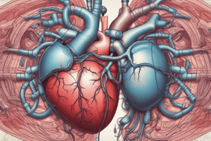
Coronary Arteries
- Coronary arteries are the arterial blood vessels of coronary circulation, which transport oxygenated blood to the heart muscle.
- The two main coronary arteries are the left coronary artery (LCA) and the right coronary artery (RCA).
- The LCA branches into the left anterior descending artery (LAD) and the left circumflex artery (LCx).
- The LAD supplies blood to the anterior wall of the left ventricle, the interventricular septum, and part of the right ventricle.
- The LCx supplies blood to the lateral and posterior walls of the left ventricle.
- The RCA supplies blood to the right atrium, the right ventricle, the posterior part of the left ventricle, and the sinoatrial (SA) and atrioventricular (AV) nodes in most individuals.
- Blockage of the coronary arteries, often due to atherosclerosis, can lead to angina (chest pain) or myocardial infarction (heart attack).
- Coronary artery bypass grafting (CABG) and percutaneous coronary intervention (PCI) are common procedures used to restore blood flow to the heart muscle in cases of coronary artery disease.
- Variations in coronary artery anatomy are common, and the dominance of either the RCA or LCA in supplying the posterior descending artery (PDA) is a key factor in determining coronary circulation patterns.
Pericardium
- The pericardium is a double-layered sac that surrounds the heart and the roots of the great vessels.
- It consists of two main layers: the fibrous pericardium and the serous pericardium.
- The fibrous pericardium is the outer layer and is made of dense connective tissue, providing protection and anchoring the heart within the mediastinum.
- The serous pericardium is the inner layer and is further divided into two layers: the parietal pericardium (lining the inner surface of the fibrous pericardium) and the visceral pericardium (epicardium), which adheres directly to the heart.
- The space between the parietal and visceral layers of the serous pericardium is the pericardial cavity, which contains a small amount of serous fluid that lubricates the surfaces and reduces friction during heartbeats.
- Functions of the pericardium include: protecting the heart from infection, preventing excessive dilation of the heart, and providing lubrication to reduce friction.
- Conditions affecting the pericardium include pericarditis (inflammation of the pericardium), pericardial effusion (accumulation of fluid in the pericardial cavity), and cardiac tamponade (compression of the heart due to fluid accumulation).
- Pericardiocentesis is a procedure to remove fluid from the pericardial cavity, often performed in cases of cardiac tamponade.
- The pericardium is innervated by the phrenic nerve, which can cause referred pain to the shoulder or neck in cases of pericardial irritation.
Studying That Suits You
Use AI to generate personalized quizzes and flashcards to suit your learning preferences.