Podcast
Questions and Answers
What is the primary function of a chest tube?
What is the primary function of a chest tube?
- To introduce medications directly into the bloodstream
- To remove air and/or fluid from the pleural space (correct)
- To deliver oxygen directly to the lungs
- To monitor the patient's heart rate
Which condition would NOT typically require the use of a chest tube?
Which condition would NOT typically require the use of a chest tube?
- Pulmonary embolism (correct)
- Pleural effusion (fluid accumulation in the pleural space)
- Post-operative cardiac surgery
- Pneumothorax (air in the pleural space)
What type of pressure is normally maintained within the pleural cavity?
What type of pressure is normally maintained within the pleural cavity?
- Neutral pressure
- Positive pressure
- Negative pressure (correct)
- Variable pressure
Which of these is a potential use of chest tube beyond drainage?
Which of these is a potential use of chest tube beyond drainage?
Why is negative pressure crucial in the pleural space?
Why is negative pressure crucial in the pleural space?
A patient has a chest tube inserted following a hemothorax. What is being removed by the chest tube?
A patient has a chest tube inserted following a hemothorax. What is being removed by the chest tube?
What is the primary purpose of chest tube insertion following cardiac surgery?
What is the primary purpose of chest tube insertion following cardiac surgery?
In which anatomical space is a chest tube placed?
In which anatomical space is a chest tube placed?
What is the primary effect of air, blood, pus, or lymph accumulating in the pleural cavity?
What is the primary effect of air, blood, pus, or lymph accumulating in the pleural cavity?
What is the main goal of chest tube therapy in the context of pleural space issues?
What is the main goal of chest tube therapy in the context of pleural space issues?
What is the underlying cause of a primary spontaneous pneumothorax?
What is the underlying cause of a primary spontaneous pneumothorax?
Which of the following conditions is characterized by milky-white fluid in the pleural space?
Which of the following conditions is characterized by milky-white fluid in the pleural space?
What is the primary cause of a massive hemothorax?
What is the primary cause of a massive hemothorax?
Which of the following is NOT a typical cause of pleural effusion?
Which of the following is NOT a typical cause of pleural effusion?
What is the primary characteristic of empyema?
What is the primary characteristic of empyema?
Which common complication can lead to secondary pneumothorax?
Which common complication can lead to secondary pneumothorax?
What is the primary role of a registered nurse during a chest tube insertion?
What is the primary role of a registered nurse during a chest tube insertion?
In which position is a patient typically placed for chest tube insertion, with regard to the affected side?
In which position is a patient typically placed for chest tube insertion, with regard to the affected side?
If a chest tube is intended to remove air, where is it commonly inserted?
If a chest tube is intended to remove air, where is it commonly inserted?
Which action is essential before a chest tube insertion procedure?
Which action is essential before a chest tube insertion procedure?
Which documented information is important post chest tube insertion?
Which documented information is important post chest tube insertion?
If the primary function of the chest tube is to remove fluid, where should it be placed?
If the primary function of the chest tube is to remove fluid, where should it be placed?
What is the purpose of the occlusive dressing after chest tube insertion?
What is the purpose of the occlusive dressing after chest tube insertion?
Which of these is a likely step in preparing a patient for chest tube insertion?
Which of these is a likely step in preparing a patient for chest tube insertion?
Following chest tube placement, which assessment finding requires immediate reporting?
Following chest tube placement, which assessment finding requires immediate reporting?
What is the primary purpose of observing for tidaling in the water-seal chamber?
What is the primary purpose of observing for tidaling in the water-seal chamber?
Which of the following is a critical action when managing the chest tube drainage system?
Which of the following is a critical action when managing the chest tube drainage system?
A patient with a chest tube shows signs of respiratory distress. Which assessment finding is most concerning?
A patient with a chest tube shows signs of respiratory distress. Which assessment finding is most concerning?
What does the presence of subcutaneous emphysema indicate?
What does the presence of subcutaneous emphysema indicate?
What should be monitored at the chest tube insertion site? (Select all that apply)
What should be monitored at the chest tube insertion site? (Select all that apply)
After chest tube insertions, if the patient's breathing is labored, and the breath sounds are decreased, what is the priority nursing concern?
After chest tube insertions, if the patient's breathing is labored, and the breath sounds are decreased, what is the priority nursing concern?
Which intervention is essential to maintain a functional chest tube system?
Which intervention is essential to maintain a functional chest tube system?
When changing a chest tube drainage collection box, what is the next step after obtaining the new box?
When changing a chest tube drainage collection box, what is the next step after obtaining the new box?
In a wet suction system, the suction pressure is regulated by which of the following?
In a wet suction system, the suction pressure is regulated by which of the following?
What is a key visual indicator that a dry suction system is operating correctly?
What is a key visual indicator that a dry suction system is operating correctly?
Which of the following is NOT an indication for chest tube removal?
Which of the following is NOT an indication for chest tube removal?
What is the recommended taping method for a chest tube dressing if the tube becomes dislodged?
What is the recommended taping method for a chest tube dressing if the tube becomes dislodged?
What is the primary reason for avoiding clamping, milking, or stripping a chest tube?
What is the primary reason for avoiding clamping, milking, or stripping a chest tube?
Why should the chest drainage box always be kept below the patient's chest level?
Why should the chest drainage box always be kept below the patient's chest level?
When documenting chest tube drainage, what is considered a critical change that should be reported to the healthcare provider?
When documenting chest tube drainage, what is considered a critical change that should be reported to the healthcare provider?
According to the guidelines, how should the nurse manage a full chest drainage collection box?
According to the guidelines, how should the nurse manage a full chest drainage collection box?
What is the purpose of keeping rubber-tipped or padded hemostats at the bedside of a patient with a chest tube?
What is the purpose of keeping rubber-tipped or padded hemostats at the bedside of a patient with a chest tube?
According to the guidelines, what position is recommended for a patient whose chest tube has been dislodged at the chest wall?
According to the guidelines, what position is recommended for a patient whose chest tube has been dislodged at the chest wall?
In the event of a chest tube dislodgement at the insertion site, why is the dressing taped on three sides only?
In the event of a chest tube dislodgement at the insertion site, why is the dressing taped on three sides only?
If a chest tube becomes disconnected from the drainage system, what is the priority nursing action?
If a chest tube becomes disconnected from the drainage system, what is the priority nursing action?
Flashcards
What is a Chest Tube?
What is a Chest Tube?
A catheter inserted into the chest cavity, used to drain fluid or air, or to prevent complications after surgery.
What is the pleural cavity?
What is the pleural cavity?
The space between the lung and the chest wall.
What is negative pressure in the pleural cavity?
What is negative pressure in the pleural cavity?
The pressure in the pleural cavity is lower than atmospheric pressure, allowing the lungs to expand during inspiration.
How does a chest tube work?
How does a chest tube work?
Signup and view all the flashcards
What is closed chest drainage?
What is closed chest drainage?
Signup and view all the flashcards
What can be drained through a chest tube?
What can be drained through a chest tube?
Signup and view all the flashcards
What is a common use for chest tubes?
What is a common use for chest tubes?
Signup and view all the flashcards
Can chest tubes be used for medication delivery?
Can chest tubes be used for medication delivery?
Signup and view all the flashcards
What happens when the negative pressure in the pleural cavity is disrupted?
What happens when the negative pressure in the pleural cavity is disrupted?
Signup and view all the flashcards
What is pneumothorax?
What is pneumothorax?
Signup and view all the flashcards
What is a primary spontaneous pneumothorax?
What is a primary spontaneous pneumothorax?
Signup and view all the flashcards
What is a secondary pneumothorax?
What is a secondary pneumothorax?
Signup and view all the flashcards
What is chylothorax?
What is chylothorax?
Signup and view all the flashcards
What is hemothorax?
What is hemothorax?
Signup and view all the flashcards
What is pleural effusion?
What is pleural effusion?
Signup and view all the flashcards
What is empyema?
What is empyema?
Signup and view all the flashcards
Chest Tube Placement
Chest Tube Placement
Signup and view all the flashcards
Analgesic or Sedating Agent
Analgesic or Sedating Agent
Signup and view all the flashcards
Sitting or Lying Position, Affected Side Elevated
Sitting or Lying Position, Affected Side Elevated
Signup and view all the flashcards
Explain Procedure to Patient
Explain Procedure to Patient
Signup and view all the flashcards
Informed Consent
Informed Consent
Signup and view all the flashcards
Provide Supplemental Oxygen
Provide Supplemental Oxygen
Signup and view all the flashcards
Chest Tube Insertion
Chest Tube Insertion
Signup and view all the flashcards
Chest Tube Placement: Location and Function
Chest Tube Placement: Location and Function
Signup and view all the flashcards
Tidaling
Tidaling
Signup and view all the flashcards
Continuous bubbling
Continuous bubbling
Signup and view all the flashcards
Subcutaneous emphysema
Subcutaneous emphysema
Signup and view all the flashcards
Keeping drainage system below chest level
Keeping drainage system below chest level
Signup and view all the flashcards
Water-seal chamber fluid level
Water-seal chamber fluid level
Signup and view all the flashcards
Chest tube dressing
Chest tube dressing
Signup and view all the flashcards
Decreased or Absent Breath Sounds
Decreased or Absent Breath Sounds
Signup and view all the flashcards
Respiratory Distress
Respiratory Distress
Signup and view all the flashcards
What is the main difference between wet and dry suction systems?
What is the main difference between wet and dry suction systems?
Signup and view all the flashcards
When can a chest tube be safely removed?
When can a chest tube be safely removed?
Signup and view all the flashcards
How should a chest tube dressing be applied?
How should a chest tube dressing be applied?
Signup and view all the flashcards
What pre-procedure steps are necessary before removing a chest tube?
What pre-procedure steps are necessary before removing a chest tube?
Signup and view all the flashcards
What is the proper patient positioning for chest tube removal?
What is the proper patient positioning for chest tube removal?
Signup and view all the flashcards
Clamping Chest Tubes
Clamping Chest Tubes
Signup and view all the flashcards
Positioning the Drainage Box
Positioning the Drainage Box
Signup and view all the flashcards
Monitoring Drainage
Monitoring Drainage
Signup and view all the flashcards
Encouraging Lung Expansion
Encouraging Lung Expansion
Signup and view all the flashcards
Managing Full Drainage Boxes
Managing Full Drainage Boxes
Signup and view all the flashcards
Dislodged Chest Tube
Dislodged Chest Tube
Signup and view all the flashcards
Disconnected Chest Tube
Disconnected Chest Tube
Signup and view all the flashcards
Emergency Equipment
Emergency Equipment
Signup and view all the flashcards
Study Notes
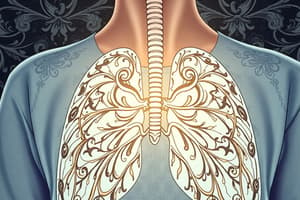
Chest Tubes
- Chest tubes are catheters inserted through the thorax to remove air and/or fluid from the pleural space.
- Purpose of chest tubes/closed chest drainage: Treat conditions that disrupt the pleural space, drain pleural cavity (air, blood, pus, lymph fluid), prevent/diminish postoperative complications (cardiac surgery), and instill fluids (chemotherapy or sclerosing agents) into the pleural space.
Indications for a Chest Tube
- The potential space around the lungs is called the pleural cavity.
- Under normal conditions, the pleural cavity is maintained by negative pressure, crucial for lung expansion during breathing.
- When air, blood, pus, or lymph collects in the pleural cavity, negative pressure is lost, hindering lung expansion.
Overall Goal of Chest Tube Therapy
- Promote lung re-expansion
- Re-establish normal negative pleural space pressure
- Restore adequate oxygenation and ventilation
- Prevent complications
Conditions That Disrupt the Pleural Space
- Pneumothorax: A collection of air in the pleural space. Loss of negative intrapleural pressure causes lung collapse.
- Primary: Occurs in the absence of lung disease or injury. Spontaneous pneumothorax is a genetic condition with unexpected occurrences in healthy individuals who develop blebs on the visceral pleura. Tall, young men are at increased risk.
- Secondary: Develops due to chest trauma (stabbing, gunshot wound, rib fractures), rupture of an emphysematous bleb, or tearing of the pleura from an invasive procedure (surgery, insertion of a subclavian line, mechanical ventilation).
- Chylothorax: Lymphatic fluid accumulation in the pleural space, often from chest trauma, tumors, or surgery within the mediastinum. Milky-white pleural fluid.
- Hemothorax: Blood in the pleural space, due to blunt or penetrating trauma or chest surgery. A massive hemothorax is when blood rapidly accumulates in the chest cavity, resulting from penetrating or blunt trauma that disrupts systemic vessels.
- Pleural Effusion: Excessive fluid in the pleural space, caused by left ventricular failure, pulmonary embolism, pneumonia, cancer, or conditions that impede pleural fluid drainage (such as a tumor blocking the lymphatic system) or complications from surgery or fluid shifts(e.g., liver or renal failure).
- Empyema: Purulent pleural fluid, potentially from a lung abscess or pneumonia.
Risks and Complications of Chest Tube Placement
- Malposition: The chest tube is not in the proper space. This is the most common complication leading to persistent air or fluid in the pleural space
- Bleeding: Often minor and resolves on its own. Bleeding into the lung may require surgical intervention, common during insertion.
- Infection: Increases with the duration of tube placement. Sterile technique is crucial. Watch for oozing, drainage, or erythema at the insertion site. High risk category includes immunocompromised patients.
- Lung Trauma and Diaphragm Perforation: During insertion, lung trauma and diaphragm perforation can occur if the chest tube is inserted too low.
- Subcutaneous Emphysema: Air leaks from the pleural space into the subcutaneous tissue after chest tube placement. Tissues of the neck, face, axilla, and chest swell, and there may be crepitus on palpation.
Preparing the Patient for Chest Tube Placement
- Monitor vital signs, electronic monitoring equipment
- Administer analgesics or sedatives as needed
- Patient positioning (sitting or lying, affected side elevated)
- Arm positioning (brought over head and secured)
- Area cleansing with an antiseptic solution.
- Explain procedure to patient, obtain informed consent (if possible)
- Provide supplemental oxygen
- Local anesthetic is given to the area
- A small incision is made
- The chest tube is inserted and sutured to the chest wall.
- Chest tube is connected to the drainage system.
Preparing a Patient for Chest Tube Placement cont.
- Steps are illustrated by pictures that show incision at midaxillary line, between 4th and 5th ribs, lateral to nipple.
- Clamping of a rib over vessels needs to be avoided.
- Insertion site to be explored by a finger.
- Using clamps, chest tube will be guided into the place.
Dependent on the Function that the Chest Tube Performs
- Air Removal: Tube insertion at the mid-clavicular, second intercostal space, near the apex of the lung
- Fluid Removal: Tube placement inferior and posterior in the pleural space, mid-axillary line, 7th or 8th intercostal space
Documentation after Initial Chest Tube Placement
- Vital signs before and after procedure
- Chest tube size and insertion site
- Physician inserting the tube
- Drainage description (type and amount)
- Drainage system type and suction pressure
- Cultures sent
- Patient response to procedure
- Medications used
- Chest x-ray results
Nursing Care After Chest Tube Placement - Ongoing Patient Assessments
- General: Vital signs, pain, respiratory rate and pattern, respiratory status, respiratory depth, ease of respiration, oxygen saturation
- Specific to Chest Tube: Assess for re-accumulation of air or fluid in the chest; decreased or absent breath sounds; signs of respiratory distress; tachypnea, dyspnea, shortness of breath, tachycardia, decreased breathing, absent breath sounds, use of accessory muscles.
- Tube Insertion Site for Drainage and Signs of Infection: Drainage, erythema, subcutaneous emphysema, signs of infection (fever, elevated WBC count).
Ongoing Patient Assessments (cont)
- Pain/discomfort (medicate as needed)
- Subcutaneous emphysema (crackling sensation under the skin)
- Skin color
Management of the Drainage System
- Ensure that the dressing is occlusive, dry, and intact, secured on all 4 sides.
- Maintain tubing and drainage system below chest level.
- Do not let patient lie on tubing, prevent kinks and occlusions
- Tightly secure all tubing connections
- Observe for tidaling (fluctuation, movement up/down in the water-seal chamber as the patient breaths)
- Observe for intermittent bubbling in the water-seal chamber
Management of Chest Drainage - (cont)
- Do not clamp, milk, or strip the tube
- Clamping to temporarily change drainage box is OK.
- Do not elevate drainage box above patient's chest level.
- Monitor and document drainage amount and color
- Document drainage values
- Assist patient with position changes, coughing, and deep breaths to facilitate fluid drainage.
- Collection box to be changed if necessary for increased quantity and drainage type.
Tube Dislodgement or Disconnection
- Keep rubber-tipped or padded hemostats, Vaseline gauze, dry sterile dressing, and tape at bedside.
- Sterile water is also crucial.
- Instructions for patient performing Valsalva Maneuver or submerge distal end of chest tube (2cm) in sterile water.
- Cover insertion site with new Vaseline gauze or sterile dressing taped on 3 sides only. Taping on 3 sides only prevents tension pneumothorax
- Patient positioning (High Fowler's).
- Ask another nurse to contact provider
Tube Disconnected (cont)
- Immediately submerge the distal end of the tube (2 cm) in sterile water for a temporary water seal.
- New drainage collection box to be brought in by another nurse.
- Tube connection to the new drainage box
- Turn on suction system
- Notify the provider
Wet and Dry Suction Systems
- Wet System: Regulates suction pressure by the water column height in the suction control chamber.
- Dry System: Mechanically regulates suction pressure. No water column; orange bellows are the visual indicator of suction operation.
Chest Tube Removal
- Indicated when lungs are fully expanded, fluid drainage has stopped, improved chest x-rays & improved respiratory status.
- Occlusive dressing (4 sides taped)
- Comprehensive Respiratory Assessments
- Patient Comfort
- Post-removal chest x-ray is typically done.
Assisting the Healthcare Provider During Chest Tube Removal
- Educate patient and provide support
- Premedicate to relieve pain and reduce anxiety
- Gather supplies (sterile gloves, suture removal kit, hemostats, occlusive dressing)
- Position patient in semi-Fowler's and pad the area
- Remove dressing and sutures
- Practitioner clamps the tube
- Patient performs Valsalva maneuver
- Tube is quickly removed
- Immediately cover wound with an occlusive dressing.
Post Chest Tube Removal Patient Care
- Cover site with Vaseline gauze and an occlusive dressing (4 sides taped)
- Follow up chest x-ray
- Monitor wound for drainage
- Comprehensive respiratory assessments
Studying That Suits You
Use AI to generate personalized quizzes and flashcards to suit your learning preferences.