Podcast
Questions and Answers
Which of the following best describes the primary abnormality in cerebrovascular disorders?
Which of the following best describes the primary abnormality in cerebrovascular disorders?
- Any functional abnormality of the central nervous system (CNS) that occurs when the normal blood supply to the brain is disrupted. (correct)
- Progressive demyelination of nerve fibers in the brain and spinal cord.
- Degeneration of neurons in specific brain regions.
- Infection of the meninges surrounding the brain and spinal cord.
A patient presents with a blood clot that formed in their leg, traveled to their brain, and caused a stroke. This is an example of what?
A patient presents with a blood clot that formed in their leg, traveled to their brain, and caused a stroke. This is an example of what?
- Aneurysm
- Embolism (correct)
- Thrombosis
- Hemorrhage
Which of the following is a non-modifiable risk factor for stroke?
Which of the following is a non-modifiable risk factor for stroke?
- Advanced age (correct)
- Diabetes mellitus
- Hyperlipidemia
- Hypertension
Which of the following is an early sign of ischemic stroke?
Which of the following is an early sign of ischemic stroke?
Which of the following physiological processes contributes to brain cell injury during ischemia?
Which of the following physiological processes contributes to brain cell injury during ischemia?
A patient with a stroke is experiencing difficulty recognizing objects by touch. What is this condition called?
A patient with a stroke is experiencing difficulty recognizing objects by touch. What is this condition called?
A patient who has had a stroke demonstrates emotional lability and hostility during rehabilitation sessions. Damage to which area of the brain might cause these symptoms?
A patient who has had a stroke demonstrates emotional lability and hostility during rehabilitation sessions. Damage to which area of the brain might cause these symptoms?
What is the primary goal of administering mannitol to a patient with increased intracranial pressure (ICP)?
What is the primary goal of administering mannitol to a patient with increased intracranial pressure (ICP)?
A patient with a head injury is suspected of having a basilar skull fracture. Which of the following assessment findings would support this suspicion?
A patient with a head injury is suspected of having a basilar skull fracture. Which of the following assessment findings would support this suspicion?
A patient with a spinal cord injury at T4 complains of a severe headache, nasal congestion, and diaphoresis. Their blood pressure is 210/110 mm Hg. Which complication is most likely occurring, and what is the initial nursing intervention?
A patient with a spinal cord injury at T4 complains of a severe headache, nasal congestion, and diaphoresis. Their blood pressure is 210/110 mm Hg. Which complication is most likely occurring, and what is the initial nursing intervention?
Flashcards
Cerebrovascular Disorders
Cerebrovascular Disorders
Umbrella term for functional abnormality of the CNS when blood supply to brain is disrupted. Stroke is primary.
Thrombus
Thrombus
A blood clot forms at the site, blocking a blood vessel.
Embolus
Embolus
A clot or substance that travels from another site blocking blood vessel.
Two Major types of Stroke?
Two Major types of Stroke?
Signup and view all the flashcards
Hypertension
Hypertension
Signup and view all the flashcards
Hemiplegia
Hemiplegia
Signup and view all the flashcards
Hemiparesis
Hemiparesis
Signup and view all the flashcards
Homonymous Hemianopsia
Homonymous Hemianopsia
Signup and view all the flashcards
Aphasia
Aphasia
Signup and view all the flashcards
Agnosia
Agnosia
Signup and view all the flashcards
Study Notes
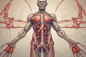
- "Cerebrovascular disorders" is an umbrella term for functional abnormalities in the central nervous system (CNS) when the normal blood supply to the brain is disrupted
- Stroke is the primary cerebrovascular disorder
Thrombosis
- A blood clot forms at the site and blocks a blood vessel
- Forms locally at the site of blockage
- It does not move, stays at the site of origin
- Formation is due to atherosclerosis or injury
- Treatment involves removing or dissolving the clot at its origin
Embolus
- A clot or other substance travels from another site and blocks a blood vessel
- It forms elsewhere, often in the heart or large veins
- Travel through the bloodstream to another part of the body
- A blood clot breaks off from the heart or deep veins and travels to the brain (embolism).
- Treatment requires removing or breaking up the clot that has traveled
2 major categories of stroke
Ischemic causes
- Large artery thrombosis
- Small penetrating artery thrombosis
- Cardiogenic embolic Cryptogenic (no known cause)
Hemorrhagic causes
- Intracerebral hemorrhage
- Subarachnoid hemorrhage
- Cerebral aneurysm
- Arteriovenous malformation
Non-modifiable risk factors for stroke
- Advanced age over 55
- Male gender
- African Americans
Modifiable risk factors for stroke
- Hypertension
- Cardiovascular disease
- Atrial fibrillation
- Coronary artery disease
- Heart failure
- Left ventricular hypertrophy
- Myocardial infarction (especially anterior)
- Rheumatic heart disease
- Elevated hematocrit (increases the risk of cerebral infarction)
- Diabetes mellitus (associated with accelerated atherogenesis)
- Oral contraceptive use (increases risk, especially with coexisting hypertension, smoking, and high estrogen levels)
- Smoking
- Drug abuse (especially cocaine)
- Excessive alcohol consumption
Ischemic stroke
- "brain attack"
- Numbness or weakness of the face, arm, or leg, especially on one side of the body
- Confusion or change in mental status
- Trouble speaking or understanding speech
- Visual disturbances
- Difficulty walking, dizziness, or loss of balance or coordination
- Sudden severe headache
Ischemia brain cell injury processes
- Occurs due to a lack of blood flow and oxygen to the brain.
- When blood flow to the brain is reduced or blocked, cells don't get enough oxygen and nutrients.
- Oxygen is depleted and brain cells can't produce energy, which can lead to dysfunction.
- A lack of energy causes:
- Acid buildup in cells
- Disruption in the balance of important ions like sodium, potassium, and calcium
Increased intracellular calcium impacts:
- Calcium levels inside cells
- Calcium levels rise abnormally and trigger harmful processes:
- Damage to cell membranes and proteins
- Formation of free radicals (damaging molecules)
- Reduced protein production (affecting cell repair and function)
- Electrical instability (depolarization)
- Excessive glutamate release damages cells
- Cell injury and death occurs causing cells to break down and die
Hemorrhagic stroke deficits
- Patients generally have more severe deficits and a longer recovery time compared to those with ischemic stroke
- Pathophysiology depends on the cause and type of cerebrovascular disorder
- Intracerebral hemorrhage is when the AVM enlarges or ruptures exposing the brain to blood
- Intracranial (cerebral) aneurysm
- Arteriovenous Malformations
- Subarachnoid hemorrhage presses nearby cranial nerves or brain tissue
- The above disorders disrupt of normal brain metabolism, increase ICP, compress and injure the brain, reduce perfusion and vasospasm which causes secondary ischemia by the brain
Major predisposing factors for stroke
- Atherosclerosis
- Emboli
- Hypertension
- Intracranial hemorrhage
- Diabetes mellitus
- Polycythemia
Initial manifestations of stroke
- Severe & sudden headache
- Trouble in speaking
- Right or left hemiparesis
- Ocular disturbances
- Confusion
- Impairment in coordination
Motor loss
- Hemiplegia is paralysis of one side of the body due to a lesion of the opposite side of the brain.
- Hemiparesis is weakness of one side of the body
- Initial clinical features may be flaccid paralysis and loss of or decrease in the deep tendon reflexes
Communication Loss
- Dysarthria (difficulty in speaking), caused by paralysis of the muscles responsible for producing speech
- Dysphasia or aphasia is a loss of speech
- There are 3 types: expressive aphasia, receptive aphasia, or global (mixed) aphasia
- Apraxia is the inability to perform a previously learned action
- Can be observed when a patient picks up a fork and attempts to comb his hair with it
Perceptual Disturbances
- Stroke can result in visual-perceptual dysfunctions, disturbances in visual-spatial relations, and sensory loss.
- Homonymous hemianopsia is loss of half of the visual field that may occur.
- Can be temporary or permanent.
- The affected side of vision corresponds to the paralyzed side of the body.
- Disturbances in visual-spatial relations frequently seen in patients with right hemispheric damage
Sensory Loss
- The sensory losses can include slight impairment of touch or may be more severe
- Severe loss may include a loss of proprioception (ability to perceive the position and motion of body parts)
- There is difficulty in interpreting visual, tactile, and auditory stimuli.
- Agnosia is the loss of being able to recognize objects through a sensory system
- It may be visual, auditory or tactile.
Cognitive impairment and psychological effects
- Learning capacity, memory, or other higher cortical intellectual functions may be impaired if damage has occurred to the frontal lobe
- Results in a limited attention span, difficulties in comprehension. and forgetfulness
- Causes patients to become frustrated in their rehabilitation program.
- Depression is common and can get exaggerated during catastrophic event responses
- Also include includes emotional lability, hostility, frustration, resentment, and a lack of cooperation.
Left hemispheric stroke
- Paralysis or weakness on right side of body
- Right visual field deficit
- Expressive, receptive, or global Aphasia
- Altered intellectual ability
- Slow, cautious behavior
Right hemispheric stroke
- Paralysis or weakness on left side of body
- Left visual field deficit
- Spatial-perceptual deficits
- Increased distractibility
- Impulsive behavior and poor judgment
- Lack of awareness of deficits
Assessment and Diagnostic Findings
- A complete physical and neurologic examination
- Includes:
- Airway patency evidenced by gag or cough reflexes
- Altered respiratory pattern
- Cardiovascular status (blood pressure, cardiac rhythm and rate, carotid bruit)
- Gross neurologic losses
- A system uses the time course to classify and guide treatment including:
- Transient ischemic attack (TIA)
- Reversible ischemic neurologic deficit
- Stroke in evolution
- Completed stroke (Hock, 1999)
- (CT) scan
- 12-lead electrocardiogram
- Carotid ultrasound
- Cerebral angiography
- Transcranial Doppler flow studies
- Transthoracic or transesophageal echocardiography,
- Magnetic resonance imaging of the brain and/or neck, xenon CT
- Single photon emission
Outcome and progression of the stage of a stroke
- Transient Ischemic Attack (TIA) symptoms disappear without lasting damage:
- Reversible Ischemic Neurologic Deficit (RIND) temporary symptoms will have a full recovery
- Stroke in Evolution brain damage will continue to develop often leading to permanent disability
- Completed Stroke causes permanent brain damage that has occurred with lasting impairments
Medical Management
- Prevention of stroke
- Prevention of recurrence
- Immediate brain resuscitation
- Stabilization and Rehabilitation
Thrombolytic Therapy
- Recombinant t-PA (tissue plasminogen activator) is a genetically engineered form of t-PA, a thrombolytic substance made naturally by the body
- Rapid diagnosis is key within 3 hours of stroke
- The initiation of thrombolytic therapy in patients with ischemic stroke leads to a decrease in the size of the stroke and an overall improvement in functional outcome after 3 months
- Delays could make patients ineligible for thrombolytic therapy because revascularization of necrotic tissue (which develops after 3 hours) increases the risk for cerebral edema and hemorrhage
- Intracranial bleeding major complication that occurs in approximately 6.5% of patients
Prevent or minimize the risk for rebleeding
- Includes:
- Bed rest with sedation to prevent agitation and stress
- Management of vasospasm
- Surgical or medical treatment to prevent rebleeding.
- Analgesics (codeine, acetaminophen) may be prescribed for head and neck pain
- The patient is fitted with elastic compression stockings to prevent deep vein thrombosis
Other Treatments
- Anticoagulant administration IV heparin or low-molecular weight heparin
- Interventions to reduce ICP
- Administer an osmotic diuretic (eg, mannitol)
- Maintain PaCO2 within the range of 30 to 35 mm Hg,
- Position to avoid hypoxia
- Elevation of the head of the bed promotes venous drainage and lowers increased ICP
- Intubation with an endotracheal tube to establish a patent airway, if necessary
- Continuous hemodynamic monitoring; systolic pressure should be maintained at less than 180 mm Hg, diastolic pressure at less than 100 mm Hg
- Maintain cardiac output
Surgical Management of Hemorrhagic Stroke
- Surgical evacuation is strongly recommended for treating a patient with a cerebellar hemorrhage if the diameter exceeds 3 cm and the Glasgow Coma Scale score is below 14
- Surgical evacuation is most frequently accomplished via a craniotomy
- An extracranial-intracranial arterial bypass may be performed to establish collateral blood supply to allow surgery on the aneurysm
- Extracranial method may be used, whereby the carotid artery is gradually occluded in the neck to reduce pressure within the blood vessel.
- Endovascular treatments can be used for aneurysms
- Two procedures are endovascular treatment (occlusion of the parent artery) and aneurysm coiling (obstruction of the aneurysm site with a coil).
Medications used for Cerebrovascular Disorders
- Given based on the type of stroke being experienced:
- Antihypertensives
- Platelet Aggregants such as Aspirin and Ticlodipine
- Anticoagulants such as Heparin and Warfarin
- Thrombolytics such as t-PA and urokinase
- Calcium channel blockers such as nimodipine mannitol
- Dexamethasone
Nursing Interventions for Cerebrovascular Disorders
- Improve mobility and preventing joint deformities
- Prevent shoulder adduction
- Position the hand and fingers
- Change positions
- Establish an exercise program
- Prepare for ambulation
- prevent shoulder pain
- Enhance self-care
- Manage sensory-perceptual difficulties
- Manage dysphagia with tube feedings
- Attain bowel and bladder control
- Improve thought processes
- Improve communication
- Maintain skin integrity
- Improve family coping skills
- Help the patient cope with sexual dysfunction
Communicating With the Aphasic Patient
- Focus on the patient
- Establish eye contact.
- Speak in a normal manner and tone.
- Use short phrases and pause between phrases to allow the patient time to understand what is being said.
- Conversations should be practical and concrete
- Utilize gestures, pictures, and objects. and model usage
- As the patient uses and handles an object, say what the object is.
- Use consistent gestures and words
- Minimize background noise and distractions
Assistive Devices to Enhance Self-Care After Stroke
- Eating Devices
- Nonskid mats to stabilize plates
- Plate guards to prevent food from being pushed off plate
- Wide-grip utensils to accommodate a weak grasp
- Bathing and Grooming Devices
- Long-handled bath sponge
- Grab bars, nonskid mats, hand-held shower heads
- Electric razors with head at 90 degrees to handle
- Shower and tub seats, stationary or on wheels
- Toileting Aids
- Raised toilet seat
- Grab bars next to toilet
- Dressing Aids
- Velcro closures
- Elastic shoelaces
- Long-handled shoe horn
- Mobility Aids
- Canes, walkers, wheelchairs
- Transfer devices such as transfer boards and belts
Comparison of Major Types of Stroke
Ischemic
- Caused by: - Large artery thrombosis - Small penetrating artery thrombosis - Cardiogenic embolic - Cryptogenic (no known cause) - Other
- Main presenting symptoms: - Numbness or weakness of the face, arm, or leg, especially on one side of the body, aphasia, vision loss (homonymous hemianopsia)
- Functional Recovery - Majority of recovery made in the first 3-6 mo, slower steps toward recovery may be made up to 1 yr and beyond with therapy
Hemorrhagic
- Caused by - Intracerebral hemorrhage - Subarachnoid hemorrhage - Cerebral aneurysm - Arteriovenous malformation
- Main presenting symptoms:
- "Worst headache of my life"
- Decreased level of consciousness
- Seizure
- Functional recovery:
- Slower recovery, typically left with more disability
Head Trauma/Injury
- Includes injury to the scalp, skull, or brain
- It’s the most common cause of death from trauma in the US
- The highest at Risk:
- Ages 15 to 24 in males
- Ages below 5 years
- Ages 75 and above
2 Forms of Traumatic Brain Injury
Primary injury
- Involves contusions, lacerations, and torn blood vessel
Secondary injury
- (hours and days after initial injury) such as cerebral edema, ischemia, and chemical changes. Brain injury differs from body injury as it resides in the skull (rigid & closed compartment)
Pathophysiology of Traumatic Brain Injury
- Brain suffers traumatic injury
- Brain swelling or bleeding increases Intracranial Pressure (ICP)
- Rigid cranium allows no room for expansion of contents so ICP increases
- Pressure on blood vessels within the brain causes blood flow to the brain to slow
- Cerebral Hypoxia and Ischemia occurs
- ICP continues to rise causing the brain to herniate
- Cerebral blood flow ceases
Scalp Injury
- Classified as a minor head injury
- The many blood vessels constrict poorly causing the scalp to bleeds profusely when injured.
- Large avulsions of the scalp are potentially life-threatening and are true emergencies.
- Scalp wounds can cause intracranial infections
- Suturing the laceration area is irrigated to remove foreign material to reduce the risk for infection.
- Subgaleal hematomas (hematomas below the outer covering of the skull) usually absorb on their own and do not require any specific treatment
Skull Fracture Signs
- Battle's sign
- Raccoon's eye
- Subconjunctival hemorrhage
- Rhinorrhea (runny nose)
- Otorrhea (mucopurulent discharge from the ear)
- Drainage of CSF is a serious problem because meningeal infection can occur
- Organisms access the cranial contents through the nose, ear, or sinus through a tear in the dura.
- Bloody CSF suggests a brain laceration or contusion
Brain Injury
- Includes open-head injuries, closed-head injuries, coup-countercoup injuries, and rotational injuries
Types of brain injury
Concussion
- A severe jarring or mild traumatic injury that causes a temporary loss of neurologic function with no apparent structural damage to the brain
- Generally involves a period of unconsciousness lasting from a few seconds to a few minutes or complete loss of consciousness
- Causes dizziness/spots before the eyes (seeing stars)
- Frontal lobe may exhibit bizarre irrational behavior
- Temporal lobe may cause temporary amnesia or disorientation
Contusion
-Bruising of the brain surface, more severe injury than concussion and may cause surface hemorrhage
- The patient is unconscious for more than a few seconds or minutes Symptoms depend on the size and the extent of Cerebral Edema
- The patient appears in motion less, presenting a faint pulse, shallow respirations, a cool and pale skin
- Also, involuntary evacuation of the bladder and bowels may occur
- The patient has a BP and temp that may be sub normal
- Brain damage, disability or death may occur
Diffuse Axonal Injury
- There is widespread damage to axons particularly in the cerebral hemispheres, corpus callosum, and brain stem
- Seen in mild, moderate, or severe head trauma and results in axonal swelling and disconnection
- With severe injuries, the patient experiences no lucid intervals and presents with immediate coma, decorticate and decerebrate posturing
Intracranial Hemorrhage
- Hematomas (collections of blood) that develop within the cranial vault are the most serious brain injuries
- Include epidural (above the dura), subdural (below the dura), or intracerebral (within the brain)
- Major symptoms can be delayed until the hematoma is large enough to cause distortion of the brain and increased ICP
- A rapidly developing hematoma, may be fatal even if small
- A larger but slowly developing collection of blood allows compensation for increases in ICP
Epidural Hematoma features
- Blood may collect in the epidural (extradural) space between the skull and the dura.
-Hemorrhage from this artery causes rapid pressure on the brain.
- Respiratory arrest may occur within minutes of injury
Subdural Hematoma features
- Is a collection of blood between the dura and the brain in a space normally occupied by a this cushion of fluid
- The most common cause is trauma, but it may also occur from coagulopathies or rupture of an aneurysm.
Intracerebral Hemorrhage and Hematoma features
- Is bleeding into the substance of the brain
- It is commonly seen in head injuries when force is exerted to the head over a small area
- Management includes:
- Supportive care
- Control of ICP
- Careful of fluid level administration, electrolytes, and antihypertensive medications
Surgical Management of hemorrhages
- Surgical intervention through craniotomy or craniectomy can remove blood clots and control further hemorrhage
- May not be possible if the bleed is in a difficult location
3 cardinal signs of Brain Death
- Coma
- Absence of brain stem reflexes evidenced by a lack of pupil dilation or oculocephalic reflex (quick movement of head to the side causes no movement of the eyes)
- Apnea meaning there is no spontaneous breathing
Assessment and Diagnostic Findings for Brain Injury
- Physical and neurologic exam
- CT Scan indicating the nature and location of lesions.
- MRI is a more accurate anatomic picture of the injury
- Cerebral angiography identifying hematomas and contusions
To Manage Brain injuries:
- Perform physical and neurologic exams
- Presume the patient has with head injury has a cervical spine injury until proven otherwise
- Maintain head and neck alignment during transport with a cervical and spinal board
- Prevent secondary injury of cerebral edema, hypotension and respiratory depression
- Treat increased ICP
- Utilizes supportive ventilatory support, seizure prevention, fluid/ electrolyte balance maintenance, nutritional support, pain and anxiety measures
Surgical Intervention for Brain Injuries
- Surgery involves decompression
- Including craniectomy which is an excision of the cranial bone without replacing it
- Burr hole decompression involving evacuation of clot & abscess
- Craniotomy: Opening to the cranium
Nursing Intervention for Brain Injuries
- Airway support
- Regular neurological assessments
- ICP treatment
- Implementing Supportive measures -Maintain electrolyte & fluid balance
- Administer nutrition as appropriate
- Implement and supervise injury prevention measure
- Support skin integrity
Clinical Manifestations of Brain Injury
- Altered level of consciousness
- Confusion
- Pupillary abnormalities (changes in shape, size, and response to light)
- Altered or absent gag reflex
- Absent corneal reflex
- Sudden onset of neurologic deficits
- Changes in vital signs (altered respiratory pattern, hypertension, bradycardia, tachycardia, hypothermia or hyperthermia)
- Vision and hearing impairment
- Sensory dysfunction
- Spasticity
- Headache
- Vertigo
- Movement disorders
- Seizures
Spinal Cord Injury
- Is generally the result of trauma to the vertebral column
- Causes
- Vehicular accidents
- Violence related injuries
- Falls
- Risk factors
- Age (15 to 35 years)
- Gender (males 80%)
- Alcohol
- Drug use
- Injury common sites
5th, 6th and 7th cervical
- (neck, vertebrae C5-7)
- 12th thoracic
- 1st lumbar
Pathophysiology of Spinal Cord Injuries:
- Spinal cord damage typically caused by concussion, contusion, laceration, compression and transection(severing)
Two categories of injuries
- Primary: as a result of direct trauma
- Secondary: contusion or tear, inducing, hypoxia, ischemia, Edema , and destruction of myelin and axons
- Important to note that secondary can be reversible in the first 4-6 hours.
Spinal Cord Injury Clinical manifestations
- Depends on level of injury
- Tetraplegia (Quadriplegia) -paralysis of 4 extremities -Occurring above C6
- A Quadriplegic with intact C6 has a potential for independence Respiration complications may occur
- Paraplegia
- Any motor function in the low half body (Torso Down) is impaired Typically, the person has function from T1 and down
Spinal Cord Injury Level and Function Impacted:
- Injury Type
- Complete Cervical 1-2
- -Paralysis of diaphragm
- No Inception in the neck and below
- Respiration inpaired
- Complete Cervical 3-5
- Paraplegia Injury Thoracic 1-6:
- Lose of Motor and Function below the neck and upper Chest area
- Injury Type:
- Thoracic T6 - Thoracic T12
- Lose of Motor and Function below Midchest and waist
- Injury Type:
- Complete Lumbar 1: -- No Inception in the Low Waistline
- Injury Type: Complete Lumbar 2 - Lumbar 5:
- NO inception below the upper legs
- Injury Type: Sacral S2 - Sacral S5:
- Anal sphincter impaired No sensation of perineum.
Spinal Cord Injury Medical Management
(Goal is to prevent futher demage) -Pharamacologic therapy - Corticosteroids (Methylprednisolone given within 8 hours after an injury occurred)
-Repertory Thorapy - Oxygen, Intubation - Scelatal fracture reduction and traction - Immobilition- Cervical collar, backboard - Neurological assesment - No neck hypextension, flexion, rotation -- Logrolling
Spinal Cord Injury Medical Complications of Management:
- Two types of shock :
- Spinal - Typically is accompanied by temp paralysis of the lower extremities that results from injury
- Neurological: Same as above (Both Share:) - -S/SX : No Reflexes , No senstation , No samatic and visceral Distention, Deep Vein Thrombosis , Pressure Ulcers , Infection
What to keep in mind about Autonomic Dysteflexia:
- sudden head ache
- Hypotension
- Nausea
- Nasal congestion
- Bradycardia
- Diaphoresis
- Autonomis Dysreflexia: This Is a Medical Emergncy!*
- The above is a compilation With a T4-T6 Lesion*.
- Caused By-
-
- Overhead Distended Bodys Rectal Stimulation
Impaction To manage (1-5 ) - Management * - Elevate head of the bed * - Check the patency of catheter * - Administer Hydralazine * -Apresoline
Key Nursing interventions For SPCI
- Promoting Adequate breathing and airway clearance
- Impoving Mobility
- promoting adaptation to sensory and conceptual allrerations ,
- improving bowel/uninary functions
- Maintaining skin integrity
Studying That Suits You
Use AI to generate personalized quizzes and flashcards to suit your learning preferences.
Related Documents
Description
Overview of cerebrovascular disorders, focusing on thrombosis and embolism as causes of stroke. Thrombosis involves local blood clot formation, while embolism involves clots traveling from elsewhere to block blood vessels. Treatment strategies differ based on the origin and nature of the clot.