Podcast
Questions and Answers
What are the components of the CNS?
What are the components of the CNS?
Brain and spinal cord
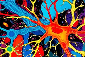
What are neurons?
What are neurons?
Specialized nerve cells that provide rapid communication
List the characteristics of neurons.
List the characteristics of neurons.
Great variation in size and shape, large cell body housing the nucleus, dendrites, and axons.
What is a synapse?
What is a synapse?
List the anatomy of the CNS.
List the anatomy of the CNS.
Describe paired cerebral hemispheres.
Describe paired cerebral hemispheres.
Describe the diencephalon.
Describe the diencephalon.
Describe the pons.
Describe the pons.
List the components of the brainstem.
List the components of the brainstem.
What is gray matter?
What is gray matter?
List the 4 types of neuroglia.
List the 4 types of neuroglia.
What is the function of oligodendrocytes?
What is the function of oligodendrocytes?
What is the function of astrocytes?
What is the function of astrocytes?
What is the function of microglia?
What is the function of microglia?
What is the function of ependymal cells?
What is the function of ependymal cells?
Describe glia limitans (astrocytes).
Describe glia limitans (astrocytes).
How many axons can an oligodendrocyte myelinate?
How many axons can an oligodendrocyte myelinate?
What does myelination do?
What does myelination do?
What is the choroid plexus?
What is the choroid plexus?
Describe the characteristics of CSF production and secretion.
Describe the characteristics of CSF production and secretion.
Do ependyma of choroid plexus rest on a basement membrane?
Do ependyma of choroid plexus rest on a basement membrane?
Describe the zona occludens of choroid plexus.
Describe the zona occludens of choroid plexus.
List the 3 meninges.
List the 3 meninges.
Describe the pia mater.
Describe the pia mater.
Describe the subarachnoid space.
Describe the subarachnoid space.
What is the perivascular space in the subarachnoid space?
What is the perivascular space in the subarachnoid space?
Describe the blood-brain barrier.
Describe the blood-brain barrier.
Describe the organization of the spinal cord.
Describe the organization of the spinal cord.
List the 4 histological regions of the spinal cord.
List the 4 histological regions of the spinal cord.
Describe the cervical features of the spinal cord.
Describe the cervical features of the spinal cord.
What are the subdivision fascicles of the dorsal columns?
What are the subdivision fascicles of the dorsal columns?
What does the medial fasciculus gracilis convey?
What does the medial fasciculus gracilis convey?
What does the lateral fasciculus cuneatus convey?
What does the lateral fasciculus cuneatus convey?
Describe the thoracic features of the spinal cord.
Describe the thoracic features of the spinal cord.
What are the ventral/anterior horns of thoracic spinal cord?
What are the ventral/anterior horns of thoracic spinal cord?
What are the dorsal/posterior horns of thoracic spinal cord?
What are the dorsal/posterior horns of thoracic spinal cord?
What are the lateral horns of thoracic spinal cord?
What are the lateral horns of thoracic spinal cord?
Describe the lumbar features of the spinal cord.
Describe the lumbar features of the spinal cord.
Where are sensory and descending nerve fibers found in lumbar region of spinal cord?
Where are sensory and descending nerve fibers found in lumbar region of spinal cord?
What are the dorsal columns in lumbar region of spinal cord?
What are the dorsal columns in lumbar region of spinal cord?
What are the ventrolateral sulci in lumbar region of spinal cord?
What are the ventrolateral sulci in lumbar region of spinal cord?
Where are nerve roots found in sacral region of spinal cord?
Where are nerve roots found in sacral region of spinal cord?
Describe the medulla oblongata.
Describe the medulla oblongata.
Describe the basal pons.
Describe the basal pons.
Describe the tegmental pons.
Describe the tegmental pons.
What does the cerebellum do?
What does the cerebellum do?
Flashcards
CNS components
CNS components
Brain and spinal cord
PNS components
PNS components
Nervous tissue outside the CNS; nerves connecting CNS to other tissues.
Neurons
Neurons
Specialized nerve cells for rapid communication.
Synapse
Synapse
Signup and view all the flashcards
CNS anatomy
CNS anatomy
Signup and view all the flashcards
Gray matter
Gray matter
Signup and view all the flashcards
White matter
White matter
Signup and view all the flashcards
Neuroglia types
Neuroglia types
Signup and view all the flashcards
Oligodendrocytes
Oligodendrocytes
Signup and view all the flashcards
Astrocytes function
Astrocytes function
Signup and view all the flashcards
Microglia
Microglia
Signup and view all the flashcards
Ependymal cells
Ependymal cells
Signup and view all the flashcards
Choroid plexus
Choroid plexus
Signup and view all the flashcards
Meninges
Meninges
Signup and view all the flashcards
Pia mater
Pia mater
Signup and view all the flashcards
Subarachnoid space
Subarachnoid space
Signup and view all the flashcards
Dura mater
Dura mater
Signup and view all the flashcards
Blood-brain barrier
Blood-brain barrier
Signup and view all the flashcards
What creates BBB?
What creates BBB?
Signup and view all the flashcards
Spinal Regions
Spinal Regions
Signup and view all the flashcards
Fasciculus Gracilis
Fasciculus Gracilis
Signup and view all the flashcards
Fasciculus Cuneatus
Fasciculus Cuneatus
Signup and view all the flashcards
Thoracic Lateral Horns
Thoracic Lateral Horns
Signup and view all the flashcards
Ventrolateral sulci of lumbar
Ventrolateral sulci of lumbar
Signup and view all the flashcards
Cerebellum Function
Cerebellum Function
Signup and view all the flashcards
Cerebellar cortex layers
Cerebellar cortex layers
Signup and view all the flashcards
Purkinje cell layer
Purkinje cell layer
Signup and view all the flashcards
Afferent Fibers
Afferent Fibers
Signup and view all the flashcards
Substantia Nigra
Substantia Nigra
Signup and view all the flashcards
Hippocampus
Hippocampus
Signup and view all the flashcards
Study Notes
- The central nervous system (CNS) consists of the brain and spinal cord.
- The peripheral nervous system (PNS) includes nervous tissue outside the CNS and nerves connecting the CNS to other body tissues.
- Neurons are specialized nerve cells that facilitate rapid communication.
- Neurons exhibit great variation in size and shape, feature a large cell body housing the nucleus, and possess dendrites and axons.
- A synapse facilitates communication between neurons through the release of neurotransmitters (NT).
- Myelination enables action potential (AP) transmission in nerve cells.
CNS Anatomy
- The CNS comprises paired cerebral hemispheres, the diencephalon, pons, brainstem, and spinal cord.
- Paired cerebral hemispheres are invested by the cerebral cortex and connected to other structures by the midbrain.
- The diencephalon contains large gray matter structures deep within the brain, including the basal ganglia and thalamus.
- The pons connects to the medulla and cerebellum via peduncles and consists of the basal pons and tegmental regions.
- The brainstem includes the pons, medulla, and midbrain.
- Gray matter consists of neuron cell bodies and their dendritic processes, with prominent nuclei in neurons.
- White matter consists of axons surrounded by a lipid-rich myelin sheath, giving it a white appearance, and prominent oligodendrocytes.
Neuroglia
- The four types of neuroglia are oligodendrocytes, astrocytes, microglia, and ependymal cells.
- Oligodendrocytes form myelin sheaths in the CNS.
- Astrocytes provide mechanical support, contribute to the blood-brain barrier, repair CNS tissue after damage, facilitate metabolite exchange, and contain bundles of intermediate filaments made of GFAP.
- Microglia constitute the CNS monocyte-macrophage system.
- Ependymal cells form the epithelium lining the ventricles and spinal cord.
- A gray matter slide shows neuropil (Np), neurons (N), oligodendrocytes (O), and astrocytes (A).
- Glia limitans, formed by astrocytes, is an impermeable barrier investing the space near the pia mater.
- An oligodendrocyte can myelinate up to 50 axons; however, one axon will need numerous cells because of the limited length of the myelin segments (internodes)
Myelination
- Myelination facilitates the action potential in nerve cells (oligodendrocytes in the CNS and Schwann cells in the PNS).
Choroid Plexus
- The choroid plexus is a vascular structure arising from the wall of the four ventricles and is responsible for CSF production.
- CSF drains from ventricular cavities via channels and the subarachnoid space.
- CSF is produced and reabsorbed at a constant rate and acts as a shock absorber.
- Sodium ions are involved in secretion from ependymal cells during CSF production.
- Each plexus contains a branching system of large blood vessels that run in the collagenous tissue.
- These are lined by cuboidal or columnar epithelium, featuring long bulbous microvilli from the luminal surface and abundant mitochondria.
- The ependyma of the choroid plexus does not rest on a basement membrane.
- The bases of cells taper and branch into fine processes that branch with underlying layer
- Zona occludens are tight junctions between epithelial cells contributing to CSF-BBB.
- Tight junctions are tightly bound at luminal surfaces and become incomplete with age.
Meninges
- The meninges consist of the pia mater, arachnoid mater, and dura mater.
- The pia mater (P) consists of collagen fibers, fine elastin fibers, and some fibroblasts, separated from underlying astrocytes by a basement membrane, and may be considered continuous with the arachnoid mater (A) as leptomeninges.
- The subarachnoid space (SS) houses meningeal vessels and CSF circulation, is connected to ventricles by 3 foramina in the 4th ventricle, is lined by flattened arachnoid cells, is loosely attached to the pia mater, and contains the periventricular space (PVS).
- The perivascular space in the subarachnoid space is the space between penetrating vessels and the pia mater.
- It is extremely narrow and its interstitial fluid drains outward to join the CSF.
- The dura mater is a dense fibroelastic layer with an internal surface lined by flat cells, closely applied but not attached to the arachnoid layer, has a potential space (subdural space if brain bleed), merges with the periosteum of the skull, and extends into brain spaces.
- The blood-brain barrier protects the brain from infective and toxic agents.
- Luminal surface membranes of the blood-brain barrier contain enzymes that destroy neurotoxic metabolites.
- Spinal cord exhibits a butterfly shape transversely and is invested in meninges with the dura being loosely attached.
Spinal Cord Regions
- The four histological regions of the spinal cord are cervical, thoracic, lumbar, and sacral.
- Cervical spinal cord features extensive gray matter, a deep ventral median fissure, a shallow dorsal midline sulcus, and each dorsal column is divided into two fascicles.
- The subdivision fascicles of the dorsal columns are the medial fasciculus gracilis and the lateral fasciculus cuneatus.
- The medial fasciculus gracilis conveys fibers from the lower limbs.
- The lateral fasciculus cuneatus conveys fibers from the upper limbs.
- A cervical features slide includes the deep ventral anterior median fissure (F), medial fasciculus gracilis (FG), lateral fasciculus cuneatus (FC), dorsolateral sulcus (S), and dorsal nerve root (R).
- Thoracic spinal cord features ventral/anterior horns, dorsal/posterior horns, and lateral horns.
- The ventral/anterior horns of thoracic spinal cord contain cell bodies of the large alpha lower motor neurons.
- The dorsal/posterior horns of thoracic spinal cord contain cell bodies of small second-order sensory neurons and relay info on pain and temperature.
- The lateral horns of thoracic spinal cord are small and contain the cell bodies of sympathetic nervous system efferent neurons.
- The thoracic features slide includes dorsal horns (D), lateral horns (L), and ventral horns (V).
- Lumbar spinal cord features a central canal in the central commissure in gray matter, dorsal columns, and ventrolateral sulci.
- Sensory and descending nerve fibers are found in the white matter of the lumbar region of the spinal cord.
- The dorsal columns in the lumbar region of the spinal cord are white matter between the dorsal horns.
- The ventrolateral sulci in the lumbar region of the spinal cord are the line of exit of the ventral nerve roots.
- A lumbar features slide shows the central commissure (C), descending dorsal columns (DC), and ventrolateral sulci (VS).
- Sacral spinal cord features nerve roots (NR) and dorsal root ganglion.
- Nerve roots are found adjacent to the spinal cord and start an oblique course in the subarachnoid space.
- The medulla oblongata is the most caudal part of the brainstem, features prominent ventral pyramids on each side, contains numerous ascending and descending tracts, and contains the 4th ventricle.
- The basal pons is a bulky ventral region consisting of crisscross bundles of longitudinal and transverse fibers.
- The tegmental pons is a smaller dorsal region.
- The cerebellum coordinates muscular activity and maintains posture and equilibrium.
Cerebellum Cortex Layers
- The three cortex layers of the cerebellum are the outer molecular layer (ML), Purkinje cell layer (PL), and inner granular cell layer (GL).
- The outer molecular layer of the cerebellum contains few neurons with many unmyelinated fibers.
- The Purkinje cell layer of the cerebellum is a single layer of huge neurons with a fine axon extending down through the inner granular layer and extensive branching dendrites in the outer molecular layer.
- The inner granular cell layer of the cerebellum is very cellular and basophilic.
- Afferent fibers in the cerebellum extend from the brainstem and pass via white matter to make connections with granular cells in the inner granular layer.
- Efferent fibers in the cerebellum are Purkinje cell axons that transverse the granular cell layer to synapse at central cerebellum.
- The substantia nigra is a large mass of gray matter extending throughout the midbrain and it allows for fine control of motor function.
- A substantia nigra slide shows multipolar neurons and cytoplasm containing neuromelanin pigment.
- The three pigmented areas in the brain are the substantia nigra, dorsal motor nucleus of the vagus, and locus coeruleus.
Cerebral Cortex Cell Types
- The characteristic cell types in the cerebral cortex are pyramidal cells, stellate (granule) cells, cells of Martinotti, fusiform cells, and horizontal cells of Cajal.
- Pyramidal cells feature pyramid-shaped cell bodies, a thick branching dendrite passing to the surface with fine dendritic branches, and an axon at the base passing into white matter.
- Stellate cells feature a star-shaped cell body, a small neuron with a short vertical axon, and several short branching neurons.
Cerebral Cortex Layers
- The six layers of the cerebral cortex are the plexiform (molecular) layer (I), outer granular layer (II), pyramidal cell layer (III), inner granular layer (IV), ganglionic layer (V), and multiform cell layer (VI).
- The efferent functions of the cortex give off branches that pass back into superficial layers and communicate with their own dendrites.
- The hippocampus is responsible for long-term memory and consists of Ammon's horn and the dentate gyrus.
- Ammon's horn is a coiled feature of the hippocampus that is inferomedial part of the temporal lobe and is divided into CA1-CA4 from outside to inside.
- The dentate gyrus is an irregular folded segment towards the middle/end.
- CA1 of the hippocampus is susceptible to sclerosis (epilepsy).
Studying That Suits You
Use AI to generate personalized quizzes and flashcards to suit your learning preferences.