Podcast
Questions and Answers
Explain how genetic defects can lead to cell injury, providing specific examples from the text.
Explain how genetic defects can lead to cell injury, providing specific examples from the text.
Genetic defects can cause cell injury by leading to a deficiency of functional proteins like enzymes. This can happen in cases of "inborn errors of metabolism," where a genetic mutation disrupts the production of a crucial enzyme needed for a specific metabolic pathway. For example, a deficiency in the enzyme phenylalanine hydroxylase, caused by a genetic defect, leads to phenylketonuria, a metabolic disorder causing mental retardation due to the accumulation of phenylalanine.
How does oxygen deprivation affect the cell and what are the consequences?
How does oxygen deprivation affect the cell and what are the consequences?
Oxygen deprivation, or hypoxia, disrupts aerobic oxidative respiration, the primary energy source for cells. This leads to a decrease in ATP production, causing various cellular dysfunctions. Failure of energy-dependent pumps leads to cell swelling, and prolonged hypoxia ultimately causes cell death.
Describe the difference between reversible and irreversible cell injury, highlighting the key morphological features of each.
Describe the difference between reversible and irreversible cell injury, highlighting the key morphological features of each.
Reversible cell injury is characterized by cellular swelling and fatty change, both of which represent adaptations of the cell to stress. In reversible injury, the cellular structures are still intact. Irreversible cell injury culminates in cell death, typically manifested by necrosis. Here, the cell membrane loses integrity, leading to leakage of cellular contents, and degradation of cellular components. Morphological features of necrosis include increased eosinophilia (pink staining) and nuclear changes such as pyknosis, karyorrhexis, and karyolysis.
Explain the concept of "cellular senescence" and its role in cell injury.
Explain the concept of "cellular senescence" and its role in cell injury.
Why is it important to understand the causes of cell injury?
Why is it important to understand the causes of cell injury?
How do immune reactions contribute to cell injury? Briefly explain the mechanism.
How do immune reactions contribute to cell injury? Briefly explain the mechanism.
What are the three patterns of nuclear changes seen in necrosis? Briefly describe each.
What are the three patterns of nuclear changes seen in necrosis? Briefly describe each.
Discuss the relevance of inflammation in the context of necrosis.
Discuss the relevance of inflammation in the context of necrosis.
Explain the concept of 'reversible cell injury' and provide two examples of cellular adaptations that could occur during this phase. Discuss the factors that determine whether a cell will undergo reversible injury or progress to irreversible injury.
Explain the concept of 'reversible cell injury' and provide two examples of cellular adaptations that could occur during this phase. Discuss the factors that determine whether a cell will undergo reversible injury or progress to irreversible injury.
Compare and contrast the two main pathways of programmed cell death: apoptosis and necrosis. Describe the morphological characteristics, biochemical features, and physiological roles of each pathway.
Compare and contrast the two main pathways of programmed cell death: apoptosis and necrosis. Describe the morphological characteristics, biochemical features, and physiological roles of each pathway.
Explain the role of reactive oxygen species (ROS) in cell injury. Describe the mechanisms by which ROS can damage cells and the cellular defense mechanisms against ROS.
Explain the role of reactive oxygen species (ROS) in cell injury. Describe the mechanisms by which ROS can damage cells and the cellular defense mechanisms against ROS.
Discuss the mechanisms by which ischemia can lead to cell injury and death. Explain the concept of 'ischemia-reperfusion injury' and its implications.
Discuss the mechanisms by which ischemia can lead to cell injury and death. Explain the concept of 'ischemia-reperfusion injury' and its implications.
Analyze the role of calcium in cell injury. Discuss how calcium dysregulation can contribute to various forms of cellular damage, including activation of enzymes, mitochondrial dysfunction, and apoptosis.
Analyze the role of calcium in cell injury. Discuss how calcium dysregulation can contribute to various forms of cellular damage, including activation of enzymes, mitochondrial dysfunction, and apoptosis.
Describe the morphological changes that occur to a cell nucleus during karyolysis. What is believed to be the underlying mechanism for this process?
Describe the morphological changes that occur to a cell nucleus during karyolysis. What is believed to be the underlying mechanism for this process?
Contrast the appearance of a cell undergoing pyknosis with one undergoing karyolysis. Explain the difference in terms of nuclear morphology.
Contrast the appearance of a cell undergoing pyknosis with one undergoing karyolysis. Explain the difference in terms of nuclear morphology.
Explain how the process of karyorrhexis contributes to the overall cellular demise during necrosis.
Explain how the process of karyorrhexis contributes to the overall cellular demise during necrosis.
Compare and contrast the characteristic features of coagulative necrosis and liquefactive necrosis. What is the key difference between these two types of necrosis in terms of tissue architecture?
Compare and contrast the characteristic features of coagulative necrosis and liquefactive necrosis. What is the key difference between these two types of necrosis in terms of tissue architecture?
Describe the likely microscopic appearance of a tissue undergoing coagulative necrosis. Include specific features relating to the cellular morphology and overall tissue structure.
Describe the likely microscopic appearance of a tissue undergoing coagulative necrosis. Include specific features relating to the cellular morphology and overall tissue structure.
Relate the involvement of leukocytes in the process of liquefactive necrosis. Explain how they contribute to the distinctive characteristics of this type of necrosis.
Relate the involvement of leukocytes in the process of liquefactive necrosis. Explain how they contribute to the distinctive characteristics of this type of necrosis.
Explain the significance of the granuloma formation in the context of caseous necrosis. What specific type of infection is typically associated with this type of necrosis?
Explain the significance of the granuloma formation in the context of caseous necrosis. What specific type of infection is typically associated with this type of necrosis?
Describe the macroscopic characteristics of caseous necrosis. How does the appearance of the necrotic tissue differ from coagulative or liquefactive necrosis?
Describe the macroscopic characteristics of caseous necrosis. How does the appearance of the necrotic tissue differ from coagulative or liquefactive necrosis?
Describe the distinguishing histological features of caseous necrosis, comparing it to fat necrosis. Discuss the relevance of this type of necrosis in tuberculosis infection.
Describe the distinguishing histological features of caseous necrosis, comparing it to fat necrosis. Discuss the relevance of this type of necrosis in tuberculosis infection.
Explain the mechanism of apoptosis, contrasting it with necrosis in terms of cellular morphology and inflammatory response. Why is apoptosis referred to as 'falling off'?
Explain the mechanism of apoptosis, contrasting it with necrosis in terms of cellular morphology and inflammatory response. Why is apoptosis referred to as 'falling off'?
List three physiological situations where apoptosis is a crucial process, providing a brief explanation for each.
List three physiological situations where apoptosis is a crucial process, providing a brief explanation for each.
Identify three pathological conditions that can trigger apoptosis. Explain the underlying mechanisms linking these conditions to apoptosis activation.
Identify three pathological conditions that can trigger apoptosis. Explain the underlying mechanisms linking these conditions to apoptosis activation.
Discuss the relevance of apoptosis in preventing excessive inflammatory response. Provide an example from the text to illustrate this concept.
Discuss the relevance of apoptosis in preventing excessive inflammatory response. Provide an example from the text to illustrate this concept.
Why is the presence of chalky-white areas on the cut surfaces of a pancreas a strong indicator of acute pancreatitis? Explain the underlying mechanism causing this appearance.
Why is the presence of chalky-white areas on the cut surfaces of a pancreas a strong indicator of acute pancreatitis? Explain the underlying mechanism causing this appearance.
Compare and contrast the appearance of necrotic fat cells with the appearance of a normal fat cell, highlighting the key changes associated with fat necrosis.
Compare and contrast the appearance of necrotic fat cells with the appearance of a normal fat cell, highlighting the key changes associated with fat necrosis.
Based on your knowledge of apoptosis, explain why it is an important mechanism for eliminating cells in organs undergoing involution. Using an example from the text, illustrate how apoptosis contributes to the process of involution in a specific organ.
Based on your knowledge of apoptosis, explain why it is an important mechanism for eliminating cells in organs undergoing involution. Using an example from the text, illustrate how apoptosis contributes to the process of involution in a specific organ.
Flashcards
Cell Injury
Cell Injury
A reversible or irreversible damage to a cell due to various stressors.
Cell Death
Cell Death
The irreversible loss of function and structure in a cell, leading to its elimination.
Reversible Injury
Reversible Injury
Type of cell injury that allows for recovery if the stress is removed.
Irreversible Injury
Irreversible Injury
Signup and view all the flashcards
Apoptosis
Apoptosis
Signup and view all the flashcards
Basophilia
Basophilia
Signup and view all the flashcards
Pyknosis
Pyknosis
Signup and view all the flashcards
Karyorrhexis
Karyorrhexis
Signup and view all the flashcards
Coagulative necrosis
Coagulative necrosis
Signup and view all the flashcards
Liquefactive necrosis
Liquefactive necrosis
Signup and view all the flashcards
Caseous necrosis
Caseous necrosis
Signup and view all the flashcards
Cerebral infarction
Cerebral infarction
Signup and view all the flashcards
Granuloma
Granuloma
Signup and view all the flashcards
Oxygen Deprivation
Oxygen Deprivation
Signup and view all the flashcards
Chemical Agents
Chemical Agents
Signup and view all the flashcards
Immunologic Reactions
Immunologic Reactions
Signup and view all the flashcards
Reversible Cell Injury
Reversible Cell Injury
Signup and view all the flashcards
Necrosis
Necrosis
Signup and view all the flashcards
Cytoplasmic Changes in Necrosis
Cytoplasmic Changes in Necrosis
Signup and view all the flashcards
Nuclear Changes in Necrosis
Nuclear Changes in Necrosis
Signup and view all the flashcards
Aging
Aging
Signup and view all the flashcards
Fat Necrosis
Fat Necrosis
Signup and view all the flashcards
Histological Feature of Fat Necrosis
Histological Feature of Fat Necrosis
Signup and view all the flashcards
Causes of Apoptosis
Causes of Apoptosis
Signup and view all the flashcards
Physiological Apoptosis Examples
Physiological Apoptosis Examples
Signup and view all the flashcards
Pathological Causes of Apoptosis
Pathological Causes of Apoptosis
Signup and view all the flashcards
Inflammatory Reaction in Apoptosis
Inflammatory Reaction in Apoptosis
Signup and view all the flashcards
Study Notes
Cell Injury and Cell Death
- Cell injury and death are categorized based on the severity and progression of the initial stimulus
- Normal cells (homeostasis) can adapt. Inability to adapt leads to cell injury.
- Reversible injury can lead to mild, transient effects. Severe, progressive injury results in cell death.
- Cell death can be through apoptosis or necrosis.
Causes of Cell Injury
- Causes range from significant physical trauma to a single gene defect creating a dysfunctional enzyme.
- One primary category is oxygen deprivation - this can stem from hypoxia or oxygen deficiency, leading to aerobic oxidative respiration interference.
Other Causes of Cell Injury
- Chemical Agents: Substances like glucose, salt, or even water, if absorbed in excess, can be injurious.
- Immunologic Reactions: Immune reactions can harm cells / tissues, even though the immune system protects the body from pathogens.
- Infectious Agents: Submicroscopic viruses to large parasites, bacteria, fungi, and protozoans are all agents able to harm cells / tissues.
- Genetic Factors: Genetic defects can cause injury through inadequacies in functional proteins, like enzymes (inborn errors of metabolism) and DNA damage.
- Nutritional Imbalances: Protein-calorie deficiency is a factor, especially in deprived populations.
- Physical Agents: Severe trauma, temperature extremes, radiation exposure, electric shocks, and sudden pressure changes are all examples.
- Aging: Cellular senescence modifies replicative and repair abilities, decreasing a cell's response to damage.
Reversible Cell Injury
- Two key morphological indicators are cellular swelling and fatty change
- Cellular swelling arises from energy-dependent ion pumps in the plasma membrane failing to maintain ionic and fluid homeostasis.
- Fatty change occurs in cases like hypoxic and toxic/metabolic injuries. Lipid vacuoles appear in the cytoplasm.
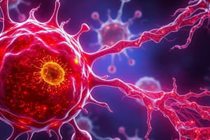
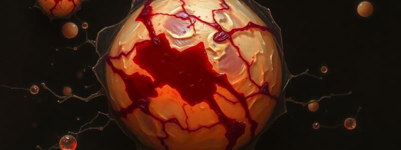
Irreversible Cell Injury (Death): Necrosis
- Necrosis is characterized by a loss of membrane integrity and leakage of cellular contents, resulting in cell dissolution.
- The released cellular products trigger a local inflammatory response.
Necrosis Morphology
- Affected cells demonstrate cytoplasmic changes
- Necrotic cells show increased eosinophilia (pink staining).
- Nuclei exhibit one of three patterns due to DNA / chromatin breakdown:
- Karyolysis: the fading of chromatin basophilia;
- Pyknosis: nuclear shrinkage, increased basophilia, and DNA condensing into a small mass.
- Karyorrhexis: the pyknotic nucleus fragments
Patterns of Tissue Necrosis
- Coagulative necrosis: Underlying tissue architecture is preserved for several days. Leukocytes break down dead cells via lysosomal enzymes; cellular debris are then removed via phagocytosis.
- Liquefactive necrosis: Tissue is digested by enzymes from leukocytes into a liquid viscous mass. Common in focal bacterial/fungal infections. Microbes stimulate inflammatory cells. Digested tissue is removed by phagocytes.
Additional Types of Necrosis
- Caseous necrosis: Often seen in tuberculous infection; characteristic of a friable, yellow-white, "cheese-like" necrotic area. Typically enclosed by a granuloma - a distinctive inflammatory border.
- Fat necrosis: Fat tissue destruction from activated pancreatic lipases. This often results from acute pancreatitis; histologically, necrotic fat cells appear with shadowy outlines and basophilic calcium deposits.
Apoptosis
- Apoptosis is a programmed cell death process where cells activate enzymes to break down components of their own cells (nuclear DNA and cytoplasmic proteins).
- Fragments detach (apoptosis - falling off)
- Does not create an inflammatory response in the host
Causes of Apoptosis
- Occurs in normal situations to eliminate excess cells to maintain a constant cell number in tissues and during development.
- Also occurs as a pathological process when cells are damaged beyond repair.
Apoptosis in Physiological Situations
- Programmed cell elimination is used in embryogenesis.
- Involution of hormone-dependent tissues (e.g., the uterus during the menstrual cycle).
- Elimination of useless cells (e.g. neutrophils in acute inflammation).
Apoptosis in Pathological Conditions
- Damaged from radiation, cytotoxic anticancer drugs, temperature extremes, and hypoxia.
- Cell injury associated with some infections, (e.g., viral infections)
- Occurs in cases of atrophy (e.g., duct obstruction, pancreas)
Studying That Suits You
Use AI to generate personalized quizzes and flashcards to suit your learning preferences.