Podcast
Questions and Answers
During which phase of mitosis does the nuclear envelope fragment, allowing microtubules to access the chromosomes?
During which phase of mitosis does the nuclear envelope fragment, allowing microtubules to access the chromosomes?
- Prophase
- Metaphase
- Prometaphase (correct)
- Telophase
What is the primary significance of 'crossing-over' during meiosis I?
What is the primary significance of 'crossing-over' during meiosis I?
- It produces identical daughter cells.
- It contributes to genetic variation among individuals. (correct)
- It repairs damaged DNA.
- It ensures the chromosome number remains constant.
How does spermatogenesis differ from oogenesis in terms of cytoplasm distribution during meiosis?
How does spermatogenesis differ from oogenesis in terms of cytoplasm distribution during meiosis?
- Both spermatogenesis and oogenesis result in unequal cytoplasm distribution.
- Spermatogenesis results in unequal cytoplasm distribution, while oogenesis results in equal distribution.
- Spermatogenesis results in equal cytoplasm distribution, while oogenesis results in unequal distribution. (correct)
- Both spermatogenesis and oogenesis result in equal cytoplasm distribution.
What crucial event is facilitated by the degradation of cohesin molecules during anaphase?
What crucial event is facilitated by the degradation of cohesin molecules during anaphase?
Why is it necessary for cells to divide into multiple smaller cells instead of remaining as one large cell?
Why is it necessary for cells to divide into multiple smaller cells instead of remaining as one large cell?
How does the chromosome number change during the spermatocyte phase of spermatogenesis?
How does the chromosome number change during the spermatocyte phase of spermatogenesis?
What marks the conclusion of prometaphase?
What marks the conclusion of prometaphase?
How does meiosis contribute to genetic diversity in sexually reproducing organisms?
How does meiosis contribute to genetic diversity in sexually reproducing organisms?
During oogenesis, at which stage does the primary oocyte arrest until ovulation?
During oogenesis, at which stage does the primary oocyte arrest until ovulation?
What is the immediate result of a spermatogonium undergoing mitosis?
What is the immediate result of a spermatogonium undergoing mitosis?
Flashcards
Cell Division
Cell Division
Cells grow and divide to create two new cells, crucial for both unicellular and multicellular organisms.
Reasons for Cell Division
Reasons for Cell Division
Critical reasons for cell division are growth, repair, and reproduction aiding organism's survival.
Mitosis Definition
Mitosis Definition
Mitosis produces new somatic cells (2n), replacing old, lost, or damaged cells via nuclear division and cytokinesis.
Prophase
Prophase
Signup and view all the flashcards
Prometaphase
Prometaphase
Signup and view all the flashcards
Metaphase
Metaphase
Signup and view all the flashcards
Anaphase
Anaphase
Signup and view all the flashcards
Telophase
Telophase
Signup and view all the flashcards
Cytokinesis
Cytokinesis
Signup and view all the flashcards
Meiosis
Meiosis
Signup and view all the flashcards
Study Notes
- All cells grow then divide to produce two new cells in both unicellular and multicellular organisms.
- Cells grow by adding materials to their cell membranes and cell walls.
- Eukaryotic cells divide by mitosis or meiosis, where prokaryotic cells divide by fission.
- Cells divide because they absorb and release nutrients through their membrane, but The larger the cell, the harder it is to get rid of all the waste that is produced.
- Additional reasons for cell division are growth, repair, and reproduction
- Mitosis results in growth.
- Humans start as one cell then increase to over 10 trillion cells as adults.
- Mitosis results in repair.
- The skin is entirely replaced every 28 days.
- Reproduction is a result of either mitosis or meiosis.
- Asexual reproduction: involves one parent, resulting in genetically identical offspring, occurs in bacteria, protists, fungi, some plants, and some animals
- Sexual reproduction: involves two parents, resulting in offspring with a combination of both parents' DNA.
Gametogenesis
- Formation of male and female gametes in the gonads (ovary and testis) is gametogenesis.
- Spermatogenesis: the production of sperm in the male testes.
- Oogenesis depends on mitosis and meiosis: the production of ova within the ovaries.
Mitosis
- A fundamental process for life, that is important in the development of embryos as well as growth and development
- Produces new somatic cells (2n), and replaces cells that are old, lost, or damaged.
- It includes nuclear division and cytokinesis.
- Before mitosis, chromosomes are copied, then they coil up so each chromosome looks like a letter X in the nucleus of the cell.
- Chromosomes consist of two sister chromatids held together by a protein complex called cohesin.
- A cell duplicates all of its contents, including its chromosomes then splits to form two identical daughter cells. Mitosis Involves Five phases, based on the physical state of the chromosomes and spindle:
- Prophase
- Prometaphase
- Metaphase
- Anaphase
- Telophase
- Cytokinesis is the final physical cell division that follows telophase, and is sometimes considered a sixth phase of mitosis.
Prophase
- Mitosis begins with prophase, where sister chromatids condense to a visible form.
- Spindle fibers start to form, extending from the centrioles.
- Microtubules make up the spindle fibers and attach to the kinetochore of the sister chromatids.
- Centrioles migrate to opposite sides of the cell. The microtubules begin to polymerize from the duplicated centrosomes.
Prometaphase
- Begins with the fragmentation of the nuclear envelope: essential step for spindle assembly
- Microtubules of the developing spindle do not have access to the chromosomes until the nuclear membrane breaks apart when the centrosomes are located outside the nucleus in animal cells.
- Microtubules disassemble and assemble rapidly as they grow out of the centrosomes, finding attachment sites at kinetochores.
- Chromosomes are pulled and tugged in opposite directions by microtubules growing out from both poles of the spindle until the pole-directed forces are balanced.
- Sister chromatids do not break apart during this since they are firmly attached by the cohesin remaining at their centromeres.
- At the end chromosomes have a bi-orientation and the kinetochores on sister chromatids are connected by microtubules to opposite poles of the spindle.
- (bipolar attachment).
Metaphase
- The stage where chromosomes can be most easily visualized.
- Chromosomes are lined up along the metaphase (equatorial) plate, an imaginary line in the center of the cell.
- The chromosomes are moved here with the help of the spindle fibers and the centrioles.
Anaphase
- The kinetochores of each chromosome are pulled apart by the spindle fibers: Separating the sister chromatids, creating two daughter chromosomes.
- Sister chromatid separation is the degradation of the cohesin molecules by a protease called “separase”.
- Following sister chromatid separation, one of the sister chromatids is pulled to one side of the cell, while the other is pulled to the opposite pole.
- This ensures that the soon-to-be daughter cells will each have full, identical sets of chromosomes,.
Telophase and Cytokinesis
- Chromosomes reach the poles in telophase.
- A new nucleus begins to form around the new sets of chromosomes at each end of the cell.
- The chromosomes unravel back into their loose form.
- Spindle fibers disassemble.
- The cell is “pinched” into two new cells.
- in cytokinesis the cell's cytoplasm divides into two, making two new cells called daughter cells, each receive one of the new nuclei, making them genetically identical to the parent cell.
- In animal cells, the cytoplasm “pinches” the cell into two daughter cells, right down the center until there are two separate daughter cells.
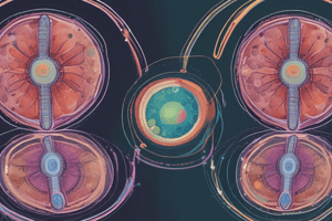
Meiosis
- Results in four gamete cells (sperm and egg).
- Needed for Sexual reproduction Offspring having a combination of DNA from both parents.
- Reduces the number of chromosomes in half which is important because when the two parent cells combine (fertilization) the offspring will end up with the same number of chromosomes as the parents.
- Humans have 46 chromosomes (23 pairs) in every body cell: Referred to as the diploid (2n) number of chromosomes.
- Sex cells, such as egg and sperm have one set of chromosomes: referred to as haploid (n) cells, and in humans this means they have 23 chromosomes total.
- Haploid cells or reproductive cells, are called gametes or sex cells. Mitosis- process which produces diploid cells, occurs in all somatic, or body cells
- Meiosis is the formation of gametes, performed by reproductive cells only resulting in a reduction of the chromosome number, forming haploid cells (n).
- Meiosis produces 4 daughter cells with half the number of chromosomes that are not identical to each other. This maintains chromosome number constant over generations, in the adult organism: When the egg (n) and the sperm (n) combine (n + n) in fertilization, the offspring is a diploid cell (2n).
- When chromosomes pair, in preparation for meiosis, they arrange with their homologous pair, also known as homologs.
- Humans have 46 chromosomes (23 pairs), each of the chromosomes in the pair are exactly alike in size, gene location and location of the centromere. The exception is the sex chromosomes. Females have a pair called XX where males are XY.
- Meiosis is how cells change from a diploid cell to a haploid cell, and in the process the homologous pairs of chromosomes are separated and end up in 4 daughter cells in two main stages: meiosis I and meiosis II.
- Pairs separate during meiosis I, and meiosis II is when the sister chromatids separate, much like in mitosis.
Meiosis I
- Meiosis I is a 4-step process, like mitosis, the cells have gone through the G1, S and G2 phases, thus the DNA has been replicated and are in the form of sister chromatids, connected at the centromere.
- The homologous chromosomes pair up.
Prophase I
- Chromosomes thicken and are visible under a microscope.
- Homologous pairs of chromosomes are tangled together and begin to move toward the equatorial plate.
- Synapsis is the side-by-side lining up of the homologous chromosomes.
- Instead of two sister chromatids like in mitosis, there are now 4 sister chromatids that move together, called a tetrad.
- Nuclear envelope disappears and spindle fibers (spindles) begin to form.
- Crossing-over can occur: homologous pairs of chromosomes are tangled together, many times, they “swap” parts of their chromosomes. This is a major source of variation between individuals.
Metaphase I
- Homologous pairs are lined up next to each other, along the equatorial plate.
Anaphase I
- Homologous pairs are separated due to the spindle fibers pulling them apart from the centromere.
- Each chromosome still has two sister chromatids.
Telophase I and Cytokinesis
- Two new cells result, each with the haploid number of chromosomes, which are still in the form of two sister chromatids during cytokinesis.
Meiosis II
- Follows after telophase I and cytokinesis.
- Daughter cells from meiosis I are what go into this phase.
- Cells divide again, but similar to mitosis rather than replication between meiosis I and meiosis II.
- There are n number of chromosomes rather than 2n.
- Four daughter cells produced.
- Follows four steps: prophase II, metaphase II, anaphase II and telophase II, followed by the final cytokinesis after two daughter cells produced in meiosis I, and each of them divide again.
- The result is 4 daughter cells, each with n number of chromosomes, which are not identical to the original parent cell or each other.
Cytoplasm Distribution Differs in Meiosis, Based on Sex of Organism
- Male end result is four spermatids, which differ in genetic information.
- Female: ovum receives an unequal share of the cytoplasm and remains functional while the polar bodies are pockets of genetic information with minimal cytoplasm and therefore non-functional.-
- Timing of meiosis is also different based on sex.
Spermatogenesis: Sperm Maturation
- Occurs continuously from puberty: spermatogonia divide into spermatozoa in the testes.
- Spermatozoa are formed in the seminiferous tubules of the testis, from basic cells called spermatogonia, which have been dormant in the tubules since the fetal period.
- Spermatozoa Production is continuous from puberty until death.
- Transformation of an immature germ cell or spermatogonium into a mature sperm takes about 64 days, producing ~300 million sperm cells per day. Spermatogenesis occurs in three phases
- Spermatogonial phase (mitosis)
- Spermatocyte phase (meiosis)
- Spermatid phase (spermiogenesis or sperm maturation).
Spermatogonial Phase:Mitotic Division
- Each spermatogonium (germ stem cell) gives rise to many spermatogonia by mitosis
- Chromosome number conserved at 46 chromosomes.
- Spermatogonia will then grow and undergo mitosis to form primary spermatocytes.
- Primary spermatocytes have 46 chromosomes.
Spermatocyte Phase: Meiotic Division
- Primary spermatocytes divide by meiosis I to reduce chromosome number and form 2 haploid secondary spermatocytes.
- Each secondary spermatocyte has 23 chromosomes.
- Secondary spermatocytes then divide by meiosis II (equational meiosis) to give rise to two spermatids.
- Each spermatid will have 23 chromosomes
Spermiogenesis Phase:Maturation
- Spermatids mature/differentiate to produce sperm
- Spermatid only changes in shape and does not divide to produce a sperm, therefore number of chromosomes will stay the same as in the spermatid
- Sperm will have 23 chromosomes.
Oogenesis: Oocyte Maturation
- Females do not make millions of ova since a female does not carry millions of fetuses. No more than one ovum is produced per month, therefore females do not have constant mitosis of their germ cells.
Prenatal Maturation
- Oogenesis begins during fetal life. During the first trimester of pregnancy the oogonia (primordial germ cells) of the female fetus divides continuously, mitotically.
- Each oogonium has 46 chromosomes, generating a population of ~7 million.
- These oogonia begin the first meiotic division but stop in prophase I of meiosis I as a primary oocyte.
- Long duration of the meiotic division may account for the high frequency of meiotic errors such as nondisjunction.
- Ovarian stromal cells surround the developing primary oocyte to form a single layer of flattened follicular cells with the primary oocyte and its follicular cells constitute the primordial follicle.
- Prior to birth there is a huge reduction in primary oocytes to only 2 million. No primary oocytes form after birth in contrast to the continuous production of primary spermatocytes in the male after puberty.
Postnatal Maturation
- Primary oocytes stay dormant in the ovaries until puberty.
- Primary oocyte increases in size with a membrane, the zona pellucida, forms around it as the follicle matures.
- Primary oocyte completes the first meiotic division just before ovulation, but is not equal, unlike males: the secondary oocyte gets almost all the cytoplasm.
- The first polar body receives little cytoplasm and is a small, nonfunctional cell that degenerates.
- At ovulation, the secondary oocyte begins the second meiotic division progressing only to metaphase II, then division arrests.
- If fertilization occurs, the second meiotic division is completed, and the ovum retains most of the cytoplasm, whereas the second polar body is small and degenerates.
- Secondary oocyte released at ovulation is surrounded by the zona pellucida and a follicular cell layer, the corona radiata.
- About 2 million primary oocytes are found in the ovaries of a newborn female, and by puberty, ~ 30-40 thousand remain.
- Only about 200-400 reach full maturity after puberty and expelled at ovulation.
Studying That Suits You
Use AI to generate personalized quizzes and flashcards to suit your learning preferences.