Podcast
Questions and Answers
What causes fat necrosis in adipose tissue?
What causes fat necrosis in adipose tissue?
- Enzyme action of lipases (correct)
- Bacterial infection
- Cellular apoptosis
- Oxygen deprivation
What is indicated by the presence of chalky white areas in fat necrosis?
What is indicated by the presence of chalky white areas in fat necrosis?
- Chronic infection
- Fat saponification (correct)
- Cellular apoptosis
- Vascular inflammation
Which condition is typically associated with fibrinoid necrosis?
Which condition is typically associated with fibrinoid necrosis?
- Chronic obstructive pulmonary disease
- Osteoarthritis
- Malignant hypertension (correct)
- Diabetes mellitus
Which feature distinguishes apoptosis from necrosis?
Which feature distinguishes apoptosis from necrosis?
How does physiological apoptosis function in tissue maintenance?
How does physiological apoptosis function in tissue maintenance?
What is a characteristic of pathological apoptosis?
What is a characteristic of pathological apoptosis?
What is a common example of physiological apoptosis during hormonal changes?
What is a common example of physiological apoptosis during hormonal changes?
Which microscopic feature is associated with fibrinoid necrosis?
Which microscopic feature is associated with fibrinoid necrosis?
What is the primary characteristic of necrosis?
What is the primary characteristic of necrosis?
Which of the following is NOT a type of necrosis?
Which of the following is NOT a type of necrosis?
What is a hallmark feature of coagulative necrosis?
What is a hallmark feature of coagulative necrosis?
What morphological feature is associated with necrotic cells?
What morphological feature is associated with necrotic cells?
Which of the following is a cause of necrosis?
Which of the following is a cause of necrosis?
During necrosis, what happens to the nuclear structure?
During necrosis, what happens to the nuclear structure?
What defines apoptosis as a process of cell death?
What defines apoptosis as a process of cell death?
Which type of necrosis is most common and occurs due to ischemia?
Which type of necrosis is most common and occurs due to ischemia?
What is a characteristic feature of coagulative necrosis in the kidney?
What is a characteristic feature of coagulative necrosis in the kidney?
In liquefactive necrosis, which of the following is most commonly seen?
In liquefactive necrosis, which of the following is most commonly seen?
Which type of necrosis is associated with a 'cottage cheese-like' appearance grossly?
Which type of necrosis is associated with a 'cottage cheese-like' appearance grossly?
What type of cellular changes are observed in necrotic cells during coagulative necrosis?
What type of cellular changes are observed in necrotic cells during coagulative necrosis?
Which cellular component predominantly infiltrates areas of necrosis during liquefactive necrosis?
Which cellular component predominantly infiltrates areas of necrosis during liquefactive necrosis?
What histological feature is commonly associated with caseous necrosis?
What histological feature is commonly associated with caseous necrosis?
What often results from bacterial infections leading to liquefactive necrosis?
What often results from bacterial infections leading to liquefactive necrosis?
What is typically observed microscopically in coagulative necrosis?
What is typically observed microscopically in coagulative necrosis?
What is a characteristic feature of apoptosis?
What is a characteristic feature of apoptosis?
Which protein is considered pro-apoptotic?
Which protein is considered pro-apoptotic?
What typically triggers apoptosis related to the accumulation of misfolded proteins?
What typically triggers apoptosis related to the accumulation of misfolded proteins?
Which of the following differences pertains to necrosis compared to apoptosis?
Which of the following differences pertains to necrosis compared to apoptosis?
Which enzymes are primarily activated during apoptosis?
Which enzymes are primarily activated during apoptosis?
In apoptosis, what happens to the nucleus?
In apoptosis, what happens to the nucleus?
What type of response occurs during necrosis?
What type of response occurs during necrosis?
What is the fate of apoptotic bodies?
What is the fate of apoptotic bodies?
Flashcards are hidden until you start studying
Study Notes
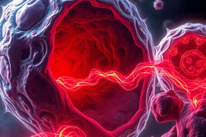
Cell Death: Necrosis and Apoptosis
- Necrosis is caused by irreversible cell injury leading to enzymatic digestion of dead cellular components.
- Apoptosis is programmed cell death and is a vital process eliminating unwanted cells through an internally programmed series of events.
Types of Necrosis
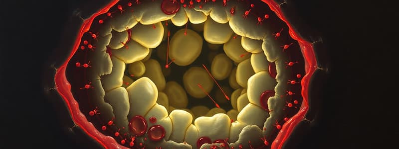
- Coagulative Necrosis is the most common type of necrosis, usually results from ischemia (loss of blood supply).
- It is most commonly seen in solid organs, such as heart, kidneys, and spleen.
- Grossly: Tissue is firm, often wedge-shaped, and pale.
- Microscopically: The outlines of the cells are retained, but cytoplasmic and nuclear details are lost.
- Liquefactive Necrosis results from enzymatic lysis of cells and proteins.
- Mainly due to the action of proteolytic enzymes released from targeted cells, liquefying the tissue.
- Most often seen in brain infarction and localized bacterial infections (abscesses).
- Grossly: Tissue is liquidy and creamy yellow due to pus formation.
- Microscopically: Lots of neutrophils and cell debris.
- Caseous Necrosis is characterized by a "cottage cheese-like" appearance.
- Seen in tuberculosis lesions and fungal infections.
- Grossly: Soft and friable necrotic tissue.
- Microscopically: Loss of cell outlines, nuclei, and acellular pink areas of necrosis with cellular debris surrounded by a granulomatous inflammatory process.
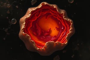
- Fat Necrosis occurs specifically in adipose tissue due to the action of lipases.
- This generates chalky white areas called fat saponification due to the deposition of calcium.
- Grossly: Chalky, white areas.
- Microscopically: Shadowy outlines of dead fat cells, surrounded by basophilic calcium deposits and an inflammatory reaction.
- Fibrinoid Necrosis is a specific type of necrosis seen in immune reactions involving blood vessels.
- Immune complexes and fibrin get deposited in vessel walls.
- Grossly: Changes are usually too small to see grossly.
- Microscopically: Vessel walls are thickened and pinkish-red.
- Seen in malignant hypertension and vasculitis.
Apoptosis
- Apoptosis is a programmed cell death where the cell membrane remains intact.
- It is an energy-dependent process eliminating unwanted cells without an inflammatory response.
- It is characterized by protein cleavage within the cell, causing cellular death.
Physiological Apoptosis
- It serves to eliminate cells that are no longer needed, maintaining a constant number of cell populations in tissue.
- Examples: Removal of excess cells during development, Involution of hormone-dependent tissues on hormone withdrawal, Homeostatic mechanism maintaining cell populations.
Pathological Apoptosis
- It eliminates cells injured beyond repair, preventing collateral tissue damage.
- Examples: DNA damage, Accumulation of misfolded proteins, Certain infections, particularly viral infections, Pathologic atrophy in parenchymal organs after duct obstruction.
Morphological Features of Apoptosis
- Involves single cells or small clusters of cells.
- Cell shrinkage.
- Membrane blebbing.
- Chromatin condensation.
- Nucleus condenses and fragments in an organized manner.
- Formation of apoptotic bodies.
- Phagocytosis of apoptotic bodies, usually by macrophages.
- No inflammatory cells.
Apoptosis Pathways
- Pro-apoptotic: Cytochrome c
- Anti-apoptotic: Bcl-2
- Activation of caspases brings about apoptosis.
- Caspases activate proteases and endonucleases that breakdown the DNA.
- Caspase-3 is an important final enzyme.
Differences and Features of Necrosis and Apoptosis
| Feature | Necrosis | Apoptosis |
|---|---|---|
| Induction | Pathological conditions | Pathological or Physiological conditions |
| Number of cells | Group of cells | Single Cells |
| Plasma Membrane | Loss of membrane integrity | Membrane remains intact |
| Cell Size | Enlarged (swelling) & lysis | Reduced (shrinkage) |
| Nucleus | Pyknosis → karyorrhexis → karyolysis | Fragmentation → formation of apoptotic bodies |
| Inflammation | Inflammatory response | NO inflammatory response |
| Fate of Cells | Phagocytosed by neutrophils and macrophages | Phagocytosed by neighbouring cells |
| Biochemical Mechanism | Energy-independent | Energy-dependent |
Studying That Suits You
Use AI to generate personalized quizzes and flashcards to suit your learning preferences.