Podcast
Questions and Answers
How do cells in direct contact primarily communicate with each other?
How do cells in direct contact primarily communicate with each other?
- Paracrine secretions
- Gap junctions (correct)
- Autocrine secretions
- Endocrine secretions
Which type of secretion affects distant cells through the bloodstream?
Which type of secretion affects distant cells through the bloodstream?
- Synaptic secretion
- Endocrine secretion (correct)
- Autocrine secretion
- Paracrine secretion
What is the primary function of connexons in gap junctions?
What is the primary function of connexons in gap junctions?
- To regulate DNA transcription
- To form channels between cells (correct)
- To synthesize proteins
- To degrade cellular waste
Which of the following substances can typically pass through gap junctions?
Which of the following substances can typically pass through gap junctions?
What is the role of neurotransmitters in cell communication?
What is the role of neurotransmitters in cell communication?
What determines whether a molecule acts as a membrane-permeable or membrane-impermeable signal?
What determines whether a molecule acts as a membrane-permeable or membrane-impermeable signal?
How do cytoplasmic effectors differ from nuclear effectors in signal transduction?
How do cytoplasmic effectors differ from nuclear effectors in signal transduction?
Which signal transduction pathway involves multiple receptors sharing common signaling proteins?
Which signal transduction pathway involves multiple receptors sharing common signaling proteins?
What is the primary characteristic of membrane-permeable ligands?
What is the primary characteristic of membrane-permeable ligands?
How do G-protein coupled receptors (GPCRs) initiate a signaling cascade?
How do G-protein coupled receptors (GPCRs) initiate a signaling cascade?
What is the primary function of ion channel receptors?
What is the primary function of ion channel receptors?
How does guanylate cyclase initiate cellular processes?
How does guanylate cyclase initiate cellular processes?
What is the key characteristic which defines protein kinase receptors?
What is the key characteristic which defines protein kinase receptors?
What process activates protein tyrosine kinase receptors (RTKs) after ligand binding?
What process activates protein tyrosine kinase receptors (RTKs) after ligand binding?
How do transmembrane scaffolds regulate signal transduction?
How do transmembrane scaffolds regulate signal transduction?
How do nuclear receptors control gene expression?
How do nuclear receptors control gene expression?
What is the role of G-proteins in signal transduction?
What is the role of G-proteins in signal transduction?
How do heterotrimeric G-proteins become activated?
How do heterotrimeric G-proteins become activated?
What is the general function of protein kinases in signal transduction?
What is the general function of protein kinases in signal transduction?
How does calmodulin mediate downstream events in response to calcium?
How does calmodulin mediate downstream events in response to calcium?
What role does adenylyl cyclase play in signal transduction?
What role does adenylyl cyclase play in signal transduction?
How do lipid kinases contribute to signal transduction?
How do lipid kinases contribute to signal transduction?
What is the function of adaptor proteins in signaling networks?
What is the function of adaptor proteins in signaling networks?
How are second messengers like cAMP and cGMP regulated within the cell?
How are second messengers like cAMP and cGMP regulated within the cell?
What is the direct effect of cAMP binding to protein kinase A (PKA)?
What is the direct effect of cAMP binding to protein kinase A (PKA)?
How does IP3 contribute to the phospholipid kinase signaling cascade?
How does IP3 contribute to the phospholipid kinase signaling cascade?
What is the immediate consequence of FGFs binding to FGFRs?
What is the immediate consequence of FGFs binding to FGFRs?
What is the role of lysosomes in handling dangerous cellular cargo?
What is the role of lysosomes in handling dangerous cellular cargo?
How are enzymes targeted to the lysosome for protein degradation?
How are enzymes targeted to the lysosome for protein degradation?
What prevents the lysosome from digesting itself?
What prevents the lysosome from digesting itself?
How are cytosolic proteins marked for degradation by the proteasome?
How are cytosolic proteins marked for degradation by the proteasome?
What is the primary function of peroxisomes in the cell?
What is the primary function of peroxisomes in the cell?
What triggers the intrinsic pathway of apoptosis?
What triggers the intrinsic pathway of apoptosis?
What visible change indicates that a cell is undergoing apoptosis?
What visible change indicates that a cell is undergoing apoptosis?
How are caspases activated during apoptosis?
How are caspases activated during apoptosis?
How is an apoptotic cell removed from the surrounding tissue?
How is an apoptotic cell removed from the surrounding tissue?
What is a key characteristic of necrosis that distinguishes it from apoptosis?
What is a key characteristic of necrosis that distinguishes it from apoptosis?
What causes necrosis?
What causes necrosis?
Flashcards
Extracellular Communication
Extracellular Communication
Communication that occurs when a signal is received from outside the cell.
Intracellular Communication
Intracellular Communication
Communication that occurs when external signals cause changes within a cell.
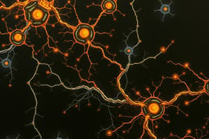
Gap Junctions
Gap Junctions
Intercellular connections directly linking the cytoplasm of adjacent cells.
Autocrine Secretions
Autocrine Secretions
Signup and view all the flashcards
Paracrine Secretions
Paracrine Secretions
Signup and view all the flashcards
Endocrine Secretions
Endocrine Secretions
Signup and view all the flashcards
Neurotransmitters
Neurotransmitters
Signup and view all the flashcards
Signal Transduction
Signal Transduction
Signup and view all the flashcards
Second Messengers
Second Messengers
Signup and view all the flashcards
Ligand
Ligand
Signup and view all the flashcards
G-Protein Coupled Receptors (GPCR)
G-Protein Coupled Receptors (GPCR)
Signup and view all the flashcards
Ion Channel Receptors
Ion Channel Receptors
Signup and view all the flashcards
Guanylate Cyclase Receptors
Guanylate Cyclase Receptors
Signup and view all the flashcards
Protein Kinase Receptors
Protein Kinase Receptors
Signup and view all the flashcards
Transmembrane Scaffolds
Transmembrane Scaffolds
Signup and view all the flashcards
Nuclear Receptors
Nuclear Receptors
Signup and view all the flashcards
G-Proteins
G-Proteins
Signup and view all the flashcards
Protein Kinases
Protein Kinases
Signup and view all the flashcards
Calcium-Binding Proteins
Calcium-Binding Proteins
Signup and view all the flashcards
Adenylyl Cyclase
Adenylyl Cyclase
Signup and view all the flashcards
Lipid Kinases
Lipid Kinases
Signup and view all the flashcards
Adaptor Proteins
Adaptor Proteins
Signup and view all the flashcards
Phosphodiesterases
Phosphodiesterases
Signup and view all the flashcards
Second Messengers
Second Messengers
Signup and view all the flashcards
Lysosomes
Lysosomes
Signup and view all the flashcards
Proteasomes
Proteasomes
Signup and view all the flashcards
Peroxisomes
Peroxisomes
Signup and view all the flashcards
Proteases
Proteases
Signup and view all the flashcards
Polyubiquitin Chain
Polyubiquitin Chain
Signup and view all the flashcards
Apoptosis
Apoptosis
Signup and view all the flashcards
Death Receptor
Death Receptor
Signup and view all the flashcards
Caspases
Caspases
Signup and view all the flashcards
Engulfment
Engulfment
Signup and view all the flashcards
Necrosis
Necrosis
Signup and view all the flashcards
Cell Lysis
Cell Lysis
Signup and view all the flashcards
Study Notes
Extracellular Communication
- Occurs when a signal is received from outside the cell.
- Cells can communicate at variable distances, either through direct contact via gap junctions or through secretions when not touching.
Intracellular Communication
- External signals lead to changes within a cell.
Direct Cell-to-Cell Communication: Gap Junctions
- Communication method for cells in direct contact.
- Connexons form channels between cells, allowing chemical signals to pass directly.
- Only small particles like ions and small signaling molecules can pass; proteins and carbohydrates are too large.
- Excitable cells, such as those in cardiac muscle, can pass electrical signals through gap junctions.
- Gap junctions are regulated and can open/close, potentially as a defense mechanism.
Cell-to-Cell Communication: Secretions
- Autocrine: Substances released affect the same cell.
- Paracrine: Substances released affect nearby cells.
- Endocrine: Substances released affect distant cells.
- Neurotransmitters: Substances released from a nerve terminal into the synapse.
Secretions: Neurotransmitters
- Occur where a nerve cell axon terminates on a target cell.
- Neurotransmitters are released into the synapse upon an excitatory signal and can bind to receptors on the target cell, be taken back by the original nerve cell, or be degraded.
- Secretions must interact with a receptor to affect the target cell.
Components of a Signaling Pathway
- Intracellular communication converts extracellular information into a cellular response via signal transduction.
- Consists of a signal (membrane permeable or impermeable), receptors, signaling proteins, and second messengers.
Structure of a Signaling Pathway
- Membrane-permeable signals bind to receptor proteins in the cytosol.
- Membrane-impermeable signals bind to transmembrane cell surface receptor proteins, activating second messengers.
- Signaling proteins and second messengers amplify and distribute signals.
- Cytoplasmic effectors lead to fast, short-lived responses.
- Nuclear effectors (transcription factors) control gene expression, resulting in slower, prolonged responses.
Signal Transduction
- Linear: One receptor interacts with one signaling protein or second messenger.
- Convergent: Several receptors share common signaling proteins or second messengers.
- Divergent: A single receptor interacts with multiple signaling proteins or second messengers.
- Multi-Branched: Combination of convergence and divergence.
Signals (Ligands)
- Originate from the extracellular space and must bind to a receptor.
- Membrane-impermeable ligands bind to proteins on the cell surface.
- Membrane-permeable ligands (primarily steroids) can interact with cytosolic receptors.
- Physical signals include pressure, temperature, and light.
Receptors Overview
- Often found on the plasma membrane but can also be found in the cytoplasm of the cell.
- Include G-Protein coupled receptors (GPCR), Ion Channels, Guanylate cyclase, Protein kinase receptors, Transmembrane scaffolds, and Nuclear receptors.
Receptors: G-Protein Coupled Receptors (GPCR)
- Structure: Seven transmembrane domains (H1 to H7) and heterotrimeric G-protein with alpha, beta, and gamma subunits.
- Function: Ligand binding causes a conformational change leading to G-protein activation.
Receptors: Ion Channel Receptors
- Also known as ‘ligand-gated channels’ in the plasma membrane.
- Transmit signal information by permitting ions to flow across the membrane.
- Ligand binding opens pores for ion flow through conformational change.
- Responsible for voluntary muscle contraction and common in nerve cell communication.
Receptors: Guanylate Cyclase
- Found bound to the membrane and soluble within the cytosol.
- Structure: Externalized ligand binding domain, transmembrane domain, and internal catalytic domain.
- Function: Membrane-bound guanylate cyclase converts GTP to cGMP upon activation, while the soluble form mediates some intracellular processes.
- Plays an important role in vision, converting light signals into electrical signals in the eye.
Receptors: Protein Kinase
- General action: Phosphorylate other proteins on serine, threonine, or tyrosine residues.
- Dysfunction is associated with cancer development.
- Two classes: RTK (phosphorylates tyrosine) and S/TKR (phosphorylates serine/threonine).
Protein Tyrosine Kinase Receptor Ligand Binding
- Inactive receptors are separate polypeptides with inactive tyrosine kinase domains.
- Ligand binding causes dimerization, activating the kinase.
- Transaurophosphorylation: One subunit phosphorylates the tyrosine amino acids on the other.
- Resulting phosphotyrosine amino acids are binding sites for additional signaling proteins.
- Ligand release and dephosphorylation by phosphoprotein phosphatases reset the kinase to its inactive state.
Receptors: Transmembrane Scaffolds
- Form large clusters to regulate signal transduction. Functions:
- Bring signaling proteins together.
- Regulate signal transduction.
- Localize signaling proteins to specific cellular areas.
- Isolate specific signalling pathways
Receptors: Nuclear Receptors (Transcription Factors)
- Found in the cytosol of cells.
- Ligand bound receptors move into the nucleus and bind to specific DNA sequences (steroid response elements) to control gene expression.
- Important in the body’s response to toxic substances.
Signaling Proteins
- Transmit and amplify signal information.
- Highly mobile in the cytosol or plasma membrane.
- Can be enzymes catalyzing chemical reactions for signal amplification or capable of binding to enzymes.
Signaling Proteins: G-Proteins
- Bind GTP and propagate signals.
- Monomeric G-Proteins: Single polypeptides with binding sites for GTP/GDP and target protein and a GTPase domain. Not coupled to GPCRs.
- Activated by GTP binding, can bind to its target protein.
- Heterotrimeric G-Proteins: Contain alpha, beta, and gamma subunits.
- Anchored to the plasma membrane and are activated by GPCRs.
- The alpha subunit binds GTP/GDP and a target protein.
- Beta/gamma subunits stabilize the inactive (GDP bound) form of the alpha subunit.
Signaling Proteins: Activity of G-Proteins
- Inactive form: Heterotrimer (alpha, beta/gamma) bound to GDP.
- Ligand binding to receptor: Changes conformation to interact with G-protein.
- GDP exchanged for GTP on alpha subunit: Heterotrimer separates into alpha and beta/gamma subunits, activating G-proteins.
- Separated subunits bind downstream targets: Propagating signal pathway.
- Alpha subunit cleaves GTP to GDP: Alpha and beta/gamma subunits reform inactive heterotrimer.
Signaling Proteins: Protein Kinases
- Most are non-receptor cytosolic signaling proteins.
- Act as intermediaries, activating other protein kinases or directly phosphorylating effector proteins.
- Phosphorylation generally activates target proteins, but some are inactivated.
Signaling Proteins: Calcium-Binding Proteins
- Intracellular calcium levels are typically low.
- When calcium levels rise, it interacts with proteins causing downstream events.
- Calmodulin: Upon calcium binding, it undergoes a conformational change, allowing it to bind to its target protein.
Signaling Proteins: Adenylyl Cyclase
- Converts ATP into cAMP perpetuating the signal.
- Binds to the alpha subunit of heterotrimeric G-proteins.
- As (alpha s): Stimulates adenylyl cyclase.
- Ai (alpha i): Inhibits adenylyl cyclase.
Signaling Proteins: Lipid Kinases
- Phosphorylate phospholipids in the cytoplasmic leaflet of the membrane.
- The added phosphate results in a conformational change, allowing the phospholipid to bind to its target protein.
Signaling Proteins: Adaptor Proteins
- Contain binding domains that recognize phosphorylated amino acids or activated structures on signaling proteins.
- These domains "glue" elements of signaling networks together.
- Important to allow cascades to be associated in the right space and time.
Features of Second Messengers
- cAMP and cGMP are degraded by phosphodiesterases, while ionic messengers like Ca2+ are sequestered into cellular organelles.
- Small in size.
- Diffuse rapidly in the cytosol or membrane.
- Amplify signals.
- Do not remain in the cytosol for long.
Heterotrimeric G-Protein Signaling Cascade
- GPCRs: Initiated by ligand binding, allowing receptor interaction with the heterotrimeric G-protein.
- cAMP: Ligand-bound receptor stimulates GDP replacement with GTP in Galpha subunit, causing dissociation into G (beta/gamma) and Galpha-GTP.
- PKA: cAMP binds to protein kinase A (PKA), releasing active catalytic subunit to phosphorylate cellular proteins.
- CREB: Common nuclear target is CREB, which, once phosphorylated by PKA, binds CBP and interacts with DNA to initiate transcription.
Phospholipid Kinase Signaling Cascade
- GPCR: Initiated by ligand binding, allowing receptor interaction with the heterotrimeric G-protein.
- The ligand-bound receptor stimulates the replacement of GDP for GTP in the Galpha subunit causing the dissociation into G (beta/gamma) and Galpha-GTP
- PLC: G Alpha-GTP binds to phospholipid kinase signaling protein phospholipase C (PLC).
- PIP2/IP3: Activated PLC breaks down phosphatidylinositol 4,5-bisphosphate to release diacylglycerol (DAG) and inositol 1,4,5-triphosphate (IP3).
- CA2+: IP3 activates its receptor on the endoplasmic reticulum, opening a calcium channel. Ca2+ then activates calcium-binding proteins.
- PKC: Diacylglycerol and Ca2+ bind to protein kinase C (PKC), activating it. Activated PKC has numerous cellular targets that it can phosphorylate.
Protein Kinase Signaling Cascade
- FGFs (Fibroblast growth factors): Stimulate mammalian cell growth.
- Bind to FGF receptors (FGFRs): Homoedimer receptor kinase. Ligand binding causes dimerization and tyrosine transautophosphorylation.
- Grb2: Example of a phosphotyrosine-binding protein. Binding causes it to undergo a conformational change to bind Sos.
- Sos: Activates Ras, leading to the replacement of GDP with GTP.
- Raf: Active Ras binds to a serine/threonine kinase called.
- MEK: Activated Raf phosphorylates MEK.
- Erk: MEK phosphorylates Erk. Dimerized Erk phosphorylates other signaling proteins in the cytosol or nucleus.
How the Cell Handles Dangerous Cargo
- Lysosomes: Break down misfolded and damaged organelles, nucleic acids, and lipids.
- Proteasomes: Break down damaged and misfolded proteins in the nucleus and cytosol.
- Peroxisomes: Handle dangerous free radicals, including reactive oxygen species.
Getting Cargo to the Lysosome
- Cargo is delivered to the lysosome in an endosome through the endomembrane system after being tagged with M6P.
- Enzymes that degrade damaged proteins (proteases) are also directed to the lysosome with the M6P tag.
- Vesicles deliver engulfed proteins and soluble proteins that are fused and digested by the proteases.
- Proteases are synthesized in the ER, tagged with M6P, and delivered to the lysosome in vesicles.
Digestion in the Lysosome
- Contains high protease concentrations for cleaving membrane and contained proteins.
- Can engulf other organelles or bacteria.
- Large molecules are broken down into basic parts (proteins → amino acids) and transported to the cytosol for reuse.
- A special coat (glycocalyx) prevents it from digesting itself.
Protein Degradation by the Lysosome
- Cytosolic Proteins: Tagged with a polyubiquitin chain for recognition by the proteasome and degradation.
- Nuclear Proteins: Degraded by nuclear proteasomes without export to the cytosol.
The Function of Peroxisomes
- Keep and use reactive oxygen species (peroxides, ions, and free radicals) safely, using enzymes including catalase.
- Contain enzymes that catalyze a variety of metabolic reactions.
- Contain essential peroxisome proteins (peroxins) synthesized in the cytosol and targeted to the peroxisome by specific signals.
- Decompose cargo such as uric acid, amino acids, and long-chain fatty acids.
Apoptosis: Programmed Cell Death
- An energy-consuming process that ends the life of a cell.
- Protects the body from damaged cells and is used in development.
Mechanism of Apoptosis
- Initiation:
- Intrinsic: From the outer membrane of the mitochondria triggered by intracellular signals like massive DNA damage, ROS, toxins, or other trauma.
- Extrinsic: Uses a plasma membrane death receptor. Neighboring cells release death ligands that bind to the death receptors of the damaged cell.
- Membrane Blebbing and Enzyme Activation:
- Cell shrinks and forms blebs
- Initiator caspases are activated by the intrinsic or extrinsic pathway, which then activate executioner caspases.
- Cell Structure Changes:
- DNA repair halts, the nuclear membrane breaks down, and the nucleus disappears.
- The cytoskeleton is disassembled and the plasma membrane phospholipid content changed with scramblases, with PS benign exposed on the exoplasmic leaflet on the plasma membrane.
- Engulfment: Phagocytes endocytose the apoptotic bodies for digestion in lysosomes, minimizing disturbance to surrounding tissues.
Necrosis: “Accidental” Cell Death
- Results from cellular injury and is the major pathway of cell death from severe damage.
Mechanism of Necrosis
- Damage: Causes include toxins, extreme heat or radiation, freezing, ischemia, pathogens, mechanical trauma.
- Swelling: Organelles lose structure and swell, vacuoles form, and DNA may be degraded.
- Destruction: The cell membrane and organelles lose integrity, spilling cellular components and triggering inflammatory signals.
Apoptosis vs Necrosis
- Causes:
- Apoptosis: DNA damage, withdrawal of growth factors or nutrients, detachment, cytotoxic lymphocytes.
- Necrosis: Trauma.
- Key Features:
- Apoptosis: Nucleus fragments, cell shrinks, apoptotic body forms.
- Necrosis: Cell swells, cell bursts, organelles break down.
- End Results:
- Apoptosis: Engulfment of fragments, no inflammation.
- Necrosis: Inflammatory response.
Studying That Suits You
Use AI to generate personalized quizzes and flashcards to suit your learning preferences.