Podcast
Questions and Answers
What is the primary advantage of using Electron Microscopy over Light Microscopy?
What is the primary advantage of using Electron Microscopy over Light Microscopy?
- It is easier to label specific proteins.
- It requires less effort to achieve a 3D view.
- It allows for live cell imaging.
- It provides a better resolution and more detail. (correct)
Which organelle can be visualized using GFP-labeled proteins?
Which organelle can be visualized using GFP-labeled proteins?
- Nucleus
- Mitochondria (correct)
- Chloroplasts
- Ribosomes
What process allows for compartmentalization of functions within eukaryotic cells?
What process allows for compartmentalization of functions within eukaryotic cells?
- Exocytosis
- Presence of internal membranes (correct)
- Cellular respiration
- Endocytosis
Which of the following statements about Electron Microscopy is FALSE?
Which of the following statements about Electron Microscopy is FALSE?
What limitation does Electron Microscopy have when comparing it to Light Microscopy?
What limitation does Electron Microscopy have when comparing it to Light Microscopy?
What is a role of internal membranes in eukaryotic cells?
What is a role of internal membranes in eukaryotic cells?
Which technique can visualize organelles through fluorescence?
Which technique can visualize organelles through fluorescence?
What is one of the challenges presented by Electron Microscopy?
What is one of the challenges presented by Electron Microscopy?
What is the primary purpose of using an antibody in the context of immunofluorescence?
What is the primary purpose of using an antibody in the context of immunofluorescence?
What wavelength does a fluorescent molecule absorb to become excited?
What wavelength does a fluorescent molecule absorb to become excited?
What is the role of detergent in the indirect immunofluorescence process?
What is the role of detergent in the indirect immunofluorescence process?
What is the significance of washing away unbound antibodies during immunofluorescence?
What is the significance of washing away unbound antibodies during immunofluorescence?
Which statement is true about the use of green fluorescent protein (GFP)?
Which statement is true about the use of green fluorescent protein (GFP)?
What is one limitation of indirect immunofluorescence?
What is one limitation of indirect immunofluorescence?
How does fluorescence sensitivity compare to other detection methods?
How does fluorescence sensitivity compare to other detection methods?
What is the result of cross-linking in the fixing process of cells for immunofluorescence?
What is the result of cross-linking in the fixing process of cells for immunofluorescence?
What is the primary function of resolving power in microscopy?
What is the primary function of resolving power in microscopy?
Which type of microscope has the highest resolving power?
Which type of microscope has the highest resolving power?
Which unit is used to measure structures at the cellular level?
Which unit is used to measure structures at the cellular level?
What is the wavelength range of visible light that is relevant for light microscopy?
What is the wavelength range of visible light that is relevant for light microscopy?
What technique is used to visualize a specific protein within a cell?
What technique is used to visualize a specific protein within a cell?
If a light microscope can resolve objects about 200 nm apart, what does this indicate about its capabilities compared to an electron microscope?
If a light microscope can resolve objects about 200 nm apart, what does this indicate about its capabilities compared to an electron microscope?
What is the abundance of different types of proteins in human cells?
What is the abundance of different types of proteins in human cells?
How does the resolving power of a microscope depend on the wavelength of illumination?
How does the resolving power of a microscope depend on the wavelength of illumination?
What is one primary function of the plasma membrane?
What is one primary function of the plasma membrane?
Which statement accurately describes the difference between cytoplasm and cytosol?
Which statement accurately describes the difference between cytoplasm and cytosol?
What is the main function of the nucleus in a cell?
What is the main function of the nucleus in a cell?
What is a key function of the endoplasmic reticulum (ER)?
What is a key function of the endoplasmic reticulum (ER)?
What defines the outer membrane of mitochondria?
What defines the outer membrane of mitochondria?
Which of the following is NOT a function of mitochondria?
Which of the following is NOT a function of mitochondria?
How do mitochondria relate to the theory of endosymbiosis?
How do mitochondria relate to the theory of endosymbiosis?
What role do nuclear pores play in the cell?
What role do nuclear pores play in the cell?
What is the primary function of the Golgi apparatus?
What is the primary function of the Golgi apparatus?
What characterizes the inner membrane of mitochondria?
What characterizes the inner membrane of mitochondria?
Where does most protein synthesis occur in a cell?
Where does most protein synthesis occur in a cell?
What component of the nucleus is involved in ribosome assembly?
What component of the nucleus is involved in ribosome assembly?
Which cellular structure is primarily responsible for lipid synthesis?
Which cellular structure is primarily responsible for lipid synthesis?
What do cristae in mitochondria enhance?
What do cristae in mitochondria enhance?
Flashcards
Resolving Power
Resolving Power
The ability to distinguish two objects that are close together. This is determined by the wavelength of the light used for illumination.
Microscopy
Microscopy
Techniques using microscopes to view objects too small to be seen with the naked eye. Light microscopy uses visible light, while electron microscopy uses a beam of electrons.
Light Microscopy
Light Microscopy
A type of microscopy that uses visible light to illuminate and visualize objects. It has a lower resolution than electron microscopy, typically resolving objects about 200nm apart.
Electron Microscopy
Electron Microscopy
Signup and view all the flashcards
Indirect Immunofluorescence (IF)
Indirect Immunofluorescence (IF)
Signup and view all the flashcards
Size Scale in Cell Biology
Size Scale in Cell Biology
Signup and view all the flashcards
What is the difference between light microscopy and electron microscopy?
What is the difference between light microscopy and electron microscopy?
Signup and view all the flashcards
How do you visualize a specific protein in a cell?
How do you visualize a specific protein in a cell?
Signup and view all the flashcards
Specificity in Immunofluorescence
Specificity in Immunofluorescence
Signup and view all the flashcards
Sensitivity in Immunofluorescence
Sensitivity in Immunofluorescence
Signup and view all the flashcards
What does 'fix' mean in immunofluorescence?
What does 'fix' mean in immunofluorescence?
Signup and view all the flashcards
What does 'permeabilize' mean in immunofluorescence?
What does 'permeabilize' mean in immunofluorescence?
Signup and view all the flashcards
What is a 'marker' in immunofluorescence?
What is a 'marker' in immunofluorescence?
Signup and view all the flashcards
How does Indirect Immunofluorescence work?
How does Indirect Immunofluorescence work?
Signup and view all the flashcards
Why is Indirect Immunofluorescence useful?
Why is Indirect Immunofluorescence useful?
Signup and view all the flashcards
What is GFP?
What is GFP?
Signup and view all the flashcards
GFP Fusion
GFP Fusion
Signup and view all the flashcards
Organelle Visualization
Organelle Visualization
Signup and view all the flashcards
Transmission Electron Microscope (TEM)
Transmission Electron Microscope (TEM)
Signup and view all the flashcards
TEM Sample Preparation
TEM Sample Preparation
Signup and view all the flashcards
Electron Microscopy Pros & Cons
Electron Microscopy Pros & Cons
Signup and view all the flashcards
Compartmentalization of Function
Compartmentalization of Function
Signup and view all the flashcards
Membrane Surface Area
Membrane Surface Area
Signup and view all the flashcards
Eukaryotic Evolution
Eukaryotic Evolution
Signup and view all the flashcards
Plasma Membrane
Plasma Membrane
Signup and view all the flashcards
Cytoplasm
Cytoplasm
Signup and view all the flashcards
Cytosol
Cytosol
Signup and view all the flashcards
Nucleus
Nucleus
Signup and view all the flashcards
Nuclear Envelope
Nuclear Envelope
Signup and view all the flashcards
Endoplasmic Reticulum (ER)
Endoplasmic Reticulum (ER)
Signup and view all the flashcards
Rough ER
Rough ER
Signup and view all the flashcards
Golgi Apparatus
Golgi Apparatus
Signup and view all the flashcards
Mitochondria
Mitochondria
Signup and view all the flashcards
Outer Membrane (Mitochondria)
Outer Membrane (Mitochondria)
Signup and view all the flashcards
Inner Membrane (Mitochondria)
Inner Membrane (Mitochondria)
Signup and view all the flashcards
Cristae (Mitochondria)
Cristae (Mitochondria)
Signup and view all the flashcards
Theory of Endosymbiosis
Theory of Endosymbiosis
Signup and view all the flashcards
Cellular DNA
Cellular DNA
Signup and view all the flashcards
Study Notes
Poll Everywhere Account Setup
- To use Poll Everywhere during lectures, download and install the mobile app or use the website, polleverywhere.com.
- For mobile use, iOS or Android devices are supported. A text-only phone can be used for some questions.
- To log in, use your Cornell Net ID email address and password.
- During class, open the app, log in, and select the course from the list (e.g., biomg1350fall24).
- Alternatively, go to pollev.com/biomg1350fall24 to log in.
- Students will respond in real-time when the instructor activates a poll through the app or website.
Cell Biology Reading: ECB6 1-39, 515-520
- The expected learning objectives include understanding size scales in cell biology and the uses of light and electron microscopy.
- Students should also be able to describe major organelles within a cell.
Size Scales Relevant to Cell Biology
- The size scales presented include 20 mm, 2mm, 0.2mm, 20 µm, 2 µm, 0.2 µm, 20 nm, 2 nm, and 0.2 nm.
- Various units of measurement are listed below:
- 1 m = 10³ mm (millimeter) = 10⁶ µm (micrometer) = 10⁹ nm (nanometer)
- 1 mm = 10⁻³ m
- 1 µm = 10⁻⁶ m
- 1 nm = 10⁻⁹ m
Resolving Power (Resolution)
- Resolving power is the ability to distinguish two close objects.
- Light microscopes use visible light (wavelengths of 400-700 nm) and can distinguish objects approximately 200 nm apart.
- Electron microscopes use a much smaller wavelength (about 0.003 nm), enabling much higher resolution.
- Higher magnification (100x, 1000x, and 100,000x) is possible with electron microscopes.
Visualizing Organelles
-
Cells contain thousands of different proteins.
-
The abundance of each type varies, from a few hundred to millions of copies per cell.
-
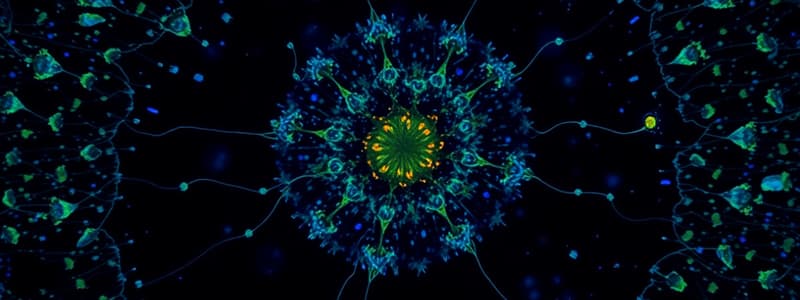
Methods like immunofluorescence (IF) can be used to visualize specific proteins within cells.
-
IF uses antibodies tagged with fluorescent molecules to target and mark the proteins of interest.
Indirect Immunofluorescence (IF)
- Specificity: The method uses antibodies which bind to a specific protein of interest.
- Sensitivity: The use of fluorescent molecules in the process aids in a higher sensitivity.
Fixing and Permeabilizing
- Cells are fixed to prevent the disruption of their structure and permeabilized to allow antibodies to enter.
Green Fluorescent Protein (GFP)
- Proteins fused with GFP can be viewed in living cells.
- GFP absorbs UV/blue light and emits green light.
Transmission Electron Microscopes (TEM)
- TEMs are used to view thin samples in a vacuum.
- The biological molecules are stained with heavy metals which scatter electrons.
Organelle Functions
- Different organelles perform specific functions within cells.
Cytosolic Components
- Cytosol contains a large proportion of RNA, proteins and ribosomes
The Nucleus
- The nucleus contains most of the cell's DNA (the ‘genome’).
- Replication and transcription occur within the nucleus.
- The nucleolus is a region within the nucleus where ribosomes are assembled.
Nuclear envelope
- The nuclear envelope surrounds the nucleus.
- It is made up of two membranes: inner & outer membrane.
Endoplasmic Reticulum (ER)
- The ER is a primary site for synthesizing lipids and membrane proteins.
- The ER secretes proteins.
Golgi Apparatus
- The Golgi apparatus modifies secreted proteins.
- The Golgi apparatus is a sorting station for vesicle trafficking.
Mitochondria
- Mitochondria are major sites for ATP production ("oxidative phosphorylation").
- They also synthesize iron-sulfur clusters.
- They are essential for producing central metabolites.
Chloroplasts
- Chloroplasts contain chlorophyll-containing membranes (thylakoids).
- Chloroplasts are responsible for photosynthesis.
Internal Membranes in Eukaryotes
- Internal membranes allow eukaryotic cells to compartmentalize different reactions
- Internal membranes increase surface area per volume.
Cytoplasm vs Cytosol
- Cytoplasm includes everything between the plasma membrane and the nucleus.
- Cytosol is the soluble portion of the cytoplasm, excluding the organelles.
Cellular Organelle Size Estimates
Values/percentages reflecting the proportion of total cell volume occupied by specific organelles are provided.
Studying That Suits You
Use AI to generate personalized quizzes and flashcards to suit your learning preferences.


