Podcast
Questions and Answers
What is the MOST LIKELY reason for Zubair experiencing right-sided weakness after 'cracking' his neck?
What is the MOST LIKELY reason for Zubair experiencing right-sided weakness after 'cracking' his neck?
- Nerve compression due to chronic degenerative changes and disc herniations. (correct)
- Exacerbation of a previous spinal cord injury from the childhood stabbing.
- Acute muscle strain due to forceful neck rotation.
- Vertebral artery dissection leading to ischemic damage.
Based on Zubair's presentation, which finding would be MOST consistent with a lesion affecting the corticospinal tract?
Based on Zubair's presentation, which finding would be MOST consistent with a lesion affecting the corticospinal tract?
- Decreased pinprick sensation on the left side.
- Increased muscle tone and exaggerated reflexes on the right side. (correct)
- Muscle atrophy in the right upper limb.
- Loss of vibration sense on the right side.
Which of the following is the MOST appropriate level to perform a lumbar puncture on Zubair, considering he is a 19-year-old?
Which of the following is the MOST appropriate level to perform a lumbar puncture on Zubair, considering he is a 19-year-old?
- L4/L5 (correct)
- L2/L3
- L3/L4
- L1/L2
In a Brown-Séquard syndrome, such as the one suspected in Zubair, which of the following sensory deficits would be expected CONTRALATERAL to the lesion?
In a Brown-Séquard syndrome, such as the one suspected in Zubair, which of the following sensory deficits would be expected CONTRALATERAL to the lesion?
Which of the listed structures passes through the intervertebral foramen?
Which of the listed structures passes through the intervertebral foramen?
The artery of Adamkiewicz is a significant feeder artery to the spinal cord. Injury to this artery during surgery can lead to what?
The artery of Adamkiewicz is a significant feeder artery to the spinal cord. Injury to this artery during surgery can lead to what?
What is the MOST immediate concern when suspecting increased intracranial pressure (ICP) before performing a lumbar puncture?
What is the MOST immediate concern when suspecting increased intracranial pressure (ICP) before performing a lumbar puncture?
After the surgery, the MRI indicated a focal signal abnormality in the right hemicord at the C4-C5 level. What is the MOST LIKELY explanation for this finding?
After the surgery, the MRI indicated a focal signal abnormality in the right hemicord at the C4-C5 level. What is the MOST LIKELY explanation for this finding?
Which of the following structures is located within the petrous part of the temporal bone?
Which of the following structures is located within the petrous part of the temporal bone?
During the surgical procedure on Zubair, the surgeon noted thickening of the posterior longitudinal ligament (PLL). What is the MOST direct consequence of PLL thickening in this case?
During the surgical procedure on Zubair, the surgeon noted thickening of the posterior longitudinal ligament (PLL). What is the MOST direct consequence of PLL thickening in this case?
A lesion affecting the superior colliculus would MOST likely impact which function?
A lesion affecting the superior colliculus would MOST likely impact which function?
Which of the following is a KEY characteristic differentiating upper motor neuron (UMN) and lower motor neuron (LMN) lesions?
Which of the following is a KEY characteristic differentiating upper motor neuron (UMN) and lower motor neuron (LMN) lesions?
What is the MOST likely reason why Mr Zubair was prescribed dexamethasone preoperatively?
What is the MOST likely reason why Mr Zubair was prescribed dexamethasone preoperatively?
Which of the listed signs or symptoms is MOST indicative of spinal shock?
Which of the listed signs or symptoms is MOST indicative of spinal shock?
Which of the following BEST describes the function of the ventral spinothalamic tract?
Which of the following BEST describes the function of the ventral spinothalamic tract?
If the anterior cerebral artery is compressed due to a subfalcine herniation, what is the MOST likely clinical presentation?
If the anterior cerebral artery is compressed due to a subfalcine herniation, what is the MOST likely clinical presentation?
Which of the following developmental anomalies directly involves a failure of the vertebral arch to close, often indicated by a tuft of hair on the skin?
Which of the following developmental anomalies directly involves a failure of the vertebral arch to close, often indicated by a tuft of hair on the skin?
In performing a lumbar puncture, which ligament is penetrated immediately prior to entering the epidural space?
In performing a lumbar puncture, which ligament is penetrated immediately prior to entering the epidural space?
Which nerve or nerve component is carried by the foramen spinosum?
Which nerve or nerve component is carried by the foramen spinosum?
A lesion affecting the red nucleus would MOST likely result in what?
A lesion affecting the red nucleus would MOST likely result in what?
Which of the following is a characteristic sign of Tabes Dorsalis?
Which of the following is a characteristic sign of Tabes Dorsalis?
Which spinal cord syndrome selectively damages the central portion of the spinal cord, often affecting upper limb function more than lower?
Which spinal cord syndrome selectively damages the central portion of the spinal cord, often affecting upper limb function more than lower?
Based on the content, where do the posterior spinal arteries originate?
Based on the content, where do the posterior spinal arteries originate?
Which functional area of the cerebral cortex is located deep to the pterion?
Which functional area of the cerebral cortex is located deep to the pterion?
Flashcards
Endosteal/Periosteal Dura Mater
Endosteal/Periosteal Dura Mater
Dura Mater (Tough mother)/ Pachymeninx: outer layer, adherent to skull, ends at foramina, separates from meningeal layer at venous sinuses.
Pterion Clinical Significance
Pterion Clinical Significance
A fracture at the pterion can lead to tearing of anterior branch of middle meningeal artery, resulting in epidural hematoma.
Middle Meningeal Artery
Middle Meningeal Artery
Middle meningeal artery is clinically the most important branch of maxillary artery.
Arachnoid Mater
Arachnoid Mater
Signup and view all the flashcards
Lumbar puncture
Lumbar puncture
Signup and view all the flashcards
Spinal Nerve Formation
Spinal Nerve Formation
Signup and view all the flashcards
Dorsal Root Function
Dorsal Root Function
Signup and view all the flashcards
Descending Tracts
Descending Tracts
Signup and view all the flashcards
Ascending Tract
Ascending Tract
Signup and view all the flashcards
Spinal Shock Symptoms
Spinal Shock Symptoms
Signup and view all the flashcards
Brown-Séquard Syndrome
Brown-Séquard Syndrome
Signup and view all the flashcards
Syringomyelia Symptoms
Syringomyelia Symptoms
Signup and view all the flashcards
Presentation
Presentation
Signup and view all the flashcards
Complete Cord Transection
Complete Cord Transection
Signup and view all the flashcards
Leptomeninges
Leptomeninges
Signup and view all the flashcards
TABES DORSALIS
TABES DORSALIS
Signup and view all the flashcards
Spinal Shock Syndrome
Spinal Shock Syndrome
Signup and view all the flashcards
Cauda Equina
Cauda Equina
Signup and view all the flashcards
Study Notes
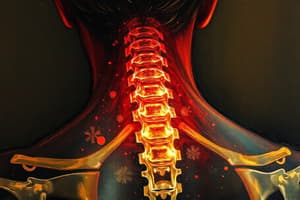
- This is a case study about Zubair a 19 year old university student complaining of right-sided weakness.
Patient History
- Zubair experienced sudden onset of right-sided weakness after "cracking" his neck
- He denies recent trauma but reports a remote history of a stabbing to the soft tissue of his neck as a child
- He has mild, persistent neck stiffness and manages discomfort by manually rotating his neck until it "cracks" multiple times a day for years
- On the day of presentation, neck rotation caused shooting pain down his neck and right-sided weakness
Expanded History
- Symptoms started the day of presentation after bending his neck right while rotating his chin to the left
- He is experiencing severe shooting pain
- He feels right sided weakness, and is unable to move his arm or leg
- He cannot stand or sit on his own
Examination
- Zubair is a yound adult, oriented in time, place, & person and unable to stand
- His temperature is 97.4° F, pulse is 72 bpm and regular, blood pressure is 120/80 mm Hg, and respiration rate is 17 breaths/min
- His right upper limb is adducted, lower limb is in adducted position, and there is no muscle mass loss
Motor exam
- Left side intact, right side impaired
- Shoulder abduction 3/5, elbow flexion 2/5, elbow extension 0/5, wrist extension 0/5, grip strength 0/5, hip flexion 3/5, knee extension 3/5, knee flexion 3/5, ankle dorsiflexion 2/5, and ankle plantarflexion 2/5
Sensory exam
- Decreased pinprick sensation on the left side at C5 and below
- Decreased vibration sensation on the right side at C4 and below
Imaging and Further Investigation
- An MRI of the cervical spine revealed large disc herniations at C4-C5 and C5-C6 with severe cord compression
- Chronic degenerative changes greater than expected are noted for his age
Differential Diagnoses
- Differential diagnoses to consider include Brown-Séquard Syndrome, Syringomyelia, Anterior Cord Syndrome, Cervical Artery Dissection, Spinal Cord Infarction, Cervical Myelopathy, and Transverse Myelitis
Consultation - The Neurosurgeon, Dr. Iqbal
- Dr. Iqbal performed a thorough neurological examination
- Moin asked if the patient is suffering from an epidural haemorrhage? Dr Iqbal asked him if he knew what pterion is?
- Dr. Iqbal told them to watch very informative about “disc herniation lumbar puncture" "vertebral column-spinal cord" & "Bones of Cranium" by scanning the following QR code
- He called Dr Hassan to confirm the diagnosis which he had in mind
Reading Material
- Skull is divided into Neurocranium (Cartilaginous and Membranous) and Vicerocranium (Cartilaginous and Membranous)
- Neural crest cells form the Prechordal, Trabeculi cranii, Ala orbitalis, Ala temporalis and Hypophyseal cartilage
- Membranous parts are make of NCC- Frontal, Temporal, Zygomatic, Paraxial, Parietal, and occipital
- Viscerocranium cartilaginous makes up 1st Arch-Dorsal part= malleus & incus, 2nd arch-Dorsal part-stapes & styloid process, and Ventral part-lesser horns & superior part of hyoid
- New born Skull has fontanelles anterior-12-18 months, Posterior-2-3months, Sphenoidal-6 months, and Mastoid-6-18 months
- Craniosynostosis is Scaphocephaly-Early closure of Sagittal suture (boat shaped) Brachiocephaly- Early closure of coronal) (Tower shaped) Plagiocephaly-early closure-unilateral coronal Trigonocephaly- early metopic closure (prominent frontal bone)
Development of Spinal Cord
- The Ventricular Zone formation is Pseudostratified columnar epithelium lining neural tube thickens and gives rise to neuroblasts and macroglia.
- The Mantle Zone formation is Neuroblasts from ventricular layer migrates to Intermediate layer which form gray matter (cell bodies)-alar (dorsal, sensory) and basal plates (ventral, Motor)
- The Marginal Zone formation is Outermost layer composed of axons of neurons in mantle layer forming white matter
- The sulcus limitans is the Boundary between the alar and basal plates, dividing sensory (alar) and motor (basal) neurons
- Neuroepithelial Layer remaining in ventricular zone gives rise to that layer, which will line the central canal
- Spinal cord length equal to vertebral column length at 8 weeks with spinal cord ending at S1 vertebral level at 28 weeks with inferior border of L1 as an adult
- Development Anomalies include Spina Bifida Occulta, Spina Bifida Cystica (Meningocele and Meningomyelocele), and Myelochisis/ Rascichisis
Cranial Fossas
- Anterior cranial fossa: Frontal Bone Forms the anterior boundary and roof of the fossa
- Middle cranial fossa: Sphenoid Bone Forms the central part of and Greater Wing of Sphenoid for lateral part of the middle cranial fossa
- Posterior cranial fossa: Occipital Bone Forms most of the posterior cranial fossa.
Meninges of the Brain
- Dura Mater (Tough mother)/ Pachymeninx is divided into endostel/periosteal and meningeal layers
- Arachnoid Matter is loosely attached to dura and connected to pia matter and contains cisterna- enlarged areas of the subarachnoid space where cerebrospinal fluid accumulates
- Dural Venous Sinuses includes superior Sagittal, inferior Sagittal, Straight Sinus, Transverse, Sigmoid, Cavernous, Superior Petrosal, Inferior Petrosal
Clinical Correlation- Types of Hemmorhages
- Extradural (Epidural) is injury to meningeal arteries/veins, often due to skull fracture at the pterion
- Subdural is tearing of superior cerebral veins at entry into the superior sagittal sinus, caused by head trauma
- Subarachnoid is rupture or leakage of a cerebral arteries, congenital aneurysm (circle of Willis) or angioma
- Cerebral Hemorrhage is Rupture of the lenticulostriate artery (branch of middle cerebral artery).
Reading Material - Dr Hassan
- Spinal Nerve is a mixed nerve of 31 pairs named according to the region of the vertebral column it is associated with
- Spinal Nerve divides into posterior Ramus (smaller) and ventral ramus (Larger), Meningeal branch, and Rami Communicantes
- Spinal Cord extends from the medulla oblongata to conus medullaris, with central canal carries of CSF
- Spinal arteries: provides the blood supply for this anterior, posterior, and segment arteries with with venous channels the drain into vertebral venous plexus
Ascending and Descending Tracts of Spinal cord
- Descending Tracts Motor consist of Lateral Corticospinal and Ventral Corticospinal Tract
- Ascending Tracts Sensory: Doral Columns (Deep touch, Propioception, Vibration), Lateral Spinothalamic Tract (Pain, Temperture), and Ventral Spinothalamic Tract (Light touch)
- The lateral corticospinal tract is responsible for rapid, skilled voluntary movements
- The spinotectal tract is responsible for spinovisual activity
- Pyramidal tract lesions have present Babinski's sign and Extrapyramidal tract lesions have Absent Babinski's Sign
-Tabes Dorsalis:
- Syphilis can cause selective destruction of nerve fibers in TABES DORSALIS MORPHOLOGY
- Cause - DORSALIS
- Pain can be experienced in lower limbs, paresthesia, and hypersensitivity to touch/heat or cold
- Problems can present in Ataxia, loss of posture appreciation, and loss of deep pain sensation
Other neurological Conditions
- Syringomyelia: Developmental abnormality in central canal
- Loss of pain and temperature (shawl-like distribution).
- Tactile, vibratory, and proprioception remain intact.
- Lower motor neuron weakness in hands.
- Bilateral spastic paralysis in legs.
- Horner syndrome in advanced cases.
Spinal Cord Injury and Paralyis
- Above C3: Paralysis of all respiratory muscles, death due to diaphragm paralysis
- C4-C5: Quadriplegia (paralysis of all four limbs)
- C5 and C6 (Erb's paralysis): Loss of abduction of shoulder, flexion of elbow, supination, extension of wrist, policeman's tip deformity
- C8 and T1 (Klumpke's paralysis): Paralysis of intrinsic muscles of the hand, claw hand
- Between T2 and L1: Paraplegia (paralysis of both lower limbs)
Lumbar Puncture
- It is perform ed to obtain samples of cerebrospinal fluid (CSF) for various diagnostic and therapeutic purposes between the third and fourth lumbar vertebrae
- Estimate CSF pressure, Collect CSF samples, Introduce air or radiopaque dyes for investigative procedures, Inject drugs for treatment or anaesthesia, Spinal anaesthesia for lower body operations
- Presences of increased intracranial pressure, Use of anticoagulants (e.g., warfarin, enoxaparin, etc), Evidence of cellulitis or abscess over the area, Significant degenerative joint disease or prior back surgeries where hardware maybe in place
Dscending Tract Injury:
- Presence of Babinski sign is a side effect of a lesion in pyramidal tract
Surgery and diagnosis
- Fatima had the opportunity to directly visualize intraoperatively which showed C4–5 disc herniation, with signs of Brown Sequard Syndrome
Studying That Suits You
Use AI to generate personalized quizzes and flashcards to suit your learning preferences.