Podcast
Questions and Answers
Which type of joint is characterized by bones joined by hyaline cartilage?
Which type of joint is characterized by bones joined by hyaline cartilage?
- Symphyses
- Synovial joints
- Synchondroses (correct)
- Fibrous joints
What structure connects bones at a symphysis joint?
What structure connects bones at a symphysis joint?
- Ligament
- Hyaline cartilage
- Fibrocartilage disc (correct)
- Synovial membrane
What role does synovial fluid NOT play in a synovial joint?
What role does synovial fluid NOT play in a synovial joint?
- Reduces friction
- Connects bones together (correct)
- Supplies oxygen to cartilage
- Absorbs shocks
Which statement about the articular capsule of a synovial joint is false?
Which statement about the articular capsule of a synovial joint is false?
Which type of cartilage covers the ends of bones in a synovial joint?
Which type of cartilage covers the ends of bones in a synovial joint?
What structure is located on the anterior side of the proximal tibia where the patellar ligament inserts?
What structure is located on the anterior side of the proximal tibia where the patellar ligament inserts?
Which part of the tibia is known for being a sharp edge that is palpable under the skin?
Which part of the tibia is known for being a sharp edge that is palpable under the skin?
Which of the following functions is NOT associated with the tibia?
Which of the following functions is NOT associated with the tibia?
What type of fracture typically requires surgical intervention like intramedullary nailing?
What type of fracture typically requires surgical intervention like intramedullary nailing?
Which part of the fibula articulates with the tibia?
Which part of the fibula articulates with the tibia?
Which type of joint allows for multiaxial movement including flexion, extension, adduction, abduction, and circumduction?
Which type of joint allows for multiaxial movement including flexion, extension, adduction, abduction, and circumduction?
What is the primary socket of the shoulder joint?
What is the primary socket of the shoulder joint?
Which ligaments are associated with the hinge type elbow joint?
Which ligaments are associated with the hinge type elbow joint?
What type of movement does the proximal radio-ulnar joint primarily permit?
What type of movement does the proximal radio-ulnar joint primarily permit?
Which type of joint is exemplified by the first carpometacarpal joint?
Which type of joint is exemplified by the first carpometacarpal joint?
What is the primary function of bones in the body?
What is the primary function of bones in the body?
What characterized the movement in an ellipsoidal type joint?
What characterized the movement in an ellipsoidal type joint?
What does the Intercondylar Fossa accommodate?
What does the Intercondylar Fossa accommodate?
What is the typical angle of inclination for the femur in adults?
What is the typical angle of inclination for the femur in adults?
Which type of femur fracture is most commonly associated with older adults?
Which type of femur fracture is most commonly associated with older adults?
What is the primary function of the tibia?
What is the primary function of the tibia?
What occurs at the Intercondylar Eminence of the tibia?
What occurs at the Intercondylar Eminence of the tibia?
Which structure is located at the proximal end of the tibia?
Which structure is located at the proximal end of the tibia?
What is the consequence of a Neck of Femur Fracture?
What is the consequence of a Neck of Femur Fracture?
What characterizes an Intertrochanteric Fracture of the femur?
What characterizes an Intertrochanteric Fracture of the femur?
Which part of the tibia articulates at the knee joint?
Which part of the tibia articulates at the knee joint?
What is the primary treatment method for a Shaft Fracture of the femur?
What is the primary treatment method for a Shaft Fracture of the femur?
Study Notes
Cartilaginous Joints
- Synchondroses: Bones bound by hyaline cartilage.
- Example: Growth plate in long bones
- Example: First rib attached to the sternum
- Symphyses: Bones joined by fibrocartilage
- Example: Pubic bone in pelvis
- Example: Between vertebrae
Synovial Joints
- Freely movable due to synovial cavity
- Ligaments hold bones together within the joint
- Articular Capsule:
- Encloses the synovial cavity
- Composed of two layers:
- Outer fibrous capsule
- Inner synovial membrane
- Synovial Fluid:
- Secreted by the synovial membrane
- Functions:
- Lubricates the joint
- Absorbs shocks
- Supplies nutrients to cartilage
- Removes waste from cartilage
Synovial Joint Structure
- Synovial membrane:
- Secretes synovial fluid
- Outer fibrous capsule (continuous with the periosteum)
- Inner synovial membrane
- Articular Cartilage:
- Covers each bone involved in the joint
- About 2 mm thick
- Ligaments:
- Hold bones together
- Help maintain correct bone placement for proper cartilage movement
Synovial Joint Classification
- 7 Types of Synovial Joints:
- Ball and Socket:
- Example: Shoulder joint (humerus and scapula)
- Movement: Multiaxial (flex, extend, adduct, abduct, circumduction)
- Hinge:
- Example: Elbow joint (humerus, ulna, radius)
- Movement: Flexion and extension around horizontal axis
- Pivot:
- Example: Proximal radio-ulnar joint (radius and ulna)
- Movement: Rotation around vertical axis (medial and lateral rotation)
- Ellipsoidal:
- Example: Wrist (radio-carpal) joint
- Movement: Modified hinge (flex, extend, adduct, abduct)
- Plane (Gliding):
- Example: Intercarpal joints
- Movement: Gliding
- Saddle:
- Example: 1st carpometacarpal joint (1st metacarpal, trapezium)
- Movement: Flex, extend, adduct, abduct, circumduction
- Condyloid:
- Example: Metacarpophalangeal (MP) and interphalangeal (IP) joints
- Movement: Flex, extend, adduct, abduct
- Ball and Socket:
Femur Anatomy
- Intercondylar Fossa (Notch):
- Deep groove between medial and lateral condyles
- Accommodates ligaments of the knee
- Patellar Surface (Trochlear Groove):
- Smooth, anterior surface between condyles where the patella glides
- Angle of Inclination: Angle between the femoral neck and shaft (around 125°)
- Angle of Torsion: Angle between the axis of the femoral head/neck and condyles.
Femur Fractures
- Neck of Femur Fracture:
- Common in older adults due to falls and osteoporosis
- Poor blood supply, risk of nonunion and avascular necrosis
- Shaft Fractures:
- Caused by high-energy trauma
- Intertrochanteric Fractures:
- Occur between greater and lesser trochanters
- Better vascularized but often require surgery
Tibia Anatomy
- Proximal End:
- Medial and Lateral Condyles: Articulate with femoral condyles to form part of the knee joint
- Intercondylar Eminence: Ridge between condyles, attachment for ligaments and menisci
- Tibial Plateau: Flat surface articulating with femur
- Tibial Tuberosity: Bony prominence on the anterior aspect, patellar ligament attachment point
Tibia Anatomy (Continued)
- Shaft (Body):
- Anterior Border: Sharp edge on front, known as the shin
- Medial Surface: Smooth and palpable
- Lateral (Interosseous) Border: Faces fibula, where interosseous membrane connects the tibia and fibula
- Soleal Line: Ridge on posterior surface, soleus muscle attachment
Tibia Anatomy (Continued)
- Distal End:
- Medial Malleolus: Projection on the medial side, forming part of the ankle joint
- Fibular Notch: Notch on lateral side where the fibula articulates
- Inferior Articular Surface: Smooth, flat surface articulating with talus in the ankle joint
Tibia Functions
- Weight-bearing
- Lower part of the knee joint
- Medial part of the ankle joint
- Attachment points for muscles and ligaments
Tibia Fractures
- Proximal Tibial Fractures:
- High-energy trauma or low-energy injuries in osteoporotic patients
- Potential knee alignment disruption
- Treatment: Open reduction and internal fixation (ORIF)
- Tibial Shaft Fractures:
- Direct trauma or falls
- Closed or open fractures
- Treatment: Intramedullary nailing
- Distal Tibial (Pilon) Fractures:
- Occur near the ankle
- May affect articular surface
- Treatment: ORIF
Fibula Anatomy
- Proximal End:
- Head of the Fibula: Rounded, articulates with tibia at the fibular notch
Fibula Clinical Applications
- Bone grafts from the fibula:
- Repairing bone defects
- Reconstructive surgery
- Addressing nonunions or malunions
Foot Anatomy
- Tarsal Bones:
- 7 bones in the foot
- Posterior part of the foot
- Connecting foot to leg
- Allow movement and weight-bearing
Foot Bone Quick Quiz
- How many phalanges are in the foot? 14
- How many metatarsals are in the foot? 5
- How many tarsals are in the foot? 7
- Total number of bones in the foot? 26
Key Anatomy and Bone Course Takeaways
- Lower Limb Bone Anatomy: Femur, tibia, fibula, patella, tarsals, metatarsals, phalanges
- Lower Limb Bone Function: Supporting body weight, enabling movement
- Key Landmarks:
- Femur: Greater/lesser trochanter, head, neck, condyles
- Tibia: Medial/lateral condyles, tibial tuberosity, medial malleolus
- Fibula: Head and lateral malleolus
- Patella: Articulating surfaces
- Tarsal bones: Calcaneus, talus, navicular
- Locomotion:
- Lower limb role in weight-bearing, balance, and movement
- Joint articulations supported by bone landmarks
Anatomy and Bone Course Questions:
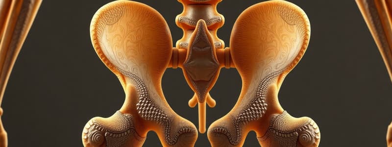
- Key features of the hip bone?
- Femur structure and major landmarks?
- Characteristics of the tibia and fibula?
- Tarsal bone contribution to foot mechanics?
Studying That Suits You
Use AI to generate personalized quizzes and flashcards to suit your learning preferences.
Related Documents
Description
This quiz covers the structure and function of cartilaginous and synovial joints in the human body. You will learn about the different types of joints, their components, and the role of synovial fluid. Test your understanding of these essential joints and their functions.


