Podcast
Questions and Answers
What is the primary function of the lymphatic system?
What is the primary function of the lymphatic system?
- Transport carbon dioxide to the lungs
- Defend the body against pathogens (correct)
- Circulate nutrients to organ systems
- Remove waste products from blood
What is lymph fluid composed of?
What is lymph fluid composed of?
- Only red blood cells
- Tissue fluid from capillaries (correct)
- Digested food from the intestines
- Oxygen and carbon dioxide
Which structure is NOT part of the lymphatic system?
Which structure is NOT part of the lymphatic system?
- Thymus
- Spleen
- Pulmonary trunk (correct)
- Lymph nodes
How does the systemic circuit primarily function?
How does the systemic circuit primarily function?
Which component of the circulatory system ejects blood into the aorta?
Which component of the circulatory system ejects blood into the aorta?
What mechanism is utilized by lymphatic capillaries to prevent backflow?
What mechanism is utilized by lymphatic capillaries to prevent backflow?
What occurs in the pulmonary capillaries?
What occurs in the pulmonary capillaries?
Which of the following describes the return pathway of blood from systemic circulation?
Which of the following describes the return pathway of blood from systemic circulation?
What primarily causes the plateau phase during an action potential in ventricular contractile fibers?
What primarily causes the plateau phase during an action potential in ventricular contractile fibers?
Which of the following waves in an ECG represents atrial depolarization?
Which of the following waves in an ECG represents atrial depolarization?
During which phase does rapid depolarization occur in a ventricular contractile fiber?
During which phase does rapid depolarization occur in a ventricular contractile fiber?
What does the depolarization of the atrial contractile fibers produce?
What does the depolarization of the atrial contractile fibers produce?
What does the QRS complex on an ECG primarily represent?
What does the QRS complex on an ECG primarily represent?
What is produced by the depolarization of ventricular contractile fibers?
What is produced by the depolarization of ventricular contractile fibers?
Which interval reflects the time taken for the action potential to travel from the SA node to the AV node?
Which interval reflects the time taken for the action potential to travel from the SA node to the AV node?
Which heart sound corresponds to the closure of the AV valves?
Which heart sound corresponds to the closure of the AV valves?
What occurs during the repolarization phase in a ventricular contractile fiber?
What occurs during the repolarization phase in a ventricular contractile fiber?
What happens during atrial systole in the cardiac cycle?
What happens during atrial systole in the cardiac cycle?
What does the T wave on an ECG signify?
What does the T wave on an ECG signify?
What is the formula for calculating cardiac output (CO)?
What is the formula for calculating cardiac output (CO)?
What phase follows depolarization in a ventricular contractile fiber action potential?
What phase follows depolarization in a ventricular contractile fiber action potential?
What does a normal R–R interval indicate?
What does a normal R–R interval indicate?
What is stroke volume defined as?
What is stroke volume defined as?
Which of the following conditions is characterized by no detectable P waves?
Which of the following conditions is characterized by no detectable P waves?
What is the main function of the coronary arteries?
What is the main function of the coronary arteries?
Which of the following structures is responsible for initiating the heartbeat?
Which of the following structures is responsible for initiating the heartbeat?
What is the role of intercalated discs in cardiac muscle?
What is the role of intercalated discs in cardiac muscle?
What type of muscle fibers are considered autorhythmic?
What type of muscle fibers are considered autorhythmic?
Which part of the cardiac conduction system transmits impulses after the AV node?
Which part of the cardiac conduction system transmits impulses after the AV node?
How does the structure of cardiac muscle fibers differ from skeletal muscle fibers?
How does the structure of cardiac muscle fibers differ from skeletal muscle fibers?
What is the effect of an artery being partially blocked in coronary circulation?
What is the effect of an artery being partially blocked in coronary circulation?
Which stage of action potential is characterized by a plateau phase?
Which stage of action potential is characterized by a plateau phase?
What is the main function of red blood cells?
What is the main function of red blood cells?
Which component of blood is primarily responsible for clotting?
Which component of blood is primarily responsible for clotting?
What is the primary characteristic of arteries?
What is the primary characteristic of arteries?
Which type of white blood cell is primarily involved in the destruction of bacteria and viruses?
Which type of white blood cell is primarily involved in the destruction of bacteria and viruses?
What process does hemostasis primarily relate to?
What process does hemostasis primarily relate to?
Which blood component is primarily liquid and makes up 55% of blood?
Which blood component is primarily liquid and makes up 55% of blood?
What is the role of calcium in cardiac muscle contraction?
What is the role of calcium in cardiac muscle contraction?
Which type of blood vessel carries blood back to the heart?
Which type of blood vessel carries blood back to the heart?
What is contained in the plasma of blood that aids in immunity?
What is contained in the plasma of blood that aids in immunity?
What type of white blood cell is primarily responsible for immunity?
What type of white blood cell is primarily responsible for immunity?
What is the effect of epinephrine and norepinephrine on heart rate?
What is the effect of epinephrine and norepinephrine on heart rate?
Which ions are crucial for maintaining effective heart pumping?
Which ions are crucial for maintaining effective heart pumping?
What is the Frank–Starling law of the heart's principle?
What is the Frank–Starling law of the heart's principle?
What factors could lead to a decrease in afterload during cardiac cycle?
What factors could lead to a decrease in afterload during cardiac cycle?
How do positive inotropic agents affect cardiac output?
How do positive inotropic agents affect cardiac output?
Which of the following is considered a positive inotropic agent?
Which of the following is considered a positive inotropic agent?
What role does the cardiovascular center in the medulla oblongata play?
What role does the cardiovascular center in the medulla oblongata play?
What group of individuals might experience increased heart rate under normal conditions?
What group of individuals might experience increased heart rate under normal conditions?
Flashcards
Lymphatic System
Lymphatic System
A system that collects and transports excess fluid (lymph) from tissues back to the circulatory system.
Lymph Nodes
Lymph Nodes
Small, bean-shaped organs that filter lymph fluid and contain white blood cells that fight infections.
Thymus
Thymus
A primary lymphoid organ where T-cells mature and learn to recognize and fight pathogens.
Spleen
Spleen
Signup and view all the flashcards
Lymph Fluid
Lymph Fluid
Signup and view all the flashcards
Circulation of Blood
Circulation of Blood
Signup and view all the flashcards
Systemic and Pulmonary Circulation
Systemic and Pulmonary Circulation
Signup and view all the flashcards
Systemic Circuit
Systemic Circuit
Signup and view all the flashcards
What is blood?
What is blood?
Signup and view all the flashcards
What is blood plasma?
What is blood plasma?
Signup and view all the flashcards
What is the function of red blood cells?
What is the function of red blood cells?
Signup and view all the flashcards
What is the function of white blood cells?
What is the function of white blood cells?
Signup and view all the flashcards
What is the function of platelets?
What is the function of platelets?
Signup and view all the flashcards
Describe the process of hemostasis.
Describe the process of hemostasis.
Signup and view all the flashcards
What are arteries and arterioles?
What are arteries and arterioles?
Signup and view all the flashcards
What are veins and venules?
What are veins and venules?
Signup and view all the flashcards
What are capillaries?
What are capillaries?
Signup and view all the flashcards
What is the lymphatic system?
What is the lymphatic system?
Signup and view all the flashcards
Circulation
Circulation
Signup and view all the flashcards
Pulmonary Circuit
Pulmonary Circuit
Signup and view all the flashcards
Coronary Circulation
Coronary Circulation
Signup and view all the flashcards
Autorhythmic Fibers
Autorhythmic Fibers
Signup and view all the flashcards
Cardiac Conduction System
Cardiac Conduction System
Signup and view all the flashcards
Sinoatrial (SA) Node
Sinoatrial (SA) Node
Signup and view all the flashcards
Atrioventricular (AV) Node
Atrioventricular (AV) Node
Signup and view all the flashcards
Rapid Depolarization
Rapid Depolarization
Signup and view all the flashcards
Plateau Phase
Plateau Phase
Signup and view all the flashcards
Repolarization
Repolarization
Signup and view all the flashcards
Electrocardiogram (ECG)
Electrocardiogram (ECG)
Signup and view all the flashcards
P wave
P wave
Signup and view all the flashcards
P-R interval
P-R interval
Signup and view all the flashcards
QRS complex
QRS complex
Signup and view all the flashcards
T wave
T wave
Signup and view all the flashcards
Cardiac Cycle
Cardiac Cycle
Signup and view all the flashcards
Atrial Systole
Atrial Systole
Signup and view all the flashcards
Ventricular Systole
Ventricular Systole
Signup and view all the flashcards
Ventricular Diastole
Ventricular Diastole
Signup and view all the flashcards
Stroke Volume
Stroke Volume
Signup and view all the flashcards
Cardiac Output
Cardiac Output
Signup and view all the flashcards
Heart Sounds
Heart Sounds
Signup and view all the flashcards
Heart Sounds
Heart Sounds
Signup and view all the flashcards
What hormones increase heart rate?
What hormones increase heart rate?
Signup and view all the flashcards
How do thyroid hormones affect heart rate?
How do thyroid hormones affect heart rate?
Signup and view all the flashcards
How do ionic imbalances affect heart function?
How do ionic imbalances affect heart function?
Signup and view all the flashcards
What is the Frank-Starling law?
What is the Frank-Starling law?
Signup and view all the flashcards
What are positive inotropic agents?
What are positive inotropic agents?
Signup and view all the flashcards
How does afterload affect stroke volume?
How does afterload affect stroke volume?
Signup and view all the flashcards
How does the nervous system influence heart rate?
How does the nervous system influence heart rate?
Signup and view all the flashcards
What factors can influence heart rate?
What factors can influence heart rate?
Signup and view all the flashcards
Study Notes
Cardiovascular System (Part I)
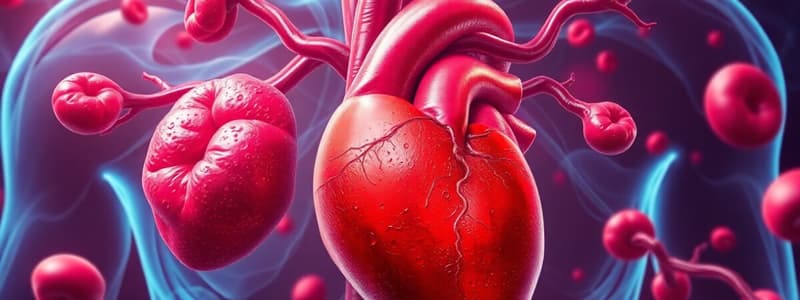
- The cardiovascular system's primary function is transporting blood, regulating various bodily functions, and protecting against diseases.
Blood Composition
- Blood is a connective tissue composed of:
- Plasma (55%): Primarily water with proteins (albumins, globulins, fibrinogen), nutrients, wastes, and gases.
- Formed elements (45%):
- Red blood cells (erythrocytes): Biconcave discs containing hemoglobin, transporting oxygen; 4.2-6.2 million per cubic mm.
- White blood cells (leukocytes): Protecting against disease-causing agents (pathogens). Different types with varying functions: neutrophils, lymphocytes, monocytes, eosinophils, basophils; 5,000-9,000 per cubic mm.
- Platelets (thrombocytes): Essential for blood clotting; 130,000-360,000 per cubic mm.
Blood Functions
- Transportation: Carries oxygen, nutrients, hormones, and waste products.
- Regulation: Maintains homeostasis, balancing acids/bases and bodily fluids.
- Protection: Combats disease-causing agents and prevents excessive blood loss.
Red Blood Cells (RBCs)
- Transport oxygen throughout the body
- Biconcave-shaped, small enough for capillary passage
- Contain hemoglobin:
- Oxyhemoglobin (bright red) carries oxygen
- Deoxyhemoglobin (dark red) lacks oxygen.
White Blood Cells (WBCs)
- Granulocytes: Neutrophils (55%) - destroy bacteria, viruses, and toxins. Eosinophils (3%) - combat parasitic infections like worms. Basophils (1%) - control inflammation and allergic reactions.
- Agranulocytes: Monocytes (8%) - Destroy bacteria, viruses, and toxins. Lymphocytes (33%) - play a crucial role in the body's immunity.
Blood Platelets
- Fragments of cells that are crucial for blood clotting.
- About 130,000-360,000 per cubic millimeter of blood.
- Essential for hemostasis (stopping bleeding).
Controlling Bleeding (Hemostasis)
- A complex of three processes:
- Blood vessel spasm
- Platelet plug formation
- Blood coagulation (formation of a clot.)
Blood Vessels
- Arteries & Arterioles: Strongest blood vessels, carry blood away from the heart under high pressure, possessing thick walls.
- Veins & Venules: Blood moves slowly in veins due to low pressure. Valves prevent backflow.
- Capillaries: Tiny blood vessels connecting arterioles to venules; sites of gas, nutrient, and waste exchange between the blood and body cells.
Lymphatic System
- Network of vessels collecting fluids between cells and returning them to the bloodstream, crucial for fluid balance and absorbing lipids.
- Also critical for defending the body against disease-causing agents (pathogens).
- Lymph nodes, thymus, and spleen are part of this system.
Lymph Fluid
- Tissue fluid that enters lymphatic capillaries
- Contains Valves preventing backflow of lymph
Circulation of Blood
- Blood circulates via two major circuits:
- Pulmonary circulation: Blood travels from the heart to the lungs for oxygen uptake and release of carbon dioxide.
- Systemic circulation: Blood travels from the heart to the rest of the body, delivering oxygen and nutrients and removing waste products.
Systemic and Pulmonary Circulation
- Systems using separate circuits allowing efficient oxygen and nutrient delivery, accompanied by waste elimination.
Coronary Circulation
- Coronary arteries supply the heart muscle with blood, providing oxygen and nutrients.
- Coronary veins collect used blood, transporting it to the right atrium.
Heart: A special muscle
- Specialized muscle with the job of pumping blood throughout the body.
Cardiac Muscle Characteristics
- Shorter, less circular than skeletal muscle fibers
- Branching structure giving a "stair-step" appearance.
- Typically one nucleus centrally located
- Ends of fibers connected by intercalated discs (desmosomes and gap junctions)
- Contains numerous mitochondria (40%).
- Actin and myosin arrangement is the same as in other muscle tissues.
Autorhythmic Fibers
- Specialized heart muscle fibers that self-excite, generating action potentials to trigger heart contractions.
- Serve as pacemakers regulating rhythmic heartbeats, forming the heart's conduction system.
Cardiac Conduction System
- Specialized conductive tissue (SA node, AV node, bundles of His, Purkinje fibers) directs electrical signals rapidly across the heart muscle, coordinating controlled and effective contractions for efficient blood pumping.
Action Potentials and Contraction
- Action potentials initiate contraction; depolarization initiates a contraction through the activation of contractile proteins.
- Depolarization occurs when there is a change in charge across the membrane, and contractile proteins are activated
ECG (Electrocardiogram)
- Composite record of action potentials generated by all heart muscle fibers.
- Wave forms (P, QRS, and T) visually represent the electrical activity during each stage of the cardiac cycle.
Cardiac Cycle
- The sequence of events where the heart alternately contracts and relaxes during blood flow; blood flows through each chamber in a rhythmic pattern.
- Atrial systole: atria contracts, blood enters relaxed ventricles.
- Ventricular systole: ventricles contract, blood is pumped into arteries.
Heart Sounds (Auscultation)
- Sounds caused by blood turbulence when heart valves close ( "lub" - atrioventricular valves, and "dup" - semilunar valves).
Cardiac Output (CO)
- Volume of blood pumped by the left ventricle per minute; calculated as heart rate multiplied by stroke volume.
Stroke Volume (SV)
- Amount of blood pumped by the left ventricle with each contraction. Regulated by factors in preload, contractility and afterload.
Factors Affecting Preload
- How much the heart chamber is stretched before it contracts; directly related to the end-diastolic volume and venous return, impacting the force of contraction.
- Frank-Starling law of the heart.
Factors Affecting Contractility
- The strength of heart contraction relating to a given volume of blood.
- Influenced by positive and negative inotropic agents (e.g., epinephrine) affecting calcium inflow.
Factors Affecting Afterload
- The pressure the ventricles must overcome to pump blood.
- Increasing afterload lowers stroke volume.
Exercise and the Heart
- Exercise significantly impacts cardiac output, Increasing heart rate and stroke volume.
Regulation of Heart Beat
- Autonomic nervous system regulates heart rate: sympathetic accelerates and parasympathetic slows it down which is regulated by the cardiovascular center in the medulla oblongata.
- Hormones (epinephrine/norepinephrine) and blood ionic levels also impact heart rate and contractility.
Questions addressed.
- How would increased sympathetic stimulation of the heart affect stroke volume?: Increased sympathetic stimulation leads to increased heart rate and contractility, subsequently increasing stroke volume.
Studying That Suits You
Use AI to generate personalized quizzes and flashcards to suit your learning preferences.


