Podcast
Questions and Answers
What is the main function of the cardiovascular system?
What is the main function of the cardiovascular system?
- Transport oxygen and nutrients to body cells (correct)
- Produce red blood cells
- Digest food
- Remove carbon dioxide from the body
The heart is about 12 cm long, 9 cm wide at its broadest point, and 6 cm thick, weighing approximately _______ in adult females.
The heart is about 12 cm long, 9 cm wide at its broadest point, and 6 cm thick, weighing approximately _______ in adult females.
250 g
The pericardium consists of only one layer.
The pericardium consists of only one layer.
False (B)
Match the following layers of the heart wall with their descriptions:
Match the following layers of the heart wall with their descriptions:
What is the reason for the rapid transmission of the impulse in the Ventricular Purkinje System?
What is the reason for the rapid transmission of the impulse in the Ventricular Purkinje System?
What does the A-V Bundle prevent?
What does the A-V Bundle prevent?
The sinus node is the normal pacemaker of the heart.
The sinus node is the normal pacemaker of the heart.
Abnormal pacemakers, such as the Ectopic Pacemaker, may cause the ventricles to contract at a rate between __ and __ beats per minute.
Abnormal pacemakers, such as the Ectopic Pacemaker, may cause the ventricles to contract at a rate between __ and __ beats per minute.
Match the following nervous control components with their distribution in the heart:
Match the following nervous control components with their distribution in the heart:
What proportion of the energy is used to move blood from low-pressure veins to high-pressure arteries?
What proportion of the energy is used to move blood from low-pressure veins to high-pressure arteries?
What term is used for the period of time when the ventricle does not change volume because all valves are closed?
What term is used for the period of time when the ventricle does not change volume because all valves are closed?
What controls the heart's pumping effectiveness?
What controls the heart's pumping effectiveness?
Increasing the arterial pressure load always decreases cardiac output.
Increasing the arterial pressure load always decreases cardiac output.
Where does the heart get about 60% of its energy from at rest?
Where does the heart get about 60% of its energy from at rest?
Cardiac muscle is more vulnerable to an oxygen deficiency than a specific fuel deficiency.
Cardiac muscle is more vulnerable to an oxygen deficiency than a specific fuel deficiency.
What system does the sinus node control?
What system does the sinus node control?
According to Einthoven's law, if the electrical potentials of any two of the three leads are given, what can be determined?
According to Einthoven's law, if the electrical potentials of any two of the three leads are given, what can be determined?
In cardiac muscle, the action potential is caused by opening of two types of channels: voltage-activated fast sodium channels and _____ channels.
In cardiac muscle, the action potential is caused by opening of two types of channels: voltage-activated fast sodium channels and _____ channels.
Match the following phases of the cardiac muscle action potential with their descriptions:
Match the following phases of the cardiac muscle action potential with their descriptions:
In the Unipolar Limb leads system, the lead designated as aVR has the positive terminal placed on the left leg.
In the Unipolar Limb leads system, the lead designated as aVR has the positive terminal placed on the left leg.
What do the precordial (chest) leads mainly record the electrical potential of?
What do the precordial (chest) leads mainly record the electrical potential of?
Vectors can be used to represent electrical potentials with the arrowhead pointing in the ________ direction.
Vectors can be used to represent electrical potentials with the arrowhead pointing in the ________ direction.
Match the following heart conditions with their causes:
Match the following heart conditions with their causes:
Complete A-V Block (Third-Degree Block) results in the complete block of the impulse from the atria into the ventricles. The ventricles spontaneously establish their own signal, usually originating in the A-V node or A-V bundle distal to the block. Therefore, the P waves become dissociated from the _____ complexes.
Complete A-V Block (Third-Degree Block) results in the complete block of the impulse from the atria into the ventricles. The ventricles spontaneously establish their own signal, usually originating in the A-V node or A-V bundle distal to the block. Therefore, the P waves become dissociated from the _____ complexes.
What is the term for a contraction of the heart before the time that normal contraction would have been expected?
What is the term for a contraction of the heart before the time that normal contraction would have been expected?
Which condition is associated with excess amounts of antiarrhythmic drugs such as quinidine or some antibiotics like fluoroquinolones or erythromycin?
Which condition is associated with excess amounts of antiarrhythmic drugs such as quinidine or some antibiotics like fluoroquinolones or erythromycin?
Ventricular Fibrillation, if not stopped within 1 to 3 minutes, is almost invariably fatal.
Ventricular Fibrillation, if not stopped within 1 to 3 minutes, is almost invariably fatal.
Match the following heart sounds with their descriptions:
Match the following heart sounds with their descriptions:
Flashcards are hidden until you start studying
Study Notes
Cardiovascular Physiology
Introduction
- The cardiovascular system is a series of tubes filled with fluid (blood) and connected to a pump (the heart)
- Functions: delivers oxygen and nutrients, removes cellular wastes and heat, facilitates cell-to-cell communication, and defends against foreign invaders
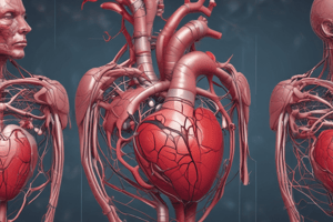
The Heart as a Pump
- Location: in the thoracic cavity, between the lungs, slightly to the left of the body's midline
- Size: about 12 cm (5 in.) long, 9 cm (3.5 in.) wide, and 6 cm (2.5 in.) thick
- Weight: 250 g (8 oz) in adult females, 300 g (10 oz) in adult males
- Composed of two separate pumps: right heart pumps blood through the lungs, left heart pumps blood through the systemic circulation
Heart Chambers
- Right heart: pumps blood through the lungs
- Left heart: pumps blood through the systemic circulation
- Each pump is composed of an atrium (upper chamber) and a ventricle (lower chamber)
- Atria act as primer pumps, helping to move blood into the ventricles
- Ventricles provide the main pumping force, propelling blood through the pulmonary circulation (right ventricle) or systemic circulation (left ventricle)
The Pericardium
- A two-layer sac surrounding the heart
- Fibrous pericardium: prevents overstretching of the heart, provides protection, and anchors the heart in the mediastinum
- Serous pericardium: composed of parietal and visceral layers, with a space called the pericardial cavity containing pericardial fluid
Layers of the Heart Wall
- Epicardium (outer layer): contains blood vessels, lymphatics, and vessels that supply the myocardium
- Myocardium (middle layer): responsible for the pumping action, composed of cardiac muscle tissue
- Endocardium (inner layer): a thin layer of endothelium overlying a thin layer of connective tissue, providing a smooth lining for the heart chambers and covering the heart valves
Heart Valves
- Atrioventricular valves: between the atria and ventricles
- Semilunar valves: between the ventricles and arteries
Physiology of Cardiac Muscle
Types of Cardiac Muscle
- Atrial muscle
- Ventricular muscle
- Specialized excitatory and conductive fibers
Cardiac Muscle Anatomy
- Fibers arranged in a latticework pattern
- Striated, with myofibrils containing actin and myosin
- Left ventricular rotation (twist) aids in left ventricular ejection and relaxation
- Subepicardial and subendocardial layers of muscle fibers spiral in different directions
Cardiac Energetics
- Cardiac muscle relies almost exclusively on aerobic respiration to make ATP
- Rich in myoglobin and glycogen
- Mitochondria occupy 25% of the cell
- Can adapt to use different organic fuels, including fatty acids, glucose, and others
Electrophysiology of the Heart
- Resting and action potentials of the myocardium
- Excitation-contraction coupling of the myocardium
- Refractory periods
- The special rhythmic, excitatory, and conduction system
Action Potentials in Cardiac Muscle
- Average 105 millivolts
- Rise from -85 millivolts to +20 millivolts during each beat
- Membrane remains depolarized for about 0.2 seconds
- Exhibits a plateau, followed by abrupt repolarization
Causes of the Long Action Potential and Plateau
- Opening of voltage-activated fast sodium channels and L-type calcium channels
- Influx of calcium and sodium ions, maintaining a prolonged period of depolarization
- Decrease in potassium permeability, decreasing the efflux of positively charged potassium ionsHere are the study notes for the text:
The Heart's Electrical Conduction System
- The left and right bundle branches of Purkinje fibers conduct cardiac impulses to all parts of the ventricles.
- The sinus (sinoatrial) node is a small, flattened, ellipsoid strip of specialized cardiac muscle located in the superior posterolateral wall of the right atrium.
Sinus Node
- Fibers of the sinus node have almost no contractile muscle filaments and are very small in diameter (3-5 micrometers).
- The sinus node has the capability of self-excitation due to leakiness of sinus nodal fibers to sodium and calcium, and a moderate number of already open sodium channels.
- The influx of Na+ through inward, "funny" currents causes the resting potential to gradually rise, eventually reaching a threshold voltage of about -40 millivolts.
- At this level, the L-type calcium channels become inactivated, and potassium channels open, resulting in hyperpolarization to about -55 to -60 millivolts.
Internodal and Interatrial Pathways
- The internodal and interatrial pathways transmit cardiac impulses through the atria, conducting at a velocity of about 1 m/sec.
- The pathways include the anterior interatrial band (Bachmann's bundle) and internodal pathways.
Atrioventricular Node
- The Atrioventricular node delays impulse conduction from the atria to the ventricles, allowing time for the atria to empty their blood into the ventricles before ventricular contraction begins.
- The total delay in the A-V nodal and bundle system is about 0.13 seconds.
Ventricular Purkinje System
- The ventricular Purkinje system leads from the A-V node through the A-V bundle into the ventricles.
- The system transmits the cardiac impulse at a velocity of 1.5 to 4.0 m/sec.
Spread of the Cardiac Impulse
- The cardiac impulse spreads at moderate velocity through the atria, but is delayed more than 0.1 second in the A-V nodal region before appearing in the ventricular septal A-V bundle.
- Once in the Purkinje fibers, the impulse spreads rapidly to the entire endocardial surfaces of the ventricles, followed by transmission through the ventricular muscle to the epicardial surfaces.
Control of Excitation and Conduction
- The sinus node is the normal pacemaker of the heart, with an discharge rate of 70 to 80 times per minute.
- The A-V nodal fibers discharge at a rate of 40 to 60 times per minute, while the Purkinje fibers discharge at a rate of 15 to 40 times per minute.
Abnormal Pacemakers—Ectopic Pacemaker
- An ectopic pacemaker is a pacemaker elsewhere than the sinus node, with a discharge rate more rapid than that of the sinus node.
- Blockage of transmission of the cardiac impulse from the sinus node to other parts of the heart can lead to abnormal pacemakers.
Purkinje System
- The Purkinje system permits synchronous contraction of the ventricular muscle.
- Rapid conduction of the Purkinje system allows the cardiac impulse to arrive at almost all portions of the ventricles within a narrow span of time.
Nervous Control of Excitation and Contraction
- The heart is supplied with both sympathetic and parasympathetic nerves.
- The parasympathetic nerves (vagi) are distributed mainly to the S-A and A-V nodes, and to a lesser extent to the muscle of the two atria.
- The sympathetic nerves are distributed to all parts of the heart.
Parasympathetic (Vagal) Stimulation
- Parasympathetic stimulation slows the cardiac rhythm and conduction by releasing acetylcholine, which decreases the rate of rhythm of the sinus node and decreases the excitability of the A-V junctional fibers.
Sympathetic Stimulation
- Sympathetic stimulation increases the cardiac rhythm and conduction by releasing norepinephrine, which increases the rate of sinus nodal discharge and the level of excitability in all portions of the heart.
Electrocardiogram (ECG)
- The ECG represents the summed electrical activity of all cells in the heart recorded from the surface of the body.
- The ECG is not the same as a single action potential, but rather represents the sum of multiple action potentials taking place in many heart muscle cells.
Waves of the ECG
- The ECG consists of three major waves: the P wave, which represents depolarization of the atria; the QRS complex, which represents ventricular depolarization; and the T wave, which represents repolarization of the ventricles.
Electrocardiographic Leads
- There are three types of ECG recording: bipolar limb leads, unipolar limb leads, and precordial (chest) leads.
- Each lead shows waves of depolarization and repolarization: P-wave, QRS complex, T-wave, and occasionally U-wave.
Vectorial Analysis
- Vectors can be used to represent electrical potentials, with the arrowhead pointing in the positive direction.
- The length of the vector is proportional to the voltage of the potential.
I hope this helps! Let me know if you have any further questions.### Atrial T Wave and Mean Electrical Axis of the Ventricular QRS
- Atrial T wave appears about 0.15 seconds after the atrial P wave, but is normally negative in the three standard bipolar limb leads
- Mean Electrical Axis of the Ventricular QRS:
- Left Axis Deviation:
- Causes: hypertension, aortic valvular stenosis or regurgitation, left ventricular hypertrophy, congenital heart conditions, left bundle branch block
- Effects: prolonged QRS complex
- Right Axis Deviation:
- Causes: congenital pulmonary valve stenosis, right ventricular hypertrophy, right bundle branch block
- Effects: prolonged QRS complex
- Left Axis Deviation:
Abnormal Voltages of the QRS Complex
- Increased Voltage:
- Causes: hypertrophy of the heart muscle, right ventricular hypertrophy due to congenital pulmonary valve stenosis
- Effects: high-voltage electrocardiogram
- Decreased Voltage:
- Causes:
- Cardiac myopathies
- Series of old myocardial infarctions
- Pericardial effusion
- Pleural effusion
- Effects: low-voltage electrocardiogram
- Causes:
Cardiac Arrhythmias
- Causes:
- Abnormal rhythmicity of the pacemaker
- Shift of the pacemaker from the sinus node to another place in the heart
- Blocks at different points in the spread of the impulse through the heart
- Abnormal pathways of impulse transmission through the heart
- Spontaneous generation of spurious impulses in almost any part of the heart
- Tachycardia:
- Defined as a heart rate greater than 100 beats/min in an adult
- Causes: increased body temperature, dehydration, blood loss anemia, sympathetic nerves stimulation, toxic conditions of the heart
- Effects: figure shows sinus tachycardia
- Bradycardia:
- Defined as a heart rate fewer than 60 beats/min
- Causes: vagal stimulation
- Effects: figure shows sinus bradycardia
- Sinus Arrhythmia:
- Caused by altered strengths of the sympathetic and parasympathetic nerve signals to the heart sinus node
- Effects: increased heart rate and decreased heart rate with each respiratory cycle
Heart Block Within the Intracardiac Conduction Pathways
- Sinoatrial Block:
- Causes: myocardial ischemia affecting the sinus node, inflammation or infection of the heart, side effects from certain medications, well-trained athletes
- Atrioventricular Block:
- Causes: ischemia of the A-V node or A-V bundle fibers, compression of the A-V bundle by scar tissue, inflammation of the A-V node or A-V bundle, extreme stimulation of the heart by the vagus nerves, degeneration of the A-V conduction system, medications such as digitalis or beta-adrenergic antagonists
- Types:
- First-Degree Block: prolonged P-R interval, defined as a delay of conduction from the atria to the ventricles
- Second-Degree Block:
- Type I (Wenckebach periodicity): progressive prolongation of the P-R interval until a ventricular beat is dropped
- Type II: a fixed number of non-conducted P waves for every QRS complex
- Third-Degree Block (Complete A-V Block): complete block of the impulse from the atria into the ventricles, ventricles spontaneously establish their own rhythm
Premature Contractions
- Caused by factors such as hypokalemia, hypomagnesemia, or hypocalcemia, excess amounts of antiarrhythmic drugs, mutations of sodium or potassium channel genes
- Effects: development of arrhythmias in long QT syndrome
Paroxysmal Tachycardia
- Caused by re-entrant circus movement feedback pathways
- Effects: rapid heart rate in paroxysms
Ventricular Fibrillation
- Caused by sudden electrical shock of the heart, ischemia of the heart muscle, or ischemia of the specialized conducting system
- Effects: most serious of all cardiac arrhythmias, can be fatal if not stopped within 1 to 3 minutes
Atrial Fibrillation and Atrial Flutter
- Atrial Fibrillation: ventricular QRS and T waves are seen
- Atrial Flutter: atrial rate at 250 beats/min, ventricular rate at 125 beats/min
Studying That Suits You
Use AI to generate personalized quizzes and flashcards to suit your learning preferences.