Podcast
Questions and Answers
What is the primary area that the precordium covers on the anterior chest?
What is the primary area that the precordium covers on the anterior chest?
- From the third to the sixth intercostal space
- From the second to the eighth intercostal space
- From the second to the fifth intercostal space (correct)
- From the first to the fourth intercostal space
During systole, which valves are closed?
During systole, which valves are closed?
- Pulmonary and aortic valves
- Aortic and pulmonic valves
- Tricuspid and mitral valves (correct)
- Semilunar valves
What is the description of the apex of the heart?
What is the description of the apex of the heart?
- The pointed end of the heart (correct)
- The muscular wall of the heart
- The base of the heart
- The widest part of the heart
What happens to the heart during diastole?
What happens to the heart during diastole?
How is cardiac output calculated?
How is cardiac output calculated?
What does preload refer to in the context of cardiac physiology?
What does preload refer to in the context of cardiac physiology?
What defines afterload in cardiac physiology?
What defines afterload in cardiac physiology?
Which of the following types of chest pain occur at rest?
Which of the following types of chest pain occur at rest?
Which type of dyspnea is characterized by pink frothy sputum?
Which type of dyspnea is characterized by pink frothy sputum?
What is a common symptom shared by mitral stenosis, mitral regurgitation, and aortic stenosis?
What is a common symptom shared by mitral stenosis, mitral regurgitation, and aortic stenosis?
What condition is characterized by loose mitral valves that prolapse back into the left atrium?
What condition is characterized by loose mitral valves that prolapse back into the left atrium?
Which of the following gastrointestinal causes can lead to chest pain?
Which of the following gastrointestinal causes can lead to chest pain?
Which statement accurately describes the role of the AV valves during diastole?
Which statement accurately describes the role of the AV valves during diastole?
Which of the following pulmonary conditions causes chest pain?
Which of the following pulmonary conditions causes chest pain?
Which types of cardiac pain are primarily associated with ischemia?
Which types of cardiac pain are primarily associated with ischemia?
Which symptoms are associated with paroxysmal nocturnal dyspnea (PND)?
Which symptoms are associated with paroxysmal nocturnal dyspnea (PND)?
What is a common cause of chest pain related to gastrointestinal issues?
What is a common cause of chest pain related to gastrointestinal issues?
Which condition is characterized by loose mitral valves leading to regurgitation?
Which condition is characterized by loose mitral valves leading to regurgitation?
What type of chest pain does NOT typically occur due to ischemia?
What type of chest pain does NOT typically occur due to ischemia?
Which type of dyspnea is likely to present with pink frothy sputum?
Which type of dyspnea is likely to present with pink frothy sputum?
What sound is associated with blood being forced into a stiff hypertrophic ventricle during late diastole?
What sound is associated with blood being forced into a stiff hypertrophic ventricle during late diastole?
Which conditions are typically associated with the presence of S3 heart sounds?
Which conditions are typically associated with the presence of S3 heart sounds?
What does a gallop rhythm indicate during early diastole?
What does a gallop rhythm indicate during early diastole?
Which heart sound is described as a blowing, swooshing sound that occurs with turbulent blood flow?
Which heart sound is described as a blowing, swooshing sound that occurs with turbulent blood flow?
During which position is the left ventricular alignment most pronounced in patients with S3 heart sounds?
During which position is the left ventricular alignment most pronounced in patients with S3 heart sounds?
What indicates possible valve stenosis or regurgitation in heart auscultation?
What indicates possible valve stenosis or regurgitation in heart auscultation?
Which heart sound marks the beginning of diastole?
Which heart sound marks the beginning of diastole?
What results in jugular venous distention?
What results in jugular venous distention?
What is a normal measurement finding for Jugular Venous Pressure (JVP)?
What is a normal measurement finding for Jugular Venous Pressure (JVP)?
What characterizes S4 heart sounds?
What characterizes S4 heart sounds?
What condition is indicated by an increased force and duration of the PMI (point of maximal impulse)?
What condition is indicated by an increased force and duration of the PMI (point of maximal impulse)?
What type of heart sound is S3 classified as?
What type of heart sound is S3 classified as?
What change typically leads to the development of heart murmurs?
What change typically leads to the development of heart murmurs?
In which position are S3 sounds best heard?
In which position are S3 sounds best heard?
Flashcards are hidden until you start studying
Study Notes
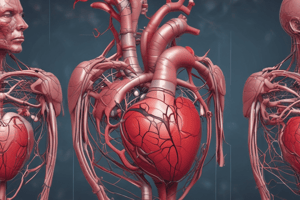
Precordium and Heart Anatomy
- The precordium covers the anterior chest area, extending from the second to the fifth intercostal space. It includes the sternum and medial parts of the left ribs.
- The superior portion of the heart is referred to as the base.
- The apex of the heart designates its pointed end.
Heart Sounds and Cardiac Cycle
- Significant intercostal spaces for auscultation of heart sounds are the 2nd to 5th intercostal spaces.
- The apical impulse results from the left ventricle's pulsation against the chest wall during systole.
Valves and Their Function
- During systole, the atrioventricular (AV) valves, comprising the tricuspid and mitral valves, close to prevent backflow of blood into the atria.
- The semilunar valves (aortic and pulmonic valves) open during systole, allowing blood to exit the ventricles.
Cardiac Contraction
- The ventricles are the parts of the heart that contract during systole, coinciding with the first heart sound (S1).
Diastole and Cardiac Output
- In diastole, the semilunar valves close, and the ventricles relax, preparing for the next cycle.
- Cardiac output is calculated as stroke volume multiplied by heart rate (CO = SV x HR).
Blood Pressure and Preload
- Blood pressure is determined by the equation: Blood Pressure = Cardiac Output x Systemic Vascular Resistance (CO x SVR).
- Preload refers to the degree of stretch on the ventricles at the end of diastole, critical for determining stroke volume and cardiac output.
Circulatory System Concepts
- Afterload: The pressure the heart must overcome to eject blood into systemic circulation.
- Pulse: Generated by pressure waves traveling through the arteries as blood is pumped from the heart.
Chest Pain Related to Ischemia
- Types of ischemic chest pain include stable angina, unstable angina, variant (Prinzmetal's) angina, and myocardial infarction.
- Chest pains occurring at rest: unstable angina and variant (Prinzmetal's) angina.
- Non-ischemic cardiac pain can arise from mitral valve prolapse, pericarditis, and dissecting aneurysms.
Mitral Valve Prolapse
- Characterized by loose mitral valves that can prolapse back into the left atrium, potentially leading to mitral regurgitation.
Causes of Chest Pain
- Pulmonary Causes: Include pulmonary embolism, pleurisy, pulmonary hypertension, pneumothorax, and mediastinal emphysema.
- Gastrointestinal Causes: Can involve esophageal spasms, esophageal reflux, and gallstone colic.
Dyspnea
- Common types of dyspnea: exertional dyspnea, orthopnea, paroxysmal nocturnal dyspnea, and continuous dyspnea.
- Pink, frothy sputum is typically associated with pulmonary edema.
- Sudden awakening feeling unable to breathe is indicative of paroxysmal nocturnal dyspnea, often relieved by movement.
Cardiac Conditions
- Mitral stenosis, mitral regurgitation, and aortic stenosis all exhibit symptoms of exertional dyspnea.
Heart Structure and Function
- The heart’s apex extends from the second to the fifth intercostal space, central to the sternum and the left ribs.
- Apical impulse is the heartbeat felt at the apex of the heart.
- Cardiac cycle:
- Closure of the AV valves (tricuspid and mitral) occurs during ventricular systole.
- Opening of the semilunar valves (aortic and pulmonic) allows blood ejection during ventricular contraction.
- Following contraction, the semilunar valves close, marked by ventricular relaxation.
Hemodynamics
- Cardiac output (CO) is calculated as Stroke Volume (SV) multiplied by Heart Rate (HR).
- Blood pressure is determined by CO multiplied by Systemic Vascular Resistance (SVR).
- Preload represents the stretch of ventricles at the end of diastole.
Ischemic Chest Pain Types
- Stable Angina: Predictable chest pain triggered by physical activity or stress.
- Unstable Angina: Occurs unexpectedly, often at rest, indicating a high risk of heart attack.
- Variant (Prinzmetal's) Angina: Caused by coronary artery spasms, can occur at rest and often cyclically.
- Myocardial Infarction: Heart attack resulting from prolonged ischemia.
Chest Pain at Rest
- Unstable Angina: Characterized by abrupt and unpredictable chest pain at rest.
- Variant (Prinzmetal's) Angina: Chest pain at rest due to vasospasm of coronary arteries.
Cardiac Pain Not Caused by Ischemia
- Mitral Valve Prolapse: Involves loose mitral valves prolapsing back into the left atrium.
- Pericarditis: Inflammation of the pericardium causing sharp chest pain.
- Dissecting Aneurysm: Sudden severe chest pain due to a tear in the aorta.
Chest Pain from Pulmonary Causes
- Pulmonary Embolism: Blockage in a pulmonary artery, leading to sharp chest pain and difficulty breathing.
- Pleurisy: Inflammation of the pleura causing pain with breathing.
- Pulmonary Hypertension: Elevated blood pressure in the lungs leading to chest discomfort.
- Pneumothorax: Air leaks into the pleural space causing sudden sharp pain and shortness of breath.
- Mediastinal Emphysema: Air trapped in the mediastinum leading to chest pain.
Chest Pain from Gastrointestinal Causes
- Esophageal Spasms: Irregular muscle contractions of the esophagus causing severe pain.
- Esophageal Reflux: Acid reflux leading to burning chest pain.
- Gallstone Colic: Pain due to gallstones, often felt in the upper abdomen and may radiate to the chest.
Dyspnea Types
- Orthopnea: Difficulty breathing while lying down, often relieved by sitting.
- Paroxysmal Nocturnal Dyspnea (PND): Sudden nighttime breathlessness that awakens the patient.
- Pulmonary Edema: Fluid accumulation in the lungs causing severe shortness of breath.
- Valvular Heart Disease: Chest pressure and shortness of breath due to heart valve abnormalities.
Dyspnea with Specific Characteristics
- Pink Frothy Sputum: Typically associated with pulmonary edema, indicating severe fluid accumulation.
- PND Episodes: Often lead to sudden awakenings during sleep due to difficulty breathing, resolved by pacing.
Similar Symptom in Mitral Conditions
- Mitral Stenosis, Mitral Regurgitation, Aortic Stenosis: These conditions commonly present with dyspnea as a shared symptom due to impaired heart function.
Physical Assessment Questions
- Systolic blood pressure (SBP) is generated by the left ventricle during ventricular contraction.
- Diastolic blood pressure (DBP) remains in the arterial tree while the heart is at rest.
- Orthostatic hypotension is characterized by a drop in SBP of ≥ 20 mm Hg or DBP of ≥ 10 mm Hg within 3 minutes of standing up.
- Jugular venous distention can be caused by right ventricular failure, pulmonary hypertension, pulmonary embolism, or cardiac tamponade.
- Normal jugular venous pressure (JVP) measurement is 2-3 cm H2O above the sternal angle or 6-8 cm H2O when measured in relation to the right atrium.
- In left ventricular dilation (volume overload), the point of maximal impulse (PMI) is displaced laterally and to a lower intercostal space.
- Left ventricular hypertrophy (pressure overload) leads to an increased force and duration of the PMI.
- Auscultatory areas of the heart follow the mnemonic APE To MAN: Aortic, Pulmonary, Erb’s point, Tricuspid, Mitral.
- The diaphragm of the stethoscope is best for detecting high-pitched sounds, while the bell is optimal for low-pitched sounds.
Heart Sounds Questions
- S1 is loudest at the apex of the heart and marks the beginning of systole.
- S2 marks the beginning of diastole.
- Abnormal heart sounds include gallops, specifically S3 and S4 sounds.
- S3 sound occurs due to blood from the left atrium slamming into an already filled ventricle, classified as a physiologic or pathological finding.
- S3 is best heard when the patient is in the left lateral recumbent position.
- The earliest physical sign of heart failure is the presence of S3 heart sounds.
- True, S3 heart sounds can be normal in children, young adults, and pregnant women.
- False, S3 heart sounds in elderly individuals typically indicate heart failure or volume overload.
- S4 heart sounds occur during late diastole, just before S1, during atrial contraction.
- S4 is characterized by blood being forced into a stiff, hypertrophic noncompliant ventricle.
- The rhythmic pattern of S4 heart sounds can be described as "A-Stiff-Wall" (S4-S1-S2).
- The palpable pulse is best felt after the S1 heart sound.
- Non-pathologic or functional murmurs are most common during conditions such as pregnancy, fever, and anemia.
- Murmurs are described as blowing or swooshing sounds indicating turbulent blood flow in the heart or great vessels.
- Heart valve changes leading to murmurs include valve stenosis and regurgitation, mitral valve prolapse, and congenital heart defects.
- In pediatric patients, heart sounds are typically found in the 4th intercostal space.
Studying That Suits You
Use AI to generate personalized quizzes and flashcards to suit your learning preferences.