Podcast
Questions and Answers
Which of the following conditions is characterized by an abnormal rhythm of the heart?
Which of the following conditions is characterized by an abnormal rhythm of the heart?
- Heart block
- Tachycardia
- Cardiac dysrhythmia (correct)
- Bradycardia
What does the P wave on an ECG represent?
What does the P wave on an ECG represent?
- Depolarization of the ventricles
- Repolarization of the atria
- Depolarization of the atria (correct)
- Repolarization of the ventricles
What is the primary treatment for heart block?
What is the primary treatment for heart block?
- Implanting an artificial pacemaker (correct)
- Surgical intervention to repair heart valves
- Medication to regulate heart rate
- Electrical cardioversion
How is cardiac output (CO) determined?
How is cardiac output (CO) determined?
What does an increase in venous return directly influence?
What does an increase in venous return directly influence?
What is ventricular fibrillation?
What is ventricular fibrillation?
Which system can alter the heart rate to increase or decrease it?
Which system can alter the heart rate to increase or decrease it?
What does the T wave on an ECG indicate?
What does the T wave on an ECG indicate?
What is the role of the coronary arteries in the heart?
What is the role of the coronary arteries in the heart?
What is primarily caused by a blockage in coronary arteries?
What is primarily caused by a blockage in coronary arteries?
What condition is characterized by the hardening of arteries due to lipid buildup?
What condition is characterized by the hardening of arteries due to lipid buildup?
What is the average heartbeat rate referred to in the cardiac cycle?
What is the average heartbeat rate referred to in the cardiac cycle?
What structure in the heart acts as the primary pacemaker?
What structure in the heart acts as the primary pacemaker?
Which part of the heart is responsible for sending electrical signals to the ventricles?
Which part of the heart is responsible for sending electrical signals to the ventricles?
What does angina pectoris signify in relation to heart health?
What does angina pectoris signify in relation to heart health?
What is the duration of a complete cardiac cycle?
What is the duration of a complete cardiac cycle?
How many layers does the pericardium have and what are their names?
How many layers does the pericardium have and what are their names?
What is cardiac tamponade?
What is cardiac tamponade?
What is the role of valves in the heart?
What is the role of valves in the heart?
What type of valve is found at the opening between the right atrium and right ventricle?
What type of valve is found at the opening between the right atrium and right ventricle?
What causes the 'dup' sound in heartbeats?
What causes the 'dup' sound in heartbeats?
What is rheumatic heart disease primarily associated with?
What is rheumatic heart disease primarily associated with?
What is a common characteristic of heart murmurs?
What is a common characteristic of heart murmurs?
How do the right and left sides of the heart differ in function?
How do the right and left sides of the heart differ in function?
Study Notes
The Pericardium
- The pericardium has two layers: the visceral pericardium (epicardium) and the parietal pericardium.
- Inflammation of the pericardium is called pericarditis.
- Cardiac tamponade is caused by fluid buildup between the visceral and parietal pericardium, compressing the heart.
Heart Contractions
- Systole is the contraction phase of the heart.
- Diastole is the relaxation phase of the heart.
Heart Valves
- Valves prevent blood backflow and ensure unidirectional blood flow.
- There are two types of valves: atrioventricular (AV) valves and semilunar (SL) valves.
- The AV valves, tricuspid and bicuspid (mitral) valves, are located at the openings of the right and left atria into their respective ventricles.
- The SL valves, pulmonary and aortic, are located at the beginnings of the pulmonary artery and aorta, respectively.
- Valve leak can lead to backflow into the previous chamber.
- Stenosed valves are narrower valves that reduce blood flow, leading to valve disorder.
Valve Disorders
- Rheumatic heart disease damages the heart due to a delayed inflammatory response to streptococcal infection.
- Mitral valve prolapse is an incompetent mitral valve, where the edges extend into the left atrium during left ventricle contraction.
Heart Sounds
- The two heart sounds, "Lub" and "Dup," are caused by the vibrations and closure of heart valves.
- "Lub" is caused by the closure of AV valves during ventricular contraction.
- "Dup" is caused by the closure of SL valves during ventricular relaxation.
- Abnormal heart sounds, often caused by abnormal valves, are called heart murmurs.
Right vs. Left Heart
- The heart acts as two separate pumps, with the right and left sides performing different functions.
Blood Flow through the Heart
- Venous blood enters the right atrium through the superior and inferior venae cavae.
- It flows through the tricuspid valve into the right ventricle, then through the pulmonary semilunar valve into the pulmonary artery to the lungs.
- Oxygenated blood from the lungs enters the left atrium, flows through the bicuspid (mitral) valve into the left ventricle.
- Lastly, the left ventricle pumps blood through the aortic semilunar valve into the aorta for distribution throughout the body.
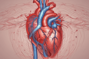
Coronary Circulation
- Coronary circulation refers to the blood flow through the right and left coronary arteries, supplying oxygen and nutrients to the myocardium (heart muscle).
Blockage of Coronary Arteries
- Blockage of blood flow through coronary arteries can cause myocardial infarction (heart attack).
- Coronary thrombosis and coronary embolism are caused by blood clots in the coronary arteries.
Coronary Heart Disease (CHD)
- CHD is a condition where blood cannot flow through the heart, leading to cell death and myocardial infarction.
- Atherosclerosis, a type of hardening of the arteries, is a common cause of CHD, where lipids build up on the inside walls of blood vessels, potentially blocking coronary blood flow.
- Angina pectoris is chest pain caused by lack of oxygen to the heart, and is a type of CHD.
Coronary Bypass Surgery
- This procedure treats patients with severely restricted coronary artery blood flow.
- Veins or vessels from other parts of the body are used to bypass blockages in the coronary artery.
Cardiac Cycle
- Each complete heartbeat is called a cardiac cycle, which typically lasts 0.8 seconds.
- The cardiac cycle is divided into systole (contraction) and diastole (relaxation).
- The average heart rate (bpm) is 72 beats per minute.
Pacemaker of the Heart
- The SA node, located in the wall of the right atrium near the superior vena cava opening, is the pacemaker of the heart.
Conduction Pathway of Electrical Signals
- The AV node is located in the right atrium along the lower part of the interatrial septum.
- The Atrial bundle (bundle of His) is located in the septum of the ventricle.
- Purkinje fibers are located in the walls of the ventricles (subendocardial).
Electrocardiography (ECG)
- ECG records electrical signals in the heart, displayed as visible tracings.
- The P wave represents atrial depolarization.
- The QRS complex represents ventricular depolarization.
- The T wave represents ventricular repolarization.
Cardiac Dysrhythmia
- This refers to any abnormality of heart rhythm.
- Heart block is a type of dysrhythmia where impulse conduction is blocked.
- Heart block can be treated by implanting an artificial pacemaker.
Heart Rate Variations
- Bradycardia is a slow heart rate (less than 60 bpm).
- Tachycardia is a rapid heart rate (more than 100 bpm).
- Sinus dysrhythmia refers to variations in heart rate during the breathing cycle.
- Premature contraction (extra systole) is a contraction that occurs sooner than expected in a normal heart rhythm.
Fibrillation
- Fibrillation occurs when cardiac muscle fibers are out of sync, leading to ineffective pumping action.
- Atrial fibrillation (AF or A-Fib) can occur in mitral stenosis, rheumatic heart disease, and atrial myocardial infarction.
- Ventricular fibrillation (VF or V-fib) is life-threatening, as it stops blood flow to vital tissues.
Treatments for Arrhythmias
- Defibrillation involves delivering an electric shock to restore normal rhythm in the heart.
- Atrial ablation destroys heart muscle in a specific location to eliminate the pathway of abnormal electrical signals, used to treat atrial fibrillation.
Blood Volume and Cardiac Output
- Stroke volume is the volume of blood ejected from one ventricle with each heartbeat.
- Cardiac output (CO) is the amount of blood pumped by one ventricle each minute (about 5 L/min at rest).
- CO is determined by the heart rate (HR) and stroke volume (SV): CO = HR x SV.
- The autonomic nervous system regulates heart rhythm to increase or decrease CO.
Factors Affecting Heart Rate
- Rising blood CO2 levels during exercise increase heart rate to restore homeostasis.
- Sudden drops in blood pressure increase heart rate to restore normal blood flow.
Stroke Volume Factors
- The volume of blood ejected by the ventricles is determined by the volume of blood returned to the heart by the veins (venous return).
- Higher venous return leads to higher stroke volume.
Studying That Suits You
Use AI to generate personalized quizzes and flashcards to suit your learning preferences.
Related Documents
Description
This quiz covers key concepts about the pericardium, heart contractions, and heart valves. Understand the structure, functions, and common conditions affecting the heart, including pericarditis and valve disorders. Test your knowledge on how the heart operates through its contraction and relaxation phases.