Podcast
Questions and Answers
Which symptom is a common manifestation of cardiac tamponade?
Which symptom is a common manifestation of cardiac tamponade?
- Hypothermia
- Cyanosis
- Dyspnea (correct)
- Bradycardia
Jugular Venous Distension is a sign of decreased pressures in the venous system.
Jugular Venous Distension is a sign of decreased pressures in the venous system.
False (B)
What procedure is both diagnostic and therapeutic for cardiac tamponade?
What procedure is both diagnostic and therapeutic for cardiac tamponade?
Pericardiocentesis
__________ is characterized by a drop in systolic blood pressure greater than 10 mmHg during inspiration.
__________ is characterized by a drop in systolic blood pressure greater than 10 mmHg during inspiration.
Match the clinical manifestations with their descriptions:
Match the clinical manifestations with their descriptions:
Which of the following is NOT a component of Beck's triad?
Which of the following is NOT a component of Beck's triad?
Electrical alternans is a key EKG finding in cardiac tamponade.
Electrical alternans is a key EKG finding in cardiac tamponade.
What is the primary imaging method used to diagnose cardiac tamponade?
What is the primary imaging method used to diagnose cardiac tamponade?
Signs to stop administering fluids include Kussmaul's sign, where jugular venous pressure remains elevated during __________.
Signs to stop administering fluids include Kussmaul's sign, where jugular venous pressure remains elevated during __________.
Which medication can be used to improve cardiac output in patients with cardiac tamponade?
Which medication can be used to improve cardiac output in patients with cardiac tamponade?
What is the primary difference between cardiac tamponade and pericardial effusion?
What is the primary difference between cardiac tamponade and pericardial effusion?
Cardiac tamponade can result in cardiogenic shock.
Cardiac tamponade can result in cardiogenic shock.
Name one potential cause of hemopericardium.
Name one potential cause of hemopericardium.
The condition that results from rapid fluid accumulation in the pericardial cavity is called __________.
The condition that results from rapid fluid accumulation in the pericardial cavity is called __________.
Match the causes of serous and serosanguinous effusions with their descriptions:
Match the causes of serous and serosanguinous effusions with their descriptions:
Which of the following is NOT a cause of cardiac tamponade?
Which of the following is NOT a cause of cardiac tamponade?
Pericardial effusion can occur slowly, allowing for pericardial stretching.
Pericardial effusion can occur slowly, allowing for pericardial stretching.
What effect does cardiac tamponade have on stroke volume?
What effect does cardiac tamponade have on stroke volume?
Rapid fluid accumulation in the pericardial cavity increases __________ on the heart.
Rapid fluid accumulation in the pericardial cavity increases __________ on the heart.
Which infectious agent is known to cause pericarditis?
Which infectious agent is known to cause pericarditis?
What is the primary physiological consequence of cardiac tamponade?
What is the primary physiological consequence of cardiac tamponade?
Cardiac tamponade can be caused by aortic dissection.
Cardiac tamponade can be caused by aortic dissection.
What condition results from gradual fluid accumulation in the pericardial cavity?
What condition results from gradual fluid accumulation in the pericardial cavity?
The accumulation of blood in the pericardial cavity is referred to as __________.
The accumulation of blood in the pericardial cavity is referred to as __________.
Match the cause of cardiac tamponade with its description:
Match the cause of cardiac tamponade with its description:
Which of the following can lead to serous and serosanguinous effusions?
Which of the following can lead to serous and serosanguinous effusions?
Cardiac tamponade is a less severe condition than pericardial effusion.
Cardiac tamponade is a less severe condition than pericardial effusion.
Name a viral agent that can cause pericarditis.
Name a viral agent that can cause pericarditis.
Rapid fluid accumulation leads to elevated __________ on the heart.
Rapid fluid accumulation leads to elevated __________ on the heart.
Which of the following describes the distinction between cardiac tamponade and pericardial effusion?
Which of the following describes the distinction between cardiac tamponade and pericardial effusion?
What is the primary goal of initial management for cardiac tamponade?
What is the primary goal of initial management for cardiac tamponade?
Electrical alternans in the QRS complexes indicates a stable hemodynamic status.
Electrical alternans in the QRS complexes indicates a stable hemodynamic status.
What is the gold standard for diagnosing cardiac tamponade?
What is the gold standard for diagnosing cardiac tamponade?
Pulsus __________ is indicated by a drop in systolic blood pressure during inspiration.
Pulsus __________ is indicated by a drop in systolic blood pressure during inspiration.
Match the following clinical manifestations with their descriptions:
Match the following clinical manifestations with their descriptions:
Which of the following tests is useful for identifying changes in QRS voltage due to fluid interference?
Which of the following tests is useful for identifying changes in QRS voltage due to fluid interference?
Pericardiocentesis can be both diagnostic and therapeutic for cardiac tamponade.
Pericardiocentesis can be both diagnostic and therapeutic for cardiac tamponade.
What is a potential sign to stop administering fluids in the case of cardiac tamponade?
What is a potential sign to stop administering fluids in the case of cardiac tamponade?
Patients may exhibit rapid, deep breaths, seeking to improve __________.
Patients may exhibit rapid, deep breaths, seeking to improve __________.
What may be observed on a chest X-ray in cases of pericardial effusion?
What may be observed on a chest X-ray in cases of pericardial effusion?
What is the primary cause of hemopericardium resulting from myocardial infarction?
What is the primary cause of hemopericardium resulting from myocardial infarction?
Cardiac tamponade allows the heart to fill effectively due to gradual fluid accumulation.
Cardiac tamponade allows the heart to fill effectively due to gradual fluid accumulation.
Name one infectious agent that can cause pericarditis.
Name one infectious agent that can cause pericarditis.
The accumulation of __________ in the pericardial cavity can lead to cardiac tamponade.
The accumulation of __________ in the pericardial cavity can lead to cardiac tamponade.
Match the conditions causing hemopericardium with their descriptions:
Match the conditions causing hemopericardium with their descriptions:
Which of the following is a potential cause of serous effusions?
Which of the following is a potential cause of serous effusions?
Cardiac tamponade can lead to decreased stroke volume and cardiac output.
Cardiac tamponade can lead to decreased stroke volume and cardiac output.
What is the primary physiological consequence of rapid fluid accumulation in the pericardial cavity?
What is the primary physiological consequence of rapid fluid accumulation in the pericardial cavity?
Pericardial effusion can progress gradually, allowing for __________ of the pericardium.
Pericardial effusion can progress gradually, allowing for __________ of the pericardium.
Which condition can result from chronic kidney disease leading to effusions?
Which condition can result from chronic kidney disease leading to effusions?
What is the primary purpose of performing a pericardiocentesis in a patient with cardiac tamponade?
What is the primary purpose of performing a pericardiocentesis in a patient with cardiac tamponade?
Electrical alternans in the QRS complexes indicates instability in hemodynamic status.
Electrical alternans in the QRS complexes indicates instability in hemodynamic status.
What is a typical clinical presentation of a patient with cardiac tamponade?
What is a typical clinical presentation of a patient with cardiac tamponade?
The condition characterized by fluid accumulation in the pericardial space is called __________.
The condition characterized by fluid accumulation in the pericardial space is called __________.
Match the following clinical manifestations with their descriptions:
Match the following clinical manifestations with their descriptions:
Which of the following is a sign of elevated right-sided pressures seen in cardiac tamponade?
Which of the following is a sign of elevated right-sided pressures seen in cardiac tamponade?
Chest X-ray is definitive for diagnosing cardiac tamponade.
Chest X-ray is definitive for diagnosing cardiac tamponade.
What is the role of norepinephrine in the management of cardiac tamponade?
What is the role of norepinephrine in the management of cardiac tamponade?
Patients showing signs to stop fluids may present with __________ during inspiration.
Patients showing signs to stop fluids may present with __________ during inspiration.
Which echocardiogram finding suggests cardiac tamponade?
Which echocardiogram finding suggests cardiac tamponade?
Which of the following is a common cause of hemopericardium?
Which of the following is a common cause of hemopericardium?
Cardiac tamponade always develops gradually.
Cardiac tamponade always develops gradually.
What condition results from elevated pressure in the pericardial cavity that affects the heart's filling capacity?
What condition results from elevated pressure in the pericardial cavity that affects the heart's filling capacity?
The accumulation of blood in the pericardial cavity is called __________.
The accumulation of blood in the pericardial cavity is called __________.
Match the causes of cardiac tamponade with their descriptions:
Match the causes of cardiac tamponade with their descriptions:
Which infectious agent is known to cause pericarditis?
Which infectious agent is known to cause pericarditis?
Pericardial effusion allows for gradual stretching of the pericardium.
Pericardial effusion allows for gradual stretching of the pericardium.
Increased intrapericardial pressure compression diminishes __________ and cardiac output.
Increased intrapericardial pressure compression diminishes __________ and cardiac output.
Name one factor that can lead to serous effusions.
Name one factor that can lead to serous effusions.
Match the following conditions causing hemopericardium with their descriptions:
Match the following conditions causing hemopericardium with their descriptions:
What is the most common initial treatment for cardiac tamponade?
What is the most common initial treatment for cardiac tamponade?
Muffled heart sounds are a sign of cardiac tamponade.
Muffled heart sounds are a sign of cardiac tamponade.
Name one key clinical feature associated with pulsus paradoxus.
Name one key clinical feature associated with pulsus paradoxus.
The accumulation of __________ in the pericardial cavity can lead to cardiac tamponade.
The accumulation of __________ in the pericardial cavity can lead to cardiac tamponade.
Match the clinical findings to their descriptions:
Match the clinical findings to their descriptions:
Which imaging method is considered the gold standard for diagnosing cardiac tamponade?
Which imaging method is considered the gold standard for diagnosing cardiac tamponade?
JVD indicates decreased blood pressure in the venous system.
JVD indicates decreased blood pressure in the venous system.
What is the significance of electrical alternans in EKG findings?
What is the significance of electrical alternans in EKG findings?
Fluid removal that improves hemodynamics confirms the diagnosis of __________.
Fluid removal that improves hemodynamics confirms the diagnosis of __________.
Which of the following medications is used to improve cardiac output in cardiac tamponade?
Which of the following medications is used to improve cardiac output in cardiac tamponade?
What is a common cause of hemopericardium following myocardial infarction?
What is a common cause of hemopericardium following myocardial infarction?
Cardiac tamponade can occur due to penetrating trauma to the heart.
Cardiac tamponade can occur due to penetrating trauma to the heart.
What physiological effect does cardiac tamponade have on diastolic filling of the heart?
What physiological effect does cardiac tamponade have on diastolic filling of the heart?
A rapid increase in intrapericardial pressure leads to decreased __________ and cardiac output.
A rapid increase in intrapericardial pressure leads to decreased __________ and cardiac output.
Match the following conditions with their potential causes:
Match the following conditions with their potential causes:
Which of the following is a cause of serous effusions?
Which of the following is a cause of serous effusions?
Cardiac tamponade results from a slow accumulation of fluid in the pericardial cavity.
Cardiac tamponade results from a slow accumulation of fluid in the pericardial cavity.
Name one infectious agent that may cause pericarditis.
Name one infectious agent that may cause pericarditis.
The condition characterized by fluid accumulation in the pericardial cavity that can lead to cardiogenic shock is known as __________.
The condition characterized by fluid accumulation in the pericardial cavity that can lead to cardiogenic shock is known as __________.
Match the causes of cardiac tamponade with their effects:
Match the causes of cardiac tamponade with their effects:
Which of the following findings is indicative of cardiac tamponade?
Which of the following findings is indicative of cardiac tamponade?
Pulsus paradoxus occurs when there is an increase in systolic blood pressure during inspiration.
Pulsus paradoxus occurs when there is an increase in systolic blood pressure during inspiration.
What is the preferred imaging technique for diagnosing cardiac tamponade?
What is the preferred imaging technique for diagnosing cardiac tamponade?
In cardiac tamponade, blood pressure changes during inspiration and expiration confirm the presence of __________.
In cardiac tamponade, blood pressure changes during inspiration and expiration confirm the presence of __________.
Match the following symptoms with their descriptions:
Match the following symptoms with their descriptions:
What is the primary initial management strategy for a patient with cardiac tamponade?
What is the primary initial management strategy for a patient with cardiac tamponade?
Beck's triad consists of hypotension, elevated jugular venous pressure, and muffled heart sounds.
Beck's triad consists of hypotension, elevated jugular venous pressure, and muffled heart sounds.
Name a potential sign that may indicate the need to stop administering fluids in a patient with cardiac tamponade.
Name a potential sign that may indicate the need to stop administering fluids in a patient with cardiac tamponade.
__________ serves as both a diagnostic and therapeutic procedure for cardiac tamponade.
__________ serves as both a diagnostic and therapeutic procedure for cardiac tamponade.
Match the clinical features with their implications:
Match the clinical features with their implications:
Flashcards are hidden until you start studying
Study Notes
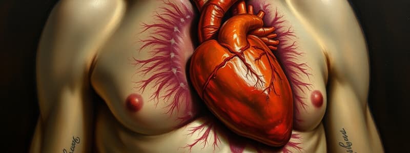
Cardiac Tamponade Overview
- Cardiac tamponade is a critical condition resulting from rapid fluid accumulation in the pericardial cavity, leading to increased pressure on the heart.
- This condition hinders the heart's ability to fill effectively, potentially resulting in cardiogenic shock.
Distinction: Cardiac Tamponade vs. Pericardial Effusion
- Both conditions involve fluid accumulation in the pericardial cavity, but they differ in severity and physiological implications.
- Pericardial effusion can be gradual and allow for pericardial stretching, while tamponade results from rapid fluid buildup or excessive volume that restricts stretching.
Causes of Hemopericardium
- Myocardial Infarction (MI): Left ventricular wall rupture can occur post-MI, leading to sudden blood accumulation.
- Trauma: Penetrating trauma causing direct injury to the heart chambers allows blood to enter the pericardial space.
- Aortic Dissection: A tear in the aorta can lead to blood tracking between layers and accumulating in the pericardial cavity.
- Cardiac Surgery: Surgical procedures can inadvertently result in bleeding into the pericardial cavity.
Causes of Serous and Serosanguinous Effusions
- Infectious Agents: Viral (e.g., Coxsackie B virus, COVID-19) and bacterial infections (e.g., staphylococcus, tuberculosis) can cause pericarditis, leading to effusions.
- Neoplasias: Cancer can metastasize to pericardial tissues, resulting in inflammatory changes and fluid accumulation.
- Chronic Kidney Disease: High urea levels from kidney failure can lead to uremic pericarditis and effusions.
- Autoimmune Disorders: Conditions like systemic lupus erythematosus may cause inflammatory effusions.
Pathophysiology of Cardiac Tamponade
- Elevated intrapericardial pressure compresses the heart, severely reducing diastolic filling, which diminishes stroke volume and cardiac output.
- Symptoms such as dyspnea arise as patients attempt to increase venous return by taking deep breaths, which momentarily improves hemodynamics but fails to resolve the underlying issue.
Clinical Manifestations
- Hypotension: Due to reduced cardiac output from restricted filling.
- Tachycardia: Reflexive increase from low blood pressure.
- Jugular Venous Distension: Increased pressure in the venous system due to impaired right heart filling.
- Dyspnea: Patients may exhibit rapid, deep breaths, seeking to augment venous return.
- Muffled Heart Sounds: Caused by the presence of excess fluid in the pericardial space; part of Beck’s triad (Hypotension, JVD, decreased heart sounds).
- Pulsus Paradoxus: A drop in systolic blood pressure greater than 10 mmHg during inspiration, indicative of significant compromise in hemodynamics.
Physical Exam Findings
- Beck’s Triad: Hypotension, elevated jugular venous pressure, and muffled heart sounds are classic indicators of cardiac tamponade.
- Hepatomegaly and Ascites: Signs of systemic venous pressure elevation due to impaired filling of the right side of the heart.
- Pitting Edema: Fluid buildup in the lower extremities may occur.
Diagnostic Approach
- Obtain a 12-lead ECG: To identify tachycardia and possible low voltage QRS complexes due to pericardial effusion.
- Investigate for changes in QRS voltage: Decreased voltage can be observed due to fluid interference with electrical conduction.
- Look for signs of hemodynamic instability: Assess blood pressure changes during inspiration and expiration to confirm the presence of pulsus paradoxus.
Summary
- Cardiac tamponade represents a serious medical emergency driven by pressure on the heart, causing significant functional compromise, with critical clinical signs and vital diagnostic features that require prompt identification and management.### Electrical Activity and Cardiac Tamponade
- Electrical alternans observed in QRS complexes, leading to alternating large and small voltage patterns.
- Key EKG findings in cardiac tamponade include tachycardia, low QRS voltage, and electrical alternans.
- EKG not definitive for diagnosis, but useful for ruling out other conditions.
- Chest X-ray may show heart enlargement (cardiomegaly) and a "water bottle sign" from fluid in the pericardial cavity, but is also not diagnostic.
Diagnostic Imaging for Cardiac Tamponade
- Echocardiogram (echo) is the gold standard for diagnosing cardiac tamponade.
- Essential echo findings include identifying pericardial effusion, right ventricular (RV) collapse, and right atrial (RA) collapse during diastole.
- A plethoric inferior vena cava (IVC) indicates elevated right-sided pressures.
- Septal shift observed during inspiration as the interventricular septum bows from the right to the left ventricle, indicating compromised filling.
Clinical Presentation and Management
- Typical presentation: dyspnea, signs of shock, and potential cardiac arrest.
- Initial management focuses on stabilizing mean arterial pressure (MAP) with intravenous fluids and vasopressors.
- Signs to stop fluids include the Kussmaul's sign, where jugular venous pressure remains elevated during inspiration.
Supportive Measures
- Norepinephrine and epinephrine can increase systemic vascular resistance and improve cardiac output by stimulating alpha-1 and beta-1 receptors.
- Avoid intubation if possible, as natural inspiratory efforts support venous return; if intubated, maintain low positive end-expiratory pressure (PEEP) to reduce intrathoracic pressure.
Definitive Treatment
- Pericardiocentesis serves as both a diagnostic and therapeutic procedure.
- If hemodynamics improve after fluid removal, cardiac tamponade is diagnosed.
- Patients with rapidly reaccumulating effusions may require a pericardial drain via pericardial window.
Conclusion
- Manage cardiac tamponade by defending MAP, performing pericardiocentesis, and addressing underlying causes.
- Continuous monitoring and collaboration with cardiology are critical for optimal patient outcomes.
Cardiac Tamponade Overview
- Critical condition from rapid fluid accumulation in the pericardial cavity increases pressure on the heart.
- Impaired heart filling can lead to cardiogenic shock.
Distinction: Cardiac Tamponade vs. Pericardial Effusion
- Cardiac tamponade is acute and limits the heart’s ability to stretch due to rapid fluid accumulation.
- Pericardial effusion develops gradually, allowing the pericardium to stretch without immediate life-threatening effects.
Causes of Hemopericardium
- Myocardial Infarction: Left ventricular wall rupture leads to sudden blood accumulation.
- Trauma: Direct heart chamber injury from penetrating trauma allows blood entry into the pericardial space.
- Aortic Dissection: A tear in the aorta allows blood to accumulate in the pericardial cavity.
- Cardiac Surgery: Surgical interventions may unintentionally cause bleeding into the pericardial space.
Causes of Serous and Serosanguinous Effusions
- Infectious Agents: Viral (e.g., Coxsackie B, COVID-19) and bacterial infections (e.g., staphylococcus, tuberculosis) can lead to effusions from pericarditis.
- Neoplasias: Cancer can metastasize to pericardial tissue, resulting in fluid accumulation.
- Chronic Kidney Disease: High urea levels can cause uremic pericarditis and effusions.
- Autoimmune Disorders: Conditions like systemic lupus erythematosus may lead to fluid accumulation.
Pathophysiology of Cardiac Tamponade
- Elevated intrapericardial pressure compresses the heart, significantly reducing diastolic filling and stroke volume.
- Symptoms such as dyspnea may arise as patients attempt deep breaths to temporarily improve blood flow.
Clinical Manifestations
- Hypotension: Caused by reduced cardiac output due to filling restrictions.
- Tachycardia: Reflexive response to low blood pressure.
- Jugular Venous Distension: Elevated venous pressure due to impaired right heart filling.
- Dyspnea: Patients may take rapid, deep breaths in an attempt to increase venous return.
- Muffled Heart Sounds: Indicative of excess fluid, part of Beck’s triad (Hypotension, JVD, decreased heart sounds).
- Pulsus Paradoxus: A significant drop in systolic blood pressure (>10 mmHg) during inspiration, showcasing hemodynamic compromise.
Physical Exam Findings
- Beck’s Triad is critical: Hypotension, elevated jugular venous pressure, and muffled heart sounds.
- Hepatomegaly and ascites may indicate elevated systemic venous pressure.
- Pitting edema in lower extremities suggests fluid accumulation.
Diagnostic Approach
- 12-lead ECG: Identifies tachycardia and low voltage QRS complexes.
- Changes in QRS voltage: Decreased voltage may indicate fluid interference.
- Monitor blood pressure fluctuations during respiration to confirm pulsus paradoxus.
Electrical Activity and Cardiac Tamponade
- Electrical alternans seen in QRS complexes indicates fluctuating voltage patterns.
- Key EKG findings include tachycardia, low QRS voltage, and electrical alternans.
- A chest X-ray may show cardiomegaly and a "water bottle sign," indicating pericardial fluid.
Diagnostic Imaging for Cardiac Tamponade
- Echocardiogram is the gold standard for diagnosis.
- Essential findings: pericardial effusion, right ventricular collapse, and right atrial collapse during diastole.
- A plethoric inferior vena cava suggests elevated right-sided pressures.
Clinical Presentation and Management
- Typical symptoms include dyspnea, signs of shock, and risk of cardiac arrest.
- Initial treatment emphasizes stabilizing mean arterial pressure with IV fluids and vasopressors.
- Monitoring for Kussmaul's sign may inform when to stop fluid administration.
Supportive Measures
- Medications like norepinephrine and epinephrine improve cardiac output by enhancing systemic vascular resistance.
- Avoid intubation if feasible; if necessary, use low positive end-expiratory pressure (PEEP) to minimize thoracic pressure.
Definitive Treatment
- Pericardiocentesis is both diagnostic and therapeutic for cardiac tamponade.
- Improvement in hemodynamics post-fluid removal confirms diagnosis.
- Patients with recurrent effusions may require a pericardial drain or window.
Conclusion
- Effective management involves stabilizing MAP, performing pericardiocentesis, and addressing underlying causes.
- Continuous monitoring and collaboration with cardiology are vital for positive patient outcomes.
Studying That Suits You
Use AI to generate personalized quizzes and flashcards to suit your learning preferences.


