Podcast
Questions and Answers
What is a unique structural characteristic of cardiac muscle cells?
What is a unique structural characteristic of cardiac muscle cells?
- They are longer and more cylindrical than skeletal muscle cells.
- They are exclusively found in the atria of the heart.
- They form cellular networks through complex junctions called intercalated discs. (correct)
- They possess multiple nuclei arranged in a linear fashion.
How does the arrangement of myocardium facilitate blood flow through the heart?
How does the arrangement of myocardium facilitate blood flow through the heart?
- It separates oxygen-rich and oxygen-poor blood in different chambers.
- It consists of a spiral arrangement that squeezes blood through in specific directions. (correct)
- It allows blood to be pumped directly from the ventricles back into the veins.
- It creates a random flow direction that improves circulation efficiency.
What distinguishes cardiac muscle cells from other muscle types?
What distinguishes cardiac muscle cells from other muscle types?
- Their location, being restricted only to the heart.
- Their wide diameter and high contractile force.
- Their ability to regenerate quickly.
- The presence of intercalated discs and branching structure. (correct)
Which statement correctly describes the nuclei within cardiac muscle cells?
Which statement correctly describes the nuclei within cardiac muscle cells?
What part of the myocardium is described as being thicker and lower?
What part of the myocardium is described as being thicker and lower?
What is the primary function of intercalated discs in cardiac muscle tissue?
What is the primary function of intercalated discs in cardiac muscle tissue?
Which characteristic is NOT true about cardiac muscle cells?
Which characteristic is NOT true about cardiac muscle cells?
What does the myocardium consist of?
What does the myocardium consist of?
What orientation does the right surface of the myocardium face?
What orientation does the right surface of the myocardium face?
What surrounds the heart and the roots of the great vessels?
What surrounds the heart and the roots of the great vessels?
Which structural function is NOT associated with the pericardium?
Which structural function is NOT associated with the pericardium?
Which of the following is a primary function of the pericardium?
Which of the following is a primary function of the pericardium?
Which of the following structures provides support and attachment for the bases of valve cusps?
Which of the following structures provides support and attachment for the bases of valve cusps?
How does the pericardium contribute to the functioning of the heart?
How does the pericardium contribute to the functioning of the heart?
What could be a consequence if the pericardium is compromised?
What could be a consequence if the pericardium is compromised?
What condition results from an accumulation of more than 100 ml of fluid in the pericardial sac?
What condition results from an accumulation of more than 100 ml of fluid in the pericardial sac?
What happens to heart sounds in a patient with cardiac tamponade?
What happens to heart sounds in a patient with cardiac tamponade?
Which of the following is a symptom of cardiac tamponade?
Which of the following is a symptom of cardiac tamponade?
Which groove separates the right and left ventricles?
Which groove separates the right and left ventricles?
How does cardiac tamponade affect blood pressure?
How does cardiac tamponade affect blood pressure?
What is the significance of the pericardial fluid in maintaining heart function?
What is the significance of the pericardial fluid in maintaining heart function?
Which structure connects the heart at its base to the great blood vessels?
Which structure connects the heart at its base to the great blood vessels?
What proportion of the heart lies on the right side compared to the left side?
What proportion of the heart lies on the right side compared to the left side?
How is the heart described in terms of its shape and size?
How is the heart described in terms of its shape and size?
Where does the coronary sinus open in the right atrium?
Where does the coronary sinus open in the right atrium?
What guards the right atrioventricular orifice?
What guards the right atrioventricular orifice?
Which vein opens into the upper part of the right atrium and has no valve?
Which vein opens into the upper part of the right atrium and has no valve?
What is the function of the Eustachian valve associated with the inferior vena cava?
What is the function of the Eustachian valve associated with the inferior vena cava?
How does the coronary sinus function in relation to the myocardium?
How does the coronary sinus function in relation to the myocardium?
What is the blood return pathway from the lower half of the body to the heart?
What is the blood return pathway from the lower half of the body to the heart?
What is the significance of the tricuspid valve in the cardiovascular system?
What is the significance of the tricuspid valve in the cardiovascular system?
Which of the following statements about the inferior vena cava is correct?
Which of the following statements about the inferior vena cava is correct?
In which structure is the right atrioventricular orifice located?
In which structure is the right atrioventricular orifice located?
What is the primary function of the mitral valve?
What is the primary function of the mitral valve?
Which of the following best describes the anatomy of the aortic valve?
Which of the following best describes the anatomy of the aortic valve?
Which statement is true regarding the papillary muscles associated with the aortic fibrous ring?
Which statement is true regarding the papillary muscles associated with the aortic fibrous ring?
What defines the posterior aortic valve cusp?
What defines the posterior aortic valve cusp?
Which two arteries are primarily derived from the left coronary cusp?
Which two arteries are primarily derived from the left coronary cusp?
What is the significance of the aortic sinuses?
What is the significance of the aortic sinuses?
What determines the names of the aortic valve cusps?
What determines the names of the aortic valve cusps?
How does the anterior mitral valve cusp compare to the posterior mitral valve cusp?
How does the anterior mitral valve cusp compare to the posterior mitral valve cusp?
What is a key function of the fibrous rings associated with the heart valves?
What is a key function of the fibrous rings associated with the heart valves?
Flashcards are hidden until you start studying
Study Notes
Cardiac Tamponade
- Increased pericardial fluid (>100 ml) leads to cardiac tamponade, compressing the heart.
- Symptoms include jugular vein distension, muffled heart sounds, and hypotension.
- Jugular vein distension can occur even when seated upright due to pressure in the superior vena cava.
- Muffling of heart sounds results from fluid insulating sound travel, making it appear distant.
- Hypotension arises as the heart struggles to pump efficiently, risking shock or cardiac arrest.
Heart Structure
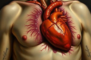
- The heart is a hollow, pyramid-shaped muscular organ located within the mediastinum and surrounded by the pericardium.
- It is roughly the size of a clenched fist, tilted left, with one-third on the right side and two-thirds on the left.
- Coronary arteries lie under the fat in the atrioventricular (AV) and interventricular grooves.
Myocardium
- The myocardium comprises cardiac muscle arranged in a spiral pattern, facilitating efficient blood movement.
- Cardiac muscle cells branch and join at intercalated discs, forming interconnected networks with a single nucleus.
- It separates the right and left ventricles, with the right side facing forward and the left side backward.
Right Atrium Features
- Contains four openings: superior vena cava, inferior vena cava, coronary sinus, and right atrioventricular orifice.
- Superior vena cava returns blood from the upper body; it has no valve.
- Inferior vena cava returns blood from the lower body and is safeguarded by a rudimentary valve (Eustachian valve), which can obstruct flow if prominent.
- The coronary sinus collects blood from the myocardium and drains into the right atrium.
Atrioventricular Valves
- The tricuspid valve (right) separates the right atrium from the right ventricle, preventing backflow during contraction.
- The mitral valve (left) guards the left atrioventricular orifice and consists of anterior and posterior cusps, with the anterior cusp being larger.
Aortic Valve
- Guards the aortic orifice with three cusps: right, left, and posterior.
- The left coronary cusp gives rise to the left main coronary artery, which splits into the circumflex and left anterior descending artery.
- The right coronary cusp originates the right coronary artery.
- Aortic sinuses behind each cusp facilitate coronary artery blood flow.
Function of Atrioventricular Rings
- Fibrous rings separate atrial and ventricular muscular walls but provide attachment points for muscle fibers.
- They prevent valve stretching and maintain open valve orifices during the heart cycle.
- These rings offer electrical insulation, ensuring electrical continuity is maintained between atria and ventricles.
Studying That Suits You
Use AI to generate personalized quizzes and flashcards to suit your learning preferences.


