Podcast
Questions and Answers
What is the primary function of the Patent Ductus Arteriosus (PDA) in the context of blood flow?
What is the primary function of the Patent Ductus Arteriosus (PDA) in the context of blood flow?
- To reduce blood pressure in the pulmonary artery
- To facilitate oxygen exchange in the left atrium
- To increase blood flow exclusively to the systemic circulation
- To allow blood from the aorta to flow back to the pulmonary artery (correct)
Which treatment option is given to help maintain the Patent Ductus Arteriosus (PDA) open in infants?
Which treatment option is given to help maintain the Patent Ductus Arteriosus (PDA) open in infants?
- Insulin
- Aspirin
- Prostoglandin E (correct)
- Epinephrine
What is a potential risk associated with surgeries like the Glenn and Fontan procedures?
What is a potential risk associated with surgeries like the Glenn and Fontan procedures?
- Risk for clots due to altered blood circulation (correct)
- Excessive blood flow to the brain
- Increased red blood cell production
- Decreased oxygen saturation in the extremities
Which symptom is NOT typically associated with Patent Ductus Arteriosus (PDA)?
Which symptom is NOT typically associated with Patent Ductus Arteriosus (PDA)?
How does a Ventricular Septal Defect (VSD) contribute to oxygenation of the blood?
How does a Ventricular Septal Defect (VSD) contribute to oxygenation of the blood?
What does the presence of a thrill during cardiac examination indicate?
What does the presence of a thrill during cardiac examination indicate?
When palpating for the apical impulse, what aspect of heart function does it reveal?
When palpating for the apical impulse, what aspect of heart function does it reveal?
What is the significance of elevated hepatic and splenic borders in a physical assessment?
What is the significance of elevated hepatic and splenic borders in a physical assessment?
During auscultation, which characteristic of heart sounds should be assessed?
During auscultation, which characteristic of heart sounds should be assessed?
What is essential to document when identifying a heart murmur?
What is essential to document when identifying a heart murmur?
In which part of the heart cycle is a diastolic murmur typically noted?
In which part of the heart cycle is a diastolic murmur typically noted?
What physical sign often accompanies a reduced capillary refill in an assessment?
What physical sign often accompanies a reduced capillary refill in an assessment?
What is the common symptom associated with Eisenmenger syndrome?
What is the common symptom associated with Eisenmenger syndrome?
What is the recommended treatment for large defects in Eisenmenger syndrome?
What is the recommended treatment for large defects in Eisenmenger syndrome?
What type of defect is most commonly associated with Down’s syndrome?
What type of defect is most commonly associated with Down’s syndrome?
Which symptom is NOT typically associated with coarctation of the aorta?
Which symptom is NOT typically associated with coarctation of the aorta?
What is a significant complication that can arise from AV canal defects post-surgery?
What is a significant complication that can arise from AV canal defects post-surgery?
Which treatment is primarily utilized for Patent Ductus Arteriosus (PDA) if it fails to close?
Which treatment is primarily utilized for Patent Ductus Arteriosus (PDA) if it fails to close?
What is a typical symptom of aortic stenosis?
What is a typical symptom of aortic stenosis?
What anatomical structure is responsible for the localized narrowing in coarctation of the aorta?
What anatomical structure is responsible for the localized narrowing in coarctation of the aorta?
Which surgical procedure is commonly used for aortic stenosis treatment?
Which surgical procedure is commonly used for aortic stenosis treatment?
What is a primary consequence of severe pulmonic stenosis?
What is a primary consequence of severe pulmonic stenosis?
Which procedure is typically used to treat pulmonic stenosis?
Which procedure is typically used to treat pulmonic stenosis?
What risk is associated with mechanical valves?
What risk is associated with mechanical valves?
Which of the following best describes the condition where the pulmonic artery entrance is narrowed?
Which of the following best describes the condition where the pulmonic artery entrance is narrowed?
Which symptom is most likely to be observed in newborns with severe pulmonic stenosis?
Which symptom is most likely to be observed in newborns with severe pulmonic stenosis?
What is the result of right ventricular failure due to pulmonic stenosis?
What is the result of right ventricular failure due to pulmonic stenosis?
Which condition involves a total fusion of the pulmonary valve?
Which condition involves a total fusion of the pulmonary valve?
What defect is NOT part of Tetralogy of Fallot?
What defect is NOT part of Tetralogy of Fallot?
What is a potential effect of the foramen ovale reopening in conditions of high right atrial pressure?
What is a potential effect of the foramen ovale reopening in conditions of high right atrial pressure?
What is likely to happen with untreated severe pulmonic stenosis in a newborn?
What is likely to happen with untreated severe pulmonic stenosis in a newborn?
What is a common symptom of Tetralogy of Fallot (TOF)?
What is a common symptom of Tetralogy of Fallot (TOF)?
Which condition requires an Atrial Septal Defect (ASD) or Patent Foramen Ovale (PFO) to facilitate blood flow?
Which condition requires an Atrial Septal Defect (ASD) or Patent Foramen Ovale (PFO) to facilitate blood flow?
What type of shunt is indicated for providing additional blood to the lungs in certain congenital heart defects?
What type of shunt is indicated for providing additional blood to the lungs in certain congenital heart defects?
In tricuspid atresia, what is the primary consequence of the absence of the tricuspid valve?
In tricuspid atresia, what is the primary consequence of the absence of the tricuspid valve?
What does the presence of a VSD (ventricular septal defect) facilitate in the context of heart defects?
What does the presence of a VSD (ventricular septal defect) facilitate in the context of heart defects?
What is a primary treatment goal for managing Tetralogy of Fallot in infants?
What is a primary treatment goal for managing Tetralogy of Fallot in infants?
What complication does complete mixing of oxygenated and deoxygenated blood lead to in congenital heart defects?
What complication does complete mixing of oxygenated and deoxygenated blood lead to in congenital heart defects?
In the context of right ventricular hypertrophy, which of the following is an anatomical characteristic?
In the context of right ventricular hypertrophy, which of the following is an anatomical characteristic?
Which anatomical structure or condition is involved in the mixing of blood due to congenital heart defects?
Which anatomical structure or condition is involved in the mixing of blood due to congenital heart defects?
What is the primary surgical intervention for managing blood flow issues in Tetralogy of Fallot?
What is the primary surgical intervention for managing blood flow issues in Tetralogy of Fallot?
Flashcards
Cardiac Thrill
Cardiac Thrill
The ability to feel a gentle vibration on the chest, indicating turbulent blood flow through the heart. It suggests a potential heart valve abnormality.
Heart Murmur
Heart Murmur
A distinctive swishing sound heard during a heart exam, often associated with turbulent blood flow through the heart valves.
Point of Maximum Intensity (PMI)
Point of Maximum Intensity (PMI)
The loudest point where you hear the heartbeat, usually located on the left side of the chest between the 5th and 6th ribs.
Apical Impulse
Apical Impulse
Signup and view all the flashcards
Left Ventricular Function
Left Ventricular Function
Signup and view all the flashcards
Pulse Quality and Symmetry
Pulse Quality and Symmetry
Signup and view all the flashcards
Warmth of Extremities and Capillary Refill
Warmth of Extremities and Capillary Refill
Signup and view all the flashcards
Ventricular Septal Defect (VSD)
Ventricular Septal Defect (VSD)
Signup and view all the flashcards
Atrioventricular Canal (AV Canal)
Atrioventricular Canal (AV Canal)
Signup and view all the flashcards
Patent Ductus Arteriosus (PDA)
Patent Ductus Arteriosus (PDA)
Signup and view all the flashcards
Coarctation of the Aorta (CoA)
Coarctation of the Aorta (CoA)
Signup and view all the flashcards
Aortic Stenosis
Aortic Stenosis
Signup and view all the flashcards
Congestive Heart Failure (CHF)
Congestive Heart Failure (CHF)
Signup and view all the flashcards
Pulmonary Hypertension
Pulmonary Hypertension
Signup and view all the flashcards
Patch Closure
Patch Closure
Signup and view all the flashcards
Stent Placement
Stent Placement
Signup and view all the flashcards
Prostaglandin E
Prostaglandin E
Signup and view all the flashcards
Blalock-Taussig
Blalock-Taussig
Signup and view all the flashcards
Bidirectional Glenn
Bidirectional Glenn
Signup and view all the flashcards
Pulmonic Stenosis
Pulmonic Stenosis
Signup and view all the flashcards
Pulmonary Atresia
Pulmonary Atresia
Signup and view all the flashcards
Foramen Ovale
Foramen Ovale
Signup and view all the flashcards
Systemic Cyanosis due to Reopened PFO
Systemic Cyanosis due to Reopened PFO
Signup and view all the flashcards
Right Ventricular Hypertrophy in Pulmonic Stenosis
Right Ventricular Hypertrophy in Pulmonic Stenosis
Signup and view all the flashcards
Balloon Angioplasty in Pulmonic Stenosis
Balloon Angioplasty in Pulmonic Stenosis
Signup and view all the flashcards
Valvotomy for Pulmonic Stenosis
Valvotomy for Pulmonic Stenosis
Signup and view all the flashcards
Tetralogy of Fallot (TOF)
Tetralogy of Fallot (TOF)
Signup and view all the flashcards
Ventricular Septal Defect (VSD) in TOF
Ventricular Septal Defect (VSD) in TOF
Signup and view all the flashcards
Overriding Aorta in TOF
Overriding Aorta in TOF
Signup and view all the flashcards
Right Ventricular Hypertrophy
Right Ventricular Hypertrophy
Signup and view all the flashcards
Tricuspid Atresia
Tricuspid Atresia
Signup and view all the flashcards
Patent Foramen Ovale (PFO)
Patent Foramen Ovale (PFO)
Signup and view all the flashcards
Atrial Septal Defect (ASD)
Atrial Septal Defect (ASD)
Signup and view all the flashcards
Complete Mixing of Oxygenated and Deoxygenated Blood
Complete Mixing of Oxygenated and Deoxygenated Blood
Signup and view all the flashcards
Blalock-Taussig Shunt
Blalock-Taussig Shunt
Signup and view all the flashcards
Pulmonary Stenosis (PS)
Pulmonary Stenosis (PS)
Signup and view all the flashcards
Subvalvular Pulmonary Stenosis (Subvalvular PS)
Subvalvular Pulmonary Stenosis (Subvalvular PS)
Signup and view all the flashcards
Complete Repair (Heart Defect)
Complete Repair (Heart Defect)
Signup and view all the flashcards
TET Spells
TET Spells
Signup and view all the flashcards
Study Notes
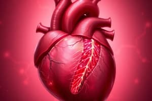
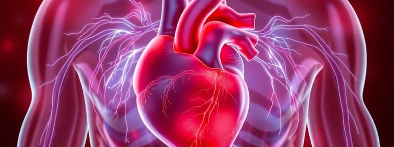
Pediatric Cardiology
- Pediatric cardiology focuses on the heart and circulatory system in children.
Fetal Circulation
- In fetal circulation, pressure is higher on the right side because pulmonary circulation resistance is higher (lungs are collapsed).
- After birth, this reverses, and the left side has higher pressure (lungs are open).
- Key structures involved in fetal circulation include the foramen ovale, ductus arteriosus, and ductus venosus.
Neonates & Infants
- Heart and great vessels develop in the first 3-8 weeks of gestation.
- Neonates and infants have higher-pitched heart sounds of greater intensity than adults.
- They rely on adequate heart rate and rhythm to maintain cardiac output because they cannot increase stroke volume easily.
- Myocardial muscle in neonates and infants is less efficient and has fewer organized myocardial fibers.
- Consequently, they depend greatly on calcium, glucose, and blood volume for adequate cardiac output.
Conduction System
- Surgeries can affect AV nodes and lead to heart blocks or arrhythmias.
- Most surgical procedures target the AV node.
- The Sinoatrial node (pacemaker) initiates the heartbeat.
- The system includes the atrioventricular node, bundle of His, right and left bundle branches, and Purkinje fibers.
Physiology
- Cardiac output is the volume of blood expelled by the heart in one minute.
- Preload is the volume of blood returning to the heart.
- Afterload is the resistance against which the ventricles pump when ejecting blood.
- Contractility is the ability of the cardiac muscle to function as an efficient pump.
Adequate Systemic Profusion
- Appropriate heart rate is essential.
- Adequate blood volume is crucial.
- Low pulmonary vascular resistance is important.
- Capillary permeability and tissue utilization of oxygen (O2) are vital.
Assessment of Cardiac Function
- History: Important aspects to consider include the mother's health (diabetes, lupus, PKU), pregnancy (assisted reproduction), and birth history (Apgar score, birth weight, complications, full-term). Family history of congenital heart disease increases risk.
- Infections (rubella, Coxsackie) and exposure to certain medications during pregnancy can increase the risk of congenital heart defects.
- Birth and family history are important details to note for assessing risk factors.
Health History of Infant
- Feeding patterns, weight gain, and development are evaluated.
- Respiratory infections and breathing problems are considered.
- Color changes, exercise intolerance (in older children), edema, chest pain, palpations, and neurological problems are assessed.
- Recent infections and exposures to toxins are also documented.
- Other health issues and medications are noted.
Physical Exam
- Vital signs, including blood pressure in all four extremities and oxygen saturation, are essential.
- General inspection of skin color, comfort level, and nutritional status are assessed.
- Heart sounds, including point of maximum intensity, apical impulse, and presence of thrill, are evaluated.
Physical Assessment Continued
- The quality and symmetry of pulses, the warmth of extremities, and capillary refill time are checked.
- Edema, and enlarged liver and/or spleen are examined.
- Auscultation of heart and lung sounds (quality, intensity, rhythm, and additional sounds) is performed.
Heart Murmurs
- Murmurs are swishing sounds detected in addition to normal heart sounds.
- Key details: location (where best heard), time (during S1 or S2 cycle), intensity (relation to child's position), and loudness are recorded.
Grading of Murmurs
- Murmur grades range from I (very faint) to VI (loud, audible without stethoscope contact).
Hemodynamics
- Blood flow originates from higher pressure areas, seeking paths of lowest resistance.
- Right-sided pressure is generally less than left-sided pressure.
- Pulmonary artery resistance is typically lower than systemic circulation resistance.
- Pulmonary artery pressure is lower than aortic pressure
Saturations
- Blood returning from the body (SVC and IVC) has lower oxygen saturation.
- Blood returning from the lungs has high oxygen saturation.
- The aorta distributes oxygenated blood to the body's organs.
Hemodynamic Classifications
- Increased pulmonary blood flow
- Decreased pulmonary blood flow
- Obstruction to blood flow out of the heart
- Mixed blood flow
Classification of Congenital Heart Disease
- Acyanotic defects (increased pulmonary blood flow)
- Cyanotic defects (decreased pulmonary blood flow, mixed blood flow, obstruction of blood flow).
- Specific defects: atrial septal defect (ASD), ventricular septal defect (VSD), patent ductus arteriosus (PDA), coarctation of the aorta, aortic stenosis, pulmonic stenosis, tetralogy of Fallot, AV canal defect, tricuspid atresia, transposition of the great arteries, total anomalous pulmonary venous connection, truncus arteriosus, hypoplastic left heart syndrome
Incidence
- Congenital heart defects (CHD) occur in approximately 5 – 8 per 1000 births.
- CHD is a significant cause of death in the first year of life.
Defects with Increased Pulmonary Blood Flow
- Atrial Septal Defect (ASD).
- Ventricular Septal Defect (VSD),
- Patent Ductus Arteriosus (PDA),
- Atrioventricular Canal Defect (AV canal).
Ventricular Septal Defect (VSD)
- Opening between right and left ventricles.
- Defects can be membranous or muscular.
- Often associated with additional defects.
- Many spontaneously close within the first year of life.
AV Canal
- Incomplete closure of endocardial cushions.
- Often accompanies a high VSD.
- Defects in mitral and tricuspid valves.
- Blood flow between all four chambers of the heart.
Patent Ductus Arteriosus (PDA)
- Failure of the ductus arteriosus to close after birth.
- Results in increased pulmonary blood flow.
- Right ventricular hypertrophy can develop.
- Symptoms: signs of heart failure (CHF), murmur.
- Treatment, medical and/or surgical.
Obstructive Defects
- Coarctation of the Aorta.
- Aortic stenosis.
- Pulmonic stenosis.
Aortic Stenosis
- Narrowing or stricture of the aortic valve.
- Results in decreased cardiac output and left ventricular hypertrophy.
- Increased pulmonary vascular congestion.
- Three main types are valvular stenosis, subvalvular stenosis, and supravalvular stenosis.
Pulmonic Stenosis
- Narrowing of entrance to pulmonary artery.
- Results in right ventricular hypertrophy and decreased pulmonary blood flow.
- Symptoms: may be asymptomatic, possible cyanosis or CHF (if severe), murmur in newborns, etc
Defects with Decreased Pulmonary Blood Flow
- Tetralogy of Fallot (TOF).
- Tricuspid atresia.
Tricuspid Atresia
- No communication between the right atrium and right ventricle.
- ASD or patent foramen ovale (PFO) are typically present.
- Complete mixing of oxygenated and deoxygenated blood.
- Patent ductus arteriosus (PDA) facilitates blood flow for oxygenation.
- VSD allows oxygenated blood to pass to the right ventricle.
- Decreased pulmonary blood flow results
Transposition of the Great Arteries (TGV)
- The pulmonary artery and aorta swap positions.
- Systemic circulation defects need to be present to allow for oxygenated blood mixing.
- Characterized by cyanosis and cardiomegaly.
- Treatment is usually an arterial switch operation to re-implant the coronary arteries.
Hypoplastic Left Heart Syndrome (HLHS)
- Underdevelopment of the left side of the heart,
- Aortic atresia.
- Requires a PFO or ASD, and systemic circulation from a PDA.
- Mild cyanosis with CHF.
- Progressive deterioration after PDA closure.
- Treatment includes Norwood procedure, bidirectional Glenn, and modified Fontan operations.
Truncus Arteriosus
- Main artery that fails to bifurcate.
Total Anomalous Pulmonary Venous Connection (TAPVC)
- Pulmonary veins emptying into the wrong circulatory system (usually the right atrium).
Congestive Heart Failure (CHF)
- Volume overload (left-to-right shunts)
- Pressure overload (obstructive defects)
- Poor contractility (cardiomyopathy)
- High cardiac output demands
Types of CHF
- Right-sided: edema, hepatomegaly
- Left-sided: respiratory distress, congestion
Clinical Manifestations
- Symptoms include tachycardia, diaphoresis, easily fatigued, tachypnea, dyspnea, mild cyanosis, retractions, grunting, wheezing, cough, poor profusion, hepatomegaly, edema, weight gain, developmental delays, and variations due to myocardiac contraction, afterload, and preload in the heart to compensate for function.
Management
- Digoxin: Increases cardiac output, reduces heart size, lowers venous pressure, and relieves edema.
- Ace inhibitors (Captopril, Enalapril): May improve heart function and reduce pressure.
- Diuretics (lasix, aldactone): Remove excess fluid and sodium.
- Nutritional support for BMR increase, caloric intake, and well-rested feeding.
Other important concepts
- Central cyanosis
- Clubbing
- Hypercyanotic spells ("tet" spells)
- Knee-chest position
Web Sites
- Websites providing information about pediatric cardiac defects.
Online Resources for Pediatric Cardiac Auscultation
- Links to websites with sounds and information for auscultation.
Studying That Suits You
Use AI to generate personalized quizzes and flashcards to suit your learning preferences.