Podcast
Questions and Answers
Why does the common extensor radialis brevis (ECRB) tendon get irritated during pronation-supination movements in individuals with tennis elbow?
Why does the common extensor radialis brevis (ECRB) tendon get irritated during pronation-supination movements in individuals with tennis elbow?
- It is excessively stretched when the forearm is in a fully pronated position.
- It experiences increased blood flow during these movements.
- It is irritated by the radial head, most commonly during these movements due to its anatomical position. (correct)
- It is directly compressed against the humerus during resisted wrist extension, leading to inflammation.
Which of the following signs would be the LEAST likely to indicate tendonitis?
Which of the following signs would be the LEAST likely to indicate tendonitis?
- Pain relief with palpation. (correct)
- Significant pain during eccentric contraction.
- Pain during resisted range of motion (RROM) of the affected muscle.
- Pain during active range of motion into the antagonist direction.
Why are eccentric resistance exercises particularly effective for strengthening tendons?
Why are eccentric resistance exercises particularly effective for strengthening tendons?
- They reduce the overall load on the tendon, preventing further injury.
- They promote rapid blood flow to the tendon, accelerating healing.
- They primarily target muscle hypertrophy, indirectly benefiting the tendon.
- They allow for controlled lengthening under tension, stimulating collagen synthesis and tendon remodeling. (correct)
A patient presents with elbow pain that worsens with resisted wrist flexion and pronation. Palpation of the medial epicondyle elicits significant tenderness. Which condition is MOST likely?
A patient presents with elbow pain that worsens with resisted wrist flexion and pronation. Palpation of the medial epicondyle elicits significant tenderness. Which condition is MOST likely?
Which of the following activities is MOST likely to aggravate olecranon bursitis?
Which of the following activities is MOST likely to aggravate olecranon bursitis?
In addition to stretching, what other intervention is MOST beneficial for a hypertonic muscle with trigger points to reduce the load on the associated tendon?
In addition to stretching, what other intervention is MOST beneficial for a hypertonic muscle with trigger points to reduce the load on the associated tendon?
How would you differentiate olecranon bursitis from triceps tendonitis based on your assessment?
How would you differentiate olecranon bursitis from triceps tendonitis based on your assessment?
What activity would be MOST likely to cause 'posterior tennis elbow'?
What activity would be MOST likely to cause 'posterior tennis elbow'?
During full elbow extension, which anatomical feature is normally NOT in contact with the trochlea?
During full elbow extension, which anatomical feature is normally NOT in contact with the trochlea?
Which nerve is MOST likely compressed within the carpal tunnel?
Which nerve is MOST likely compressed within the carpal tunnel?
What primary function does the interosseous membrane serve in relation to the radius and ulna?
What primary function does the interosseous membrane serve in relation to the radius and ulna?
In the elbow joint, considering it functions as a 3rd order lever system, what is a direct consequence of this lever arrangement?
In the elbow joint, considering it functions as a 3rd order lever system, what is a direct consequence of this lever arrangement?
If a patient presents with elbow joint effusion, where would edema be MOST evident upon palpation?
If a patient presents with elbow joint effusion, where would edema be MOST evident upon palpation?
Which statement BEST describes the relationship between tendonitis and tendinopathy for the purposes of treatment?
Which statement BEST describes the relationship between tendonitis and tendinopathy for the purposes of treatment?
An athlete reports pain during resisted wrist extension with the elbow in full extension. Which condition is MOST likely indicated?
An athlete reports pain during resisted wrist extension with the elbow in full extension. Which condition is MOST likely indicated?
If a patient is experiencing pain during pulling motions, which anatomical structure is MOST likely involved?
If a patient is experiencing pain during pulling motions, which anatomical structure is MOST likely involved?
A physical therapist is treating a patient with a tibial fracture that occurred 4 weeks ago. Radiographic imaging reveals a visible fracture line and a calcified callus formation. The patient reports tenderness upon palpation at the fracture site. Based on this information, which of the following interventions should be avoided?
A physical therapist is treating a patient with a tibial fracture that occurred 4 weeks ago. Radiographic imaging reveals a visible fracture line and a calcified callus formation. The patient reports tenderness upon palpation at the fracture site. Based on this information, which of the following interventions should be avoided?
A patient presents with a suspected radial head subluxation (Nursemaid’s elbow). Which of the following historical details would MOST strongly support this diagnosis?
A patient presents with a suspected radial head subluxation (Nursemaid’s elbow). Which of the following historical details would MOST strongly support this diagnosis?
A patient with a mid-shaft humeral fracture has undergone open reduction internal fixation (ORIF). Which of the following interventions should be avoided?
A patient with a mid-shaft humeral fracture has undergone open reduction internal fixation (ORIF). Which of the following interventions should be avoided?
Following cast removal after a distal radius fracture, a patient presents with significant hypotonia in the forearm muscles. Which of the following treatment techniques should be approached with the MOST caution?
Following cast removal after a distal radius fracture, a patient presents with significant hypotonia in the forearm muscles. Which of the following treatment techniques should be approached with the MOST caution?
A patient is referred to physical therapy with a suspected stress fracture of the tibia. Which of the following assessment techniques is MOST contraindicated during the initial evaluation?
A patient is referred to physical therapy with a suspected stress fracture of the tibia. Which of the following assessment techniques is MOST contraindicated during the initial evaluation?
A patient is being treated for a Colles' fracture. Which of the following signs indicates that the fracture has reached the 'consolidation' phase?
A patient is being treated for a Colles' fracture. Which of the following signs indicates that the fracture has reached the 'consolidation' phase?
A patient who sustained a fracture has developed significant edema in the affected extremity. Which assessment technique would be MOST appropriate for quantifying the extent of the edema?
A patient who sustained a fracture has developed significant edema in the affected extremity. Which assessment technique would be MOST appropriate for quantifying the extent of the edema?
A patient is diagnosed with a radial nerve compression at the elbow. Which anatomical structure is MOST likely involved in causing this compression?
A patient is diagnosed with a radial nerve compression at the elbow. Which anatomical structure is MOST likely involved in causing this compression?
A patient presents with elbow pain. Which combination of test and positive indication most strongly suggests lateral epicondylitis?
A patient presents with elbow pain. Which combination of test and positive indication most strongly suggests lateral epicondylitis?
Following immobilization of the elbow, which of these would be the MOST appropriate initial structure to palpate and how?
Following immobilization of the elbow, which of these would be the MOST appropriate initial structure to palpate and how?
In the early subacute phase of tendon rehabilitation, what criterion indicates that the program is progressing too quickly?
In the early subacute phase of tendon rehabilitation, what criterion indicates that the program is progressing too quickly?
A patient is in the chronic stage of lateral epicondylitis rehab. Which sign, if reported by the patient, warrants immediate modification (reduction) of their exercises?
A patient is in the chronic stage of lateral epicondylitis rehab. Which sign, if reported by the patient, warrants immediate modification (reduction) of their exercises?
When designing an exercise program focused on increasing range of motion for a patient with elbow stiffness, which principle should be prioritized?
When designing an exercise program focused on increasing range of motion for a patient with elbow stiffness, which principle should be prioritized?
Which of these exercise progressions aligns with the described principles for tendon rehabilitation?
Which of these exercise progressions aligns with the described principles for tendon rehabilitation?
A patient presents with suspected medial collateral ligament (MCL) injury. Which test is most appropriate to assess the integrity of this ligament and what positive indication is expected?
A patient presents with suspected medial collateral ligament (MCL) injury. Which test is most appropriate to assess the integrity of this ligament and what positive indication is expected?
Following an initial assessment, the patient is diagnosed with pronator teres syndrome due to median nerve entrapment. Which test would MOST likely reproduce the patient's symptoms?
Following an initial assessment, the patient is diagnosed with pronator teres syndrome due to median nerve entrapment. Which test would MOST likely reproduce the patient's symptoms?
Flashcards
Tendon Overload Cause
Tendon Overload Cause
Tendons become overloaded when the attached muscle is hypertonic or full of trigger points due to increased load.
Best way to strengthen a tendon
Best way to strengthen a tendon
Eccentric resistance exercises.
Assessing Tendonitis
Assessing Tendonitis
Pain during lengthening (AROM/PROM), resisted contraction(RROM/MMT), and palpation.
Common Extensor Tendonitis Cause
Common Extensor Tendonitis Cause
Signup and view all the flashcards
Most Common CET Tendon
Most Common CET Tendon
Signup and view all the flashcards
Common Flexor Tendonitis Cause
Common Flexor Tendonitis Cause
Signup and view all the flashcards
Most Common Affected CFT
Most Common Affected CFT
Signup and view all the flashcards
Olecranon Bursitis Cause
Olecranon Bursitis Cause
Signup and view all the flashcards
What is an end feel?
What is an end feel?
Signup and view all the flashcards
Which nerves are commonly compressed around the elbow?
Which nerves are commonly compressed around the elbow?
Signup and view all the flashcards
Why does injury to one elbow joint affect the entire elbow?
Why does injury to one elbow joint affect the entire elbow?
Signup and view all the flashcards
What is the normal carrying angle of the elbow?
What is the normal carrying angle of the elbow?
Signup and view all the flashcards
When is the interosseous membrane taut, and what does it prevent?
When is the interosseous membrane taut, and what does it prevent?
Signup and view all the flashcards
What does the oblique cord do?
What does the oblique cord do?
Signup and view all the flashcards
What is the impact of the elbow being a 3rd order lever?
What is the impact of the elbow being a 3rd order lever?
Signup and view all the flashcards
What is tendinopathy/tendonitis, and how is it treated?
What is tendinopathy/tendonitis, and how is it treated?
Signup and view all the flashcards
Ligamentous Valgus Instability Test
Ligamentous Valgus Instability Test
Signup and view all the flashcards
Ligamentous Varus Instability Test
Ligamentous Varus Instability Test
Signup and view all the flashcards
Cozen’s Test
Cozen’s Test
Signup and view all the flashcards
Mill’s Test
Mill’s Test
Signup and view all the flashcards
Maudsley’s Test
Maudsley’s Test
Signup and view all the flashcards
Pronator Teres Syndrome Test
Pronator Teres Syndrome Test
Signup and view all the flashcards
Mid-Range Loading
Mid-Range Loading
Signup and view all the flashcards
Exercise Challenge Indicator
Exercise Challenge Indicator
Signup and view all the flashcards
ROM Assessment After Fracture
ROM Assessment After Fracture
Signup and view all the flashcards
Perfusion Test After Fracture
Perfusion Test After Fracture
Signup and view all the flashcards
Girth Measurements After Fracture
Girth Measurements After Fracture
Signup and view all the flashcards
Muscle Tone Testing After Fracture
Muscle Tone Testing After Fracture
Signup and view all the flashcards
Post-Fracture Traction Contraindication
Post-Fracture Traction Contraindication
Signup and view all the flashcards
Heat Therapy Near a Cast: Contraindicated
Heat Therapy Near a Cast: Contraindicated
Signup and view all the flashcards
Nursemaid's Elbow
Nursemaid's Elbow
Signup and view all the flashcards
Nerve Compression Locations
Nerve Compression Locations
Signup and view all the flashcards
Study Notes
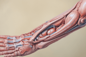
- The elbow complex consists of three synovial joints: humeroulnar, humeroradial, and proximal radioulnar.
Joint Types
- Humeroulnar joint: synovial hinge joint
- Humeroradial joint: synovial, ellipsoidal/gliding
- Proximal Radioulnar joint: synovial pivot joint
Articulating Surfaces
- Humeroulnar: Trochlea of humerus with trochlear notch of the ulna
- Humeroradial: Capitulum of the humerus with the head of the radius
- Proximal Radioulnar: Head of the radius with the radial notch of the ulna
Capsular Strength/Coaptation
- Humeroulnar joint has weak-moderate coaptation
- Humeroradial joint has weak coaptation.
- Proximal Radioulnar joint has weak coaptation.
Ligaments
- Ulnar Collateral Ligament with a triangular shape comprising of three bands.
- Anterior band stems from the medial epicondyle to the medial edge of coronoid process
- Posterior band connects the medial epicondyle and olecranon process
- Transverse band stabilizes the distal attachments of ant. & post. bands
- Ulnar Collateral Ligament resists VALGUS stress
- Radial Collateral Ligament has a V-shape and is located on the underside of the lateral epicondyle, connecting to the annular ligament and radial notch
- Radial Collateral Ligament resists VARUS stress
- Annular Ligament: primary stabilizer of the proximal radioulnar joint.
- Anchors the head of the radius to the radial notch
- Prevents inferior distraction of the radius
- Damage can occur in children from jerking movements, resulting in a 'pulled-elbow'
- Quadrate Ligament: an inferior thickening of the joint capsule.
- Posterior fibers become taut with pronation
- Anterior fibers become taut with supination
Extras
- Interosseous Membrane: dissipates force and stabilizes both radioulnar joints
- Prevents proximal radius displacement on ulna during pushing movements
- Oblique Cord: originates at the radial notch of the ulna and inserts on the medial surface of the radius neck
- The Oblique Cord Prevents radius displacement on ulna, especially during pulling movements
- Ligament of Struthers: Found in 1% of the population.
Osteokinematics
- Humeroulnar joint: 1 degree of freedom for flexion and extension
- Humeroradial joint: 2 degrees of freedom, flexion-extension, supination-pronation
- Proximal Radioulnar: 1 degree of freedom for supination and pronation
Arthrokinematics
- Humeroulnar: the trochlea of the humerus is convex in the anterior/posterior direction and concave in the medial/lateral direction
- Humeroradial: the capitulum of the humerus is convex and the radial head is concave
- Proximal Radioulnar: the head of the radius is convex and the radial notch of ulna is concave
Resting Position
- Humeroulnar: 70° flexion with 10° supination
- Humeroradial: full extension and supination
- Proximal Radioulnar: 70° flexion with 35° supination
Closed Pack Position
- Humeroulnar: Full extension and supination
- Humeroradial: 90° flexion and 5° supination
- Proximal Radioulnar: 5° supination and full extension
Capsular Pattern
- Humeroulnar: Flexion is more limited than extension
- Humeroradial: Supination and pronation are limited only if severe
- Proximal Radioulnar: supination = pronation
ROM and End Feel
- Flexion (HU and HR): 0-150°, soft
- Extension (HU and HR): 0-5°, hard
- Supination (HR and PRU): 0-90°, firm
- Pronation (HR and PRU): 0-70/90°, firm/hard
Other key points
- The three true joints of the elbow share one joint capsule
- Injury to any one joint will affect the whole elbow complex
- Joint effusion/edema is evident in the triangular space between the radial head, tip of the olecranon, and lateral epicondyle.
Biomechanics
- The elbow has a normal slight valgus carrying angle
- On full EXTENSION, the MEDIAL part of the olecranon is not in contact with the trochlea (medial gap)
- On full FLEXION the LATERAL part of the olecranon is not in contact with the trochlea (lateral gap)
Mid Radioulnar Joint
- Radius connection to ulna via the interosseous membrane
- The interosseous membrane is taut midway between supination and pronation (neutral position)
- The interosseous membrane prevents proximal displacement of the radius on the ulna, which most likely occurs with pushing movements
- The interosseous membrane Helps to dissipate forces from the hand so they are less at the elbow
- The oblique cord runs at right angles to the interosseous membrane and assists in preventing distal displacement of the radius on the ulna.
3rd Order Lever
- The elbow functions as a 3rd order lever with the fulcrum at the trochlea, effort from muscle attachments, and load as the weight in the hand
- Load in the hand is 10x more at the elbow joint
- The joint has poor mechanical advantage
- The joint is a good speed multiplier
Tendonitis (Tendinopathy)
- Tendonitis is inflammation/degeneration of a tendon due to overuse.
- Treatment involves offloading the tendon to allow recovery and then strengthening it
- Offloading can be achieved by reducing provocative movements and massage to the muscle belly.
- Eccentric resistance exercises are optimal for strengthening tendons.
Assessing for tendonitis
- Pain on AROM or PROM in the antagonist direction
- Pain with resisted contraction (RROM or MMT) of the affected muscle, specifically with an eccentric component.
- Pain with palpation
Most Commonly Affected Tendons
- Common Extensor Tendon:
- Most common; 'Tennis elbow'
- Provoked by repetitive, forceful extension, supination, radial deviation.
- ECR Brevis is most commonly affected due to irritation from the radial head during pronation-supination movements
- Can be most tender at the supracondylar ridge, epicondyle, or just distal over the bulk of the tendon
- Common Flexor Tendon:
- 'Golfer's elbow'
- Provoked by repetitive, forceful pronation and flexion
- Pronator teres most common, followed by FC radialis
- Provoked by forceful gripping.
- Triceps Brachii Tendon:
- 'Posterior tennis elbow'
Bursitis (Olecranon)
- Usually due to trauma to the elbow or overuse of surrounding structures.
- Any activity that involves a lot of time on the elbows
- Bursitis is aggravated by compression
- PROM empty end-feel
Differentiation from Tendonitis
- Pain with compression that does not get worse with load, especially eccentric load
Fractures
- Assessment includes ROM (Active, Passive, and Resisted)
- Palpation is contraindicated before consolidation.
- Further testing may include assessment of perfusion, swelling, muscle tone, and tissue health
- Precautions during immobilization include avoiding tractioning before union and avoiding heat or ice if hardware has been used
Nursemaid's Elbow
- Radial head subluxation commonly caused by pulling on a child's arm
- Caused by the conical shape of the radial head before 8 years old
- Annular ligament allows for the subluxation
- Commonly reduced by a doctor or chiropractor
Key Points for Orthopedic Tests
- Cozen's test (method 1) is for lateral epicondylitis
- A positive test consists of sudden severe pain at lat epicondyle, supracondylar ridge, or CET
- Indication: Complaints of pain in the area especially if MOI is repetitive muscle-use of forearm
Home Care Principles
- Therapy programs should:
- Not cause pain/inflammation
- Be progressive and gradual
- Start as early as possible (no inflammation presence)
- Exercise Selection Considerations:
- Loaded exercises performed in mid-range
- Exercises should increase range unweighted
- Passive before active exercises are prescribed
- Isometric exercises before eccentric exercises which should be done before plyometric exercises
- Open chain exercises before closed chain exercises
Signs of an Overly Challenging Program
- Discomfort that lasts more than 2 hours in the acute/subacute stage.
- Discomfort lasts more than 4 hours in the chronic stage.
- Reliance on pain medication to control discomfort.
- Increased pain at rest, extreme fatigue, rebound muscle spasm, or increased weakness
Studying That Suits You
Use AI to generate personalized quizzes and flashcards to suit your learning preferences.