Podcast
Questions and Answers
Which artery is primarily responsible for supplying blood to the medial aspect of the cerebral hemispheres?
Which artery is primarily responsible for supplying blood to the medial aspect of the cerebral hemispheres?
- Middle cerebral artery (MCA)
- Internal carotid artery
- Posterior cerebral artery (PCA)
- Anterior cerebral artery (ACA) (correct)
Occlusion of the middle cerebral artery (MCA) would most significantly affect which region of the cerebral cortex?
Occlusion of the middle cerebral artery (MCA) would most significantly affect which region of the cerebral cortex?
- Lateral parietal and temporal lobes (correct)
- Brainstem
- Medial frontal lobe
- Occipital lobe
The posterior cerebral artery (PCA) primarily supplies which lobe of the brain?
The posterior cerebral artery (PCA) primarily supplies which lobe of the brain?
- Frontal lobe
- Basal ganglia
- Temporal and Occipital lobes (correct)
- Parietal lobe
Which of the following arteries is NOT part of the Circle of Willis?
Which of the following arteries is NOT part of the Circle of Willis?
The Circle of Willis provides a crucial function in the cerebral vasculature. What is the primary benefit of this anatomical arrangement?
The Circle of Willis provides a crucial function in the cerebral vasculature. What is the primary benefit of this anatomical arrangement?
Which arteries directly contribute to the vertebrobasilar system?
Which arteries directly contribute to the vertebrobasilar system?
A patient presents with symptoms indicative of a stroke affecting the occipital lobe. Which artery is most likely involved?
A patient presents with symptoms indicative of a stroke affecting the occipital lobe. Which artery is most likely involved?
Lenticulostriate arteries, branches of the middle cerebral artery (MCA), are particularly vulnerable in certain types of stroke. Damage to these arteries primarily affects which structures?
Lenticulostriate arteries, branches of the middle cerebral artery (MCA), are particularly vulnerable in certain types of stroke. Damage to these arteries primarily affects which structures?
Aneurysms are a type of cerebrovascular accident (CVA) characterized by:
Aneurysms are a type of cerebrovascular accident (CVA) characterized by:
Atherosclerosis, a major risk factor for stroke, is best described as:
Atherosclerosis, a major risk factor for stroke, is best described as:
A carotid dissection, a cause of stroke, involves:
A carotid dissection, a cause of stroke, involves:
Arteriovenous malformations (AVMs) in the brain are characterized by:
Arteriovenous malformations (AVMs) in the brain are characterized by:
Infarction and ischemia in the context of cerebrovascular disease refer to:
Infarction and ischemia in the context of cerebrovascular disease refer to:
Occlusive stroke is primarily caused by:
Occlusive stroke is primarily caused by:
Hemorrhagic stroke is mainly caused by:
Hemorrhagic stroke is mainly caused by:
Which venous sinus runs along the superior midline of the falx cerebri?
Which venous sinus runs along the superior midline of the falx cerebri?
The straight sinus is formed by the confluence of which two sinuses?
The straight sinus is formed by the confluence of which two sinuses?
The confluence of sinuses, or torcula, primarily involves which sinuses?
The confluence of sinuses, or torcula, primarily involves which sinuses?
Blood from the transverse sinuses ultimately drains into which sinuses?
Blood from the transverse sinuses ultimately drains into which sinuses?
The cavernous sinus is unique among dural venous sinuses because it:
The cavernous sinus is unique among dural venous sinuses because it:
The great cerebral vein of Galen drains primarily into which venous sinus?
The great cerebral vein of Galen drains primarily into which venous sinus?
The superficial middle cerebral vein, also known as the vein of Sylvius, drains into which sinus?
The superficial middle cerebral vein, also known as the vein of Sylvius, drains into which sinus?
The primary role of pericytes in the blood-brain barrier (BBB) is to:
The primary role of pericytes in the blood-brain barrier (BBB) is to:
Astrocytes contribute to the blood-brain barrier (BBB) by:
Astrocytes contribute to the blood-brain barrier (BBB) by:
The metabolic barrier component of the blood-brain barrier (BBB) is primarily attributed to the enzyme:
The metabolic barrier component of the blood-brain barrier (BBB) is primarily attributed to the enzyme:
Which of the following substances can readily cross the blood-brain barrier (BBB) via lipid-soluble diffusion?
Which of the following substances can readily cross the blood-brain barrier (BBB) via lipid-soluble diffusion?
Receptor-mediated transport across the blood-brain barrier (BBB) is essential for the passage of:
Receptor-mediated transport across the blood-brain barrier (BBB) is essential for the passage of:
Ion channels in the blood-brain barrier (BBB) primarily regulate the passage of:
Ion channels in the blood-brain barrier (BBB) primarily regulate the passage of:
Which of the following is NOT a primary function of cerebrospinal fluid (CSF)?
Which of the following is NOT a primary function of cerebrospinal fluid (CSF)?
The blood-CSF barrier is located at the:
The blood-CSF barrier is located at the:
In bacterial meningitis, an increase in CSF protein is often observed. This is primarily due to:
In bacterial meningitis, an increase in CSF protein is often observed. This is primarily due to:
The appearance of red blood cells (RBCs) in cerebrospinal fluid (CSF) is most indicative of:
The appearance of red blood cells (RBCs) in cerebrospinal fluid (CSF) is most indicative of:
L-DOPA is used in Parkinson's disease treatment because it can:
L-DOPA is used in Parkinson's disease treatment because it can:
Carbidopa is often administered with L-DOPA in Parkinson's treatment to:
Carbidopa is often administered with L-DOPA in Parkinson's treatment to:
The anterior spinal artery supplies the:
The anterior spinal artery supplies the:
Posterior spinal arteries typically supply the:
Posterior spinal arteries typically supply the:
Watershed infarcts are typically caused by:
Watershed infarcts are typically caused by:
A patient with a stroke affecting the anterior cerebral artery (ACA) territory is most likely to present with:
A patient with a stroke affecting the anterior cerebral artery (ACA) territory is most likely to present with:
A patient exhibiting aphasia and hemiparesis predominantly in the arm and face likely has a stroke in the territory of which artery?
A patient exhibiting aphasia and hemiparesis predominantly in the arm and face likely has a stroke in the territory of which artery?
Visual field deficits, particularly homonymous hemianopia, are characteristic of stroke involving which arterial territory?
Visual field deficits, particularly homonymous hemianopia, are characteristic of stroke involving which arterial territory?
In the context of cerebral venous drainage, 'empty delta sign' on imaging is associated with:
In the context of cerebral venous drainage, 'empty delta sign' on imaging is associated with:
Flashcards
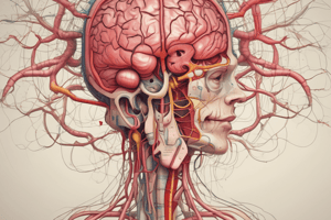
Arterial Vasculature
Arterial Vasculature
The arterial blood supply to the brain.
Examples of CVA
Examples of CVA
Conditions affecting blood supply to the brain, such as aneurysm, atherosclerosis, dissection, arteriovenous malformation and ischemia.
Aneurysm
Aneurysm
A balloon-like bulge in an artery wall.
Atherosclerosis
Atherosclerosis
Signup and view all the flashcards
Dissection
Dissection
Signup and view all the flashcards
Arterio-Venous Malformation
Arterio-Venous Malformation
Signup and view all the flashcards
Ischemia
Ischemia
Signup and view all the flashcards
Venous Vasculature
Venous Vasculature
Signup and view all the flashcards
Blood Brain Barrier
Blood Brain Barrier
Signup and view all the flashcards
Vertebral and Basilar Arteries
Vertebral and Basilar Arteries
Signup and view all the flashcards
Internal Carotid Arteries
Internal Carotid Arteries
Signup and view all the flashcards
Circle of Willis
Circle of Willis
Signup and view all the flashcards
Anastomosis of Vessels
Anastomosis of Vessels
Signup and view all the flashcards
Anterior Cerebral Artery (ACA)
Anterior Cerebral Artery (ACA)
Signup and view all the flashcards
Middle Cerebral Artery (MCA)
Middle Cerebral Artery (MCA)
Signup and view all the flashcards
Posterior Cerebral Artery (PCA)
Posterior Cerebral Artery (PCA)
Signup and view all the flashcards
Posterior Communicating Artery
Posterior Communicating Artery
Signup and view all the flashcards
Ophthalamic Artery
Ophthalamic Artery
Signup and view all the flashcards
Pontine Arteries
Pontine Arteries
Signup and view all the flashcards
Internal Auditory Arteries
Internal Auditory Arteries
Signup and view all the flashcards
Anterior Choroidal Arteries
Anterior Choroidal Arteries
Signup and view all the flashcards
Lenticulostriate Arteries
Lenticulostriate Arteries
Signup and view all the flashcards
Recurrent Artery of Heubner
Recurrent Artery of Heubner
Signup and view all the flashcards
Thalamoperforator and Posterior Choroidal
Thalamoperforator and Posterior Choroidal
Signup and view all the flashcards
Thalamo-geniculate Artery
Thalamo-geniculate Artery
Signup and view all the flashcards
Quadrigeminal Artery
Quadrigeminal Artery
Signup and view all the flashcards
Posterior Spinal Artery
Posterior Spinal Artery
Signup and view all the flashcards
Ischemia
Ischemia
Signup and view all the flashcards
Hypoxia
Hypoxia
Signup and view all the flashcards
Infarction
Infarction
Signup and view all the flashcards
Occlusive Stroke
Occlusive Stroke
Signup and view all the flashcards
Hemorrhagic Stroke
Hemorrhagic Stroke
Signup and view all the flashcards
Global Cerebral Ischemia
Global Cerebral Ischemia
Signup and view all the flashcards
Focal Cerebral Ischemia
Focal Cerebral Ischemia
Signup and view all the flashcards
Lacunar Infarction
Lacunar Infarction
Signup and view all the flashcards
Penetrating Arteries
Penetrating Arteries
Signup and view all the flashcards
Descending Motor
Descending Motor
Signup and view all the flashcards
Ascending Sensory
Ascending Sensory
Signup and view all the flashcards
Etat Lacunaire
Etat Lacunaire
Signup and view all the flashcards
Watershed Infarction
Watershed Infarction
Signup and view all the flashcards
Study Notes
- The following are study notes on the Arterial and Venous Vasculature of the brain, Cerebrovascular Accidents (CVA), Blood Brain Barrier and Cerebral Spinal Fluid (CSF)
Arterial Vasculature
- The cerebral arterial system is divided into the vertebral and basilar arteries and the internal carotid arteries.
- The cerebellar, cerebral, spinal and pontine are all arteries included in the vertebral and basilar arteries of the brain.
- Cerebral arteries are included in the internal carotid arteries.
- Circle of Willis provides an anastomosis, that provides an alternate route for blood, in case one route is blocked.
- Vertebral and Basilar arteries include the posterior cerebral artery (PCA), superior cerebellar artery (SCA), anterior inferior cerebellar artery (AICA), posterior inferior cerebellar artery (PICA), and anterior and posterior spinal arteries
Circle of Willis
- Internal Carotid artery includes the posterior communicating artery, middle cerebral artery (MCA), anterior communicating artery, anterior cerebral artery (ACA) and ophthalmic artery
- Pontine and internal auditory arteries are also components of the the Circle
Internal Carotid System
- The internal carotid system consists of the internal carotid, ophthalmic, posterior communicating, anterior communicating, anterior cerebral (A1, A2), middle cerebral (M1 proximal branch, M2 segments), lenticulostriate, and anterior choroidal arteries.
Vertebro-Basilar System
- The vertebro-basilar system includes the vertebral, basilar, spinal, posterior inferior cerebellar, anterior inferior cerebellar, superior cerebellar, and posterior cerebral (P1, P2) arteries.
Latex Filled Vasculature
- Anterior communicating artery in the brain connects the Anterior cerebral artery (ACA) to the brain
- A1 segment ACA, and the M1 segment MCA are large cerebral arteries
- Superior Cerebellar Artery, AICA and PICA make up the the blood supply of the posterior brain structures
Examples of CVA
- CVA can be caused by aneurysms, atherosclerosis, dissection, arterio-venous malformations, infarction and ischemia
Atherosclerosis
- Atherosclerosis at bifurcations of major vessels in the Circle of Willis
- Atherosclerosis, inside the circle, enables anastomosis for collateral supply
- Basilar artery has no collateral perfusion
Risk Factors of CVA
- Risk Factors that could cause Atherosclerosis are occlusive stroke, and eventually infarction, hemorrhage
- Aneurysm, dissection, hemorrhage, infarction cause tissue death
Internal Vascular Supply
- Basal ganglia, hippocampus and posterior limb of internal capsule supplied by anterior choroidal artery.
- Basal ganglia and the posterior limb of internal capsule are supplied by lenticulostriate arteries, coming off of the M1 proximal branch of MCA
- Basal ganglia, limbic structures, and anterior limb of internal capsule supplied by the recurrent artery of Heubner, a penetrating branch coming off the proximal ACA
- The thalamus and posterior limb of internal capsule is suplied by the thalamoperforator, thalamogeniculate and posterior choroidal
Stroke Symptoms
- Occlusion of lenticulo-striate branches off left M1 segment into internal capsule
Midline Cortical Supply
- The midline cortical supply is the area perfused by the ACA and PCA
- Calloso-marginal and peri-callosal artery, medial temporal, occipital are supplied by the ACA, and PCA
External Cortical Supply
- Antero-medial frontal lobe gets supply from ACA
- Lateral frontal, parietal, temporal lobe gets supply from MCA (M3 superior, M4 inferior)
- Parietal, inferior temporal, occipital gets supply from PCA
Global cerebral ischemia
- Global cerebral ischemia is a diffuse state of hypoxia or ischemia throughout the brain and can be caused by cardiac arrest, shock, and hypo-perfusion
Focal Cerebral Ischemia
- This ischemia is local and specifically caused occlusion from embolism, thrombus or vasculitis.
Intra-Cerebral Hemorrhage
- This hemorrhage occurs in lacunar infarctions and Charcot-Bouchard micro-aneurysms
- Penetrating arteries to the thalamus, basal ganglia and internal capsule are end arteries, meaning there is no anastomosis and no collateral perfusion
Small Lacunes
- Lacunes in the posterior limb of the internal capsule cause pure hemi-paresis with descending motor issues and no sensory loss
- Lacunes in the ventral posterior nucleus of the thalamus cause pure hemi-sensory loss (ALS or DCML) with ascending sensory issues
- Etat lacunaire causes progressive dementia, cranial nerve palsies, dysarthria, dysphagia, (+) gag reflex, emotional imbalance
Venous System Drainage
- Superficial, Deep Veins, Internal Cerebral Veins, Basal Veins of Rosenthal drain into Internal Jugular Vein
Superficial Veins
- Superficial veins drain into the superior sagittal and cavernous sinuses.
Deep Veins
- Deep veins drain into the Great Vein of Galen
Blood Brain Barrier (BBB)
- Astrocytes induce endothelial tight junctions / BBB.
- Metabolic barrier - MAO.
- Pericytes maintain the BBB
Exchange from Blood or Brain/CSF: -
- Lipid soluble diffusion
- Receptor mediated transport
- Ion channel diffusion
Cerebral Spinal Fluid (CSF)
- CSF are functions as a shock absorber
- Are involved in removal of metabolites
- Work as pH buffer
Blood CSF Barrier
- Capillary filtration
- Active epithelial transport
- Low in protein, low in ionic. Increased CSF protein: meningitis
- Appearance of RBCs occur due to Infarction, Infection
Getting Dopamine into Brain
- Oral L-DOPA crosses the blood brain barrier
- Combine with carbidopa to decrease L-DOPA metabolism.
- L-DOPA converted to Dopamine
Studying That Suits You
Use AI to generate personalized quizzes and flashcards to suit your learning preferences.